Abstrak
Penelitian ini bertujuan untuk nwnastikan hubungan anatomik sarafi antata organutn subfornicale dengan nucleus supraopticus dan nucleus paraventricularis hypothalani, dengan nenggwtakan metode pelacakan neuroanatonti yang nrenggunakan 4',6-diamidino-2- phenylittdol dihydrochloride (DAPI) dan salah satu metode inunoperoksidase yang n,enggunaknn kit "l-abeled StreptAvidin- Biotin" (LSAB) dan antibodi kelinci terhadap neurofsin manusia sebagai antibodi priner. Pada penelitian ini digunakan 7 ekor tikus putih dewasa galur Wistar untuk pelacakan neuroanato,ni dan 15 eknr tikus putih dewasa galur Lenbaga Maknnan Rakyat, Jakarta (LMR) untuk pengkajian inunoperoksidase. Hasil penelitian menunjukkan bahwa secara anatonûk organum subfornicale dihubungknn dengan nucleus supraopticus dan nucleus parav entricularis hypothalani.
Abstract
The obiective of this investigation is to ascertain the anatomical connections between the subfornical organ and the supraoptic and paraventricular nuclei of the hypothalanus by utilizing a neuroanatomical-tract tracing nrcthod using a single tracer 4', 6-diatttidino-2-phenylindol dihydrochloride (DAPI) and one of the inmrunoperoxidase nrcthods, using l-abeled StreptAvidin- Biorin (LSAB) kit and Rabbit anti Hunnn neurophysin as a prinnry antibody. The neuroanatonical-tract tracing study used 7 adult albino rat of the Letnbaga Makanan Ralcyat, Jakarta (LMR) strain. The results revealed that the subfornical otgan was anatonûcally connected v,ith the supraoptic and paraventricular nuclei of the hypothalanus.
Keywords : Subfornical organ, supraoptic nucleus, paraventricular nucleus, neural connections, neuroanatonical, tract tracirlg nrcthod, itnnunoperoxidase method.
136 Daryanto
Tracing
the Neural Connections
Supraoptic and Paraventricular
lJsing
Neuroanatomical-tract tracing
DaryantoINTRODUCTION
[image:1.595.67.287.439.501.2]The subfornical organ is one of the circumventricular organs (Hofer, 1958, cit. Weindl, I973)r located in the roof of the third cerebral ventricle and attached to the ventral surface of the hippocampal commissure2'3 1""" Figure 1 and 2).
The subfornical organ has been a curiosity mostly
to
embryologists and comparative anatomists. The subfornical organ-has been identifiedin
at least 131 vertebrate species.r Its topography is consistent among animals. It is a midline tubercle at the dorsal aspectof
the third ventricle nearthe
interventricular foramina of Monro.It
is only about 1 mm2 in total tissue area.and appears
to
be uniformin
size among ,p""i"r3'4 (Figure 2).Med J Univ Indon
between the Subfornical Organ and
the
Nuclei
and
Immunoperoxidase Methods
in Albino Rats
The subfornical organ is embryologically derived from the telencephalic ependyma at the point where the tela choroidea and lamina terminalis
-"rg".2
The subfornical organ has attracted considerable attention because of its unusual characteristics and its location within the cerebral ventricle space. It is a small structure possessing distinct morphological features that set apart from other neuronal nuclear systems,3 al cells and a dense plexus
of
(Figure 2).
is highly vascular,3 and lacks
Miselis and colleagues has identified a substantial
system
of
efferent_f-ibers emanating from the SFO.6 Recent anatomical?'8 and physiological studieseVol 3, No 3, July - Septenber
1994
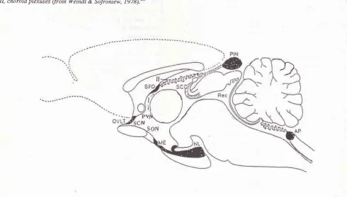
Connections between Subfornical Organ and Supraoptic and Paraventricular Nuclei r37Figure 1. Median sagittal view of the rat brain, showing the location of the subfornical organ a,,n,'tg 'the circunventricular or-gans, AP, area postretna; ME, nedian enûnence; NL, neural lobe; OVLT, organu,n vasculosuttr of the lamina terninalis; PIN, pineal organ; Rec. recessus collicularis ofthe aqueduct; SFO,_subfornical organ; SCO, subcontnissural organ; I, cerebral third ventricle; II, choroid plexuses (from Weindl & Sofroniew, 197q.2Ô
Figure 2. Median sagittal view of the rat brain, showing the structure of the subfornical organ, stained by henrutoxylin-eosin (HE) staining technique. I, dorsal coluntn; II, the bod1, ofthe SFo; III, ventral column; IV, cerebral third ventricle; V. choroid plexuses oT
the cerebral third ventricle. 'f
[image:2.595.72.563.403.682.2]138 Daryanto
cated that the SFO is in fact a nucleus with neurons that appears to innervate directly other neurons in the para-ventricular and supraoptic nuclei that release vasopres-sin from the posterior pituitary, as well as neurons in the meCian preoptic
nucleus
that
projectto
those nuclei.EThe SFO is known to participate in drinking in-duced by angiotensin-Illo'lf and the control of vaso-pressin release from the lnsterior pituitary.12
The neurons
in
the subfornical organ haveef-ferent
projections to both the paraventricular
and supraoptic nuclgi of the hypothalamus (electrophysio-logical studies)7 suggesting the importanceof these
pathwaysfor
controlling
body
fluid
balance and central cardiovascular regulation.ence of afferents and efferents to and from the subfor-nical organ to both the supraoptic (SON) and para-ventricular (PVN) nuclei of the hypothalamus, a region known to contain
the cell
bodiesof oxytocin
and vasopressin-secreting n.uronrl4'16 and relulate the circulating levels of the hormones. Electrical stimula-tion to the region of the SFO influences the excitability of the magnocellular neurosecretory neurons and then both of SON and by electrical stimu-Ërponr"t8 and the ntrationl2 observed following intravenous administration of
angiotension-il.
The majority of neurons in the mammalian hypo-thalamic supraoptic
and
paraventricular nuclei are classic magnocellular neurosecretoary cells synthesiz-ing either vasopressin or oxytocin, and releasing thesep
their axon terminals in theneurohypo-p
onseto
appropriate stimule.lT'leîïis
p
lled a
neurosecretion.lo'leFrom
an anatomical viewpoint, these neurosecretory neurons in that receive a diversity of synaptic contracts, a thirdof
which at least arise from extranuclear sources.Oxytocin and vasopressin are typical neuronal hormones, i.e. hormones secreted into the circulation by nerve cells. The term neurosecretion was originally coined to,
describe
the
secretionof
hormones by t"oaotr, l6Like other peptide
hormones, both vasopressin and oxytocin granules havea
characteristic neuro-physin associatedwith
the granulesin
the neruons which secrete them, neurophysin I in the case ofoxy-Med J Univ Indon
tocin and neurophysin II in the case of vasopressin. The neurophysins were originally thought
to be binding
polypeptides, but it now appears that they are simply partsof
the precursor molecules. The precursor for vasopressin is prepropresopressin and the precursor for oxytocin is prepro-oxyphysin.l6The
lFO
is implicated in angiotensin Il-induced drinking,20 a centraliy mediated piessor reslnnse, and the releaseof
vasopressin from the posterior pituita-rj1.t2'tt Recent ,"port. have described efferentprojec-li:ïi'o:fStupraoptic
nucleus arising from neurons in The aim of this investigation was to ascertain the anatomically nerve connections between the subforni-cal organ and the supraoptic and paraventricular nucleiof
the hypothalamus,by
utilizing
neuroanatomical-tract tracing and immunoperoxidase method.MATERIALS
AND METIIODSNeuroanatomical-tract tracing method
Seven adult albino rats
of
the Wistar strain weighing between 25O-35O g, were usedin
this experiment, Each animal was anesthesized intraperitoneally using 10% chloralhydrate and these rats were placed in the stereotaxic frame (Figure 3). One small holein the
skull was made into which a fine glass micropipette was placed, filled with a small amount of 4'-6-diami-dino-2-phenylindol dihydrochloride(DAPI)
(Figure 3,1). The location of the hole were 1,40 mm caudal to the bregma and 1,0 mm lateral to the linea sagittalis, and the glass micropipette was lowered into the sub-fornical organ (Figure 4). The depth was 4,31 mm and at an angle of 12,5o to the sterotaxic vertical plane. The location of the hole and the depth of the penetration of the glass micropipette was decided empirically, based on the animal's weight and also from the rats brain atlas by Paxinos & Watson.23 The bregma was taken as the point ofreferrence to established a fixed relation between the measurement and geometryof
the head where the hole is to be drilled.Vol 3, No 3, July - September 1994 Connections bebreen Subfornical Organ and Supraoptic and Paraventricular Nuclei 139
Figurej.Afigureshowingaposirionoftheratinthestereotaxicapparatus.I,aglassnticropipettefilledbyasingleJluorescent tràcer DAP1;-2, polyethylene tube (PE-20); 3, Hamilton's nicrosyrmge'
cî the location of the subfornical organ ailnnç the other nuclei
a Hatnilton's ntycrosyrine (Figure 3, 3) at the stereotaxic behind the bregma, and 1,0 nm lateral to the sulcus sagittalis
the stereotaric vertical plane. CC, corpus callosuttr; HC' hyp-angular septal nucleus; MS, nredial septal nucleus; AC' anterio-r cular nucleus: OT, optic tract; OVLT, organu"t vasculosun ofthe Iantina ternrinalis; SO, hypothalanric supraoptic nucleus; OC, optic chiasn; PP, posterior pituitary; AP, anlerior pituitary (fron
140 Daryanto
The brain was further immersed in lO% formalin (pH 7,3)
for
a while and then cut seriallyin
frontal slices at 20 um thickness in a cryostat. The specimen was air-driedin
a dark box and coveredwith
non-fluorescent glycerine. Two outof
every five sections were mounted on clean glycerine coated glass slides and the exact site of injections as well as the extentof
diffusion within each structure were examined. The
sections
were
microscopically examined under the fluorescent microscope (Nikon optiphot-EF) with a filter system ofU
+ 40K. Thereafter the exact location of the labeled cell was depicted in the enlarged map (18X)
of the brain section (camera lucida draw-ings).This investigation by utilizing neuroanatomical-tract tracing method was done at the Department
of
Anatomy, Kobe University School of Medicine, Kobe,Japan.
Immunoperoxidase method
This investigation used one of the immunohistochemi-cal method, i.e. the immunoperoxidase method, and was based on the assumption
of
the production and secretion of neurosecretoric substances. The immuno-peroxidase method, was based on the reactionof
an-tigen-antibody complexes. This investigation used 15 adult, male and female albino rats, primary antibody
( Rabbit anti-human N eur ophysitt), and Labe le d
Strept-Avidin-Biotin
(LSAB)Kits,
and were done at the Departmentof
Anatomical
Pathology,Faculty of
Medicine, University
of
Indonesia, Jakarta and the Laboratory of Histology, Faculty of Medicine, Gadjah Mada University, Yogyakarta.The staining procedure used in this investigation is one
of
the many immuno-enzymatic staining me-thods, i.e. the two-step indirect methoduing
labeled streptAvidin-Biotinkit
on
parafine embeddedsec-tions.
These methods utilize the high affinity of avidin or streptavidin for biotin. Avidin has four binding sites for biotin. This method require a biotinylated antibody as a
link
antibody.2aIn
tÀis investigaiion the Rabbit anti-Human-Neurophysin was used as a primary an-tibody.RESULTS
Neuroanatomical-tract tracing study
Seven animals received injection of DAPI in the sub-fornical organ.
MedJ Univ Indon
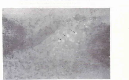
Figure 5 is a frontal section of the brain, showing the site of DAPI injection, and the SFO filled with the injected DAPI (anow).
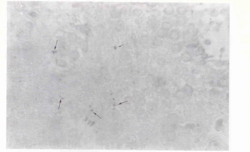
Figure 6 is a frontal section of the rat brain at the stereotaxic coordinates of IA (interaural) 7,60 mm and bregma
-
1,40 mm andIA
7,20 mmand bregma-
1,80 mm showing the sites of the DAPI labeled parent cell bodiesin
the supraoptic nucleus (a) and paraventri-cular nucleus (b). The colorofthe
labeled parent cell body is a clear white upon a blue colored background. The results revealed that the parent nerve cell bodiesi4
both paraventricular and supraoptic nucleiof
the hypothalamus are clearly labeled. This figure also depicts the topography of the neurons in the supraoptic and paraventricular nuclei of hypothalamus.The results of this investigation demonstrated that the single fluorescent tracer (DAPI) has labeled the parent cell bodies of the neurons in the paraventricular and supraoptic nuclei.
Figure '7.
A
camera lucida drawing the frontal section of the rat brain, at the stereotaxic coordinate of: (a) IA (interaural) 7,60 mm and bregma - 1,40 mm andlA
7,2O mm and bregma- 1,80
mmto
depicts the topography and the sitesof
theDAPI
labeled parent cell bodies in the supraoptic nucleus, paraventricular nucleus, the caudate-putamen nuclues and in the dorsal hippocampal nucleus.Immunoperoxidase study
The result
of
this investigation by using oneof
the immunoperoxidase methods using the LSAB kits re-vealed that the subfornical organ gave a positive reac-lion (brown reaction) and the locationof
the neuro-secretoric substances which gave the positive reactionis extracellular (see Figure 8 and 9). On the other hand, figure I
I
and 12 ab (frontal sections of the rat brain at the stereotaxic coordiates betweenIA
7,60 mm and bregma-
1,40 mm and IA 7,2O mm and bregma-
1,80mm) showed a positive reaction (brown reaction) in the cell bodies of the magnocellular neurosecretory system (intracellular)
in
the paraventricular and supraoptic nuclei of the hypothalamus. The location of the brown reaction suggested that the neurosecretoric materials were synthetized and secreted by the nerve cell bodiesof
the magnocellular neurosecretory systemin
those nuclei.DISCUSSION
Vol 3, No 3, July - September
1994
Connections berween Subfornical Organ and Supraoptic and ParavenlricularNuclei l4l
Figure 5. A frontal section of the rat brain at the stereotclric coordinates berween IA 7,60
nn
and bregna - 1,40, and IA 7,2O nnr and bregma - 1,EO mnt, showing the exantples of the sites of DAPI injection in the subfornical org,an.Figure 6. A frontal section of the rat broin at îhe stereotLxic coordinates of IA 7,60 mnt
ofthe sites ofDAPI labeled parent cell bodies in the paraventricular nucleus (a) and IÀ
exanple of the sites of DAPI labeled parent cell bodies in the supraoptic nucleus (b).
142 Daryanto Med J Univ Indon
<ll"r".
-1:î3 m.M.-
à1".,.
-1:33:":: [image:7.595.102.536.97.393.2] [image:7.595.112.547.449.696.2]-b
Figure 7. A canera Lucida drawing of the frontal section of the rat brain, at the stereotaûc coordinates of: (a) IA (interaural) 7,60 nunandbregna- l,40nunshowingthesitesof the DAP(injectioninthesubfornical organ (1) and labeled parent cell bodies in
the paraventricular nucleus (2); (b) IA 7,20 mm and bregna - l,8O mm showing the sires of the DAPI labeled parent cell bodies in the dorsal hippocatnpal nucleus (3), the supraoptic nucleus (4), the caudate-putanen (5).
Vol 3, No 3, July - September
1994
Connections between Subfornical Organ andSupraoptic and paraventricularNuclei
143144 Daryanto MedJ Univ Indon
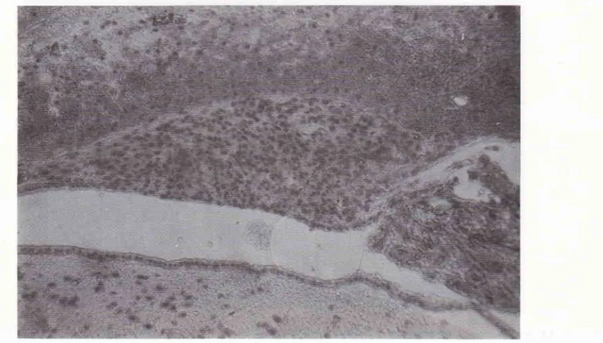
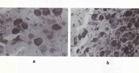
Figure 11. Photonicrograph ofafrontal section ofthe rat brain at the stereotaxic coordinates berween IA 7,60 ntnr and bregnrc -1,40 mm, and IA 7,20 mm andbregma - 1,80 nm showing an example of sites of positive reaction (brown reaction) to the lnbeled StreptAvidin-Biotin staining methods. The location of the brown reaction is in the neme cell bodies of the nngnocellulcr neurosecretory system (intracellular) in the supraoptic and paraventricular nuclei of the hypothalanws. The locaîion of the brown reaction suggested that the neurosecrerc were synthetized and prod.uced by these nerve cell bodies of the nngnocellular
neurosecretory systen in those nuclei. X 40.
Figure 12 ab. Photomicrograph of afrontal section of the rat brain at the stereotaxic coordinates between IA 7,60 ntnr and bregma 1,40 mm, and IA 7,20 mrn and bregma - I,8O mm, showing an example of sites of positive reaction (brown reaction) in the neme cell bodies of the magnocellular neurosecretory system (intracellularly) in the paraventricular (a) and supraoptic nuclei (b) of the hypothalamus. X 400.
Vol 3, No 3, JuIy - septenber
1994
connections betvveen Subfornical organ andSupraoptic and paraventricular Nuclei 145'of the neuroanatomical- tract tracing methods using a
single fluorescent tracer 4',6-
diamidino-2-phenylin-dol dihydrochloride (DAPI) (which was devàloped by
Heimer and Robards)Æ
and
oneof
theimmuno-peroxidase methods,
i.e.
the LabeledStreptAvidin-Biotin techniques (LSAB), which was developed by Naish et a/.2a
Injection of a single fluorescent tracer (DApI) in
seven adult albino rats of the Wistar strain, de monstrat-ed that DAPI had labeled the parent cell bodies of the
neuron
in
the fourteen nuclei and areasin
the brain,including the supraoptic and paraventricular nuclei
of
the hypothalamus.
In this study, the projections of nerve fibers to the
subfornical organ
from
those nuclei, especially thesupraoptic
and
the
paraventricularnuclei,
were verified by the retrogradely transported DAPI. These results that all labeled nerve fibers and cell bodies that were traced from the SFO were afferents of thesupra-optic and paraventricular nuclei, according to the ob-servations that labeled axons can be followed to retro-gradely labeled neurons that may given rise to them.
Similar findings have been reported
by
inves-tigators using other techniques,
for
examplès, using electrophysical methods, which stated thatlh"re*".é
some connections between the SFO and the suoraootic and paraventricular nuclei
of
the hypothalamus,14,15 and using the autoradiographicand
tracer'santero-grade transportation method which demonstrated the
connections between the SFO and the median preoptic, supraoptic and paraventricular nucleus, Based upon an
electrophysiological method and.r_lsing acetylcholine,
saralasin and atropin Saro et al. l
/
demonstrated that there were some connections between the subfornicalorgan and the paraventrilar nucleus.
The results of this investigation
by
utilizing theimmunoperoxidase method
on
15 ratsof
the LMRstrain, showed a positive repons by giving brown reac_
tioninthe subfornical organ. The location of the brown
reaction, however, is extracellular. So this result
re-vealed that
the
SFO contains neurosecretoric ma_terials, which was positive to the immunoperoxidase
method. This method used Rabbit anti-human neuro_
physin as a primary antibody, and LSAB kit; the biotin linked
to
the primary antibody, produces abiotiny-lated conjugate which, when added to the tissue sec-tion, localizes the sites of antigen within the section.2T
The neurophysin,
is
a precursorof
vasopresin andoxytocin
hormones.
Moreover,
the existencesof
neurosecretoric
material
extracellularly proves thatthis neurosecretoric
material
was
synthetised andproduced by another nuclei in the brain. This results
ensured that, in fact, no secretory
cell
were found inthe SFO.
The origin of the neurosecretoric material which gave
a
positive resultby
giving
a
brown reaction,turned out to be the neurosecretoric material found in
the cytoplasma of the cell bodies
of
the magnocellularneurosecretory system
of,the
supraoptic and para_ventricular
nucleiof
the hypothalamus. Theneuro-secrete's existence
in
the cell bodiesof
those nucleiproved that
this
neurosecrete was synthetised andproduced by those nuclei.
Similar findings have been reported by other
in-vestigators, i.e. Stutinsky le and Gantng l6
*iri"h
"tut"dthat the neurosecretoric material was synthetised and
produced
by
thecell
bodiesof
the magnocellular
neurosecretory system
in
the supraopticand
para_ventricular nuclei
of
the hypothalamus. Thisneuro-secretoric material was then transported to the
neuro-hypophysis via the hypothalamo-hypophysealis-tracr through the neurosecretion process (see Figure l3).
There was some neurosecrete material in the SFO
which gave positive reaction to the immunoperoxidase
staining methods, eventhough in small amounts. This
proved that the SFO sends efferent projections in small
amounts to those nuclei (see Figure 13).
This investigation using the LSAB kits,
in
fact,can be used as a tracer to trace the nerve fiber
connec-tions between the
subfornical
ticandparaventricularnucleioft
heresults were
in
concordancewith
the other result byusing the neuroanatomical-tract tracing method.
This
investigation, concluded thatby
utilizingneuroanatomical-tract tracing method and
immuno-peroxidase method (LSAB kits) the subfornical organ
was proved to be anatomically connected to the supra_ optic and paraventricular nuclei of the hypothalamus.
REFERENCES
l.
Weindl A. Neuroendocrine aspects of circumventricular organ. In: Ganong WF, Martini L, eds. Frontiers in neuroen_ docrinology. New York: Oxford University press, 1973:3_ 22.2.
fW. The subfornical organ intopographical anatomy.
I
3. Dellmann HD and Simpson JB. The subfornical organ. In_ ternational Rev Cytol 1979; 58:333-421.
4. Mark MH, Farmer PM. The human subfornical organ: and anatomic and ultrastructural study. Annals of clinical and I-aboratory Science 1984; 14(5):427 -42.
5. Dellmann HD, Simpson IB. Regional differences in the
morphology
of
the rat èubfornical organ. Brain Res.1976; I l6:389-400.
t46 Daryanto MedJ Univ Indon
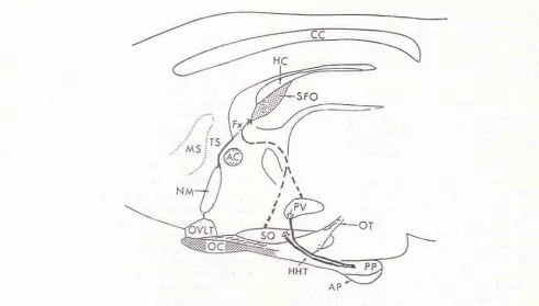
Figure 13. A schenatic sagittal drawing of the rat diencephalon depict the locatiott of the subfornical organ (SFO) lying in the of,thg^third ventricle underthe hippocanpal conunissure (HC) and
fornft
(Ft); inaccordance with recent ta,t/'te neurosecretoric ,ilaterial is to be transported through a neurosecrelion process in the hypothalanro-hypophysealis- tact (HHT) (solid heary lines) from the supraoptic and paraventricular nuclei of the hl,pothala,nus to the poste-rior pituitary (PP), some nerve fibers also projected to the SFO from those nuclei (intennittent lines). This neme fibers transportasnallamountofneurosecretoricnnterialto the SFO and release
it
to the blood capillariesvia rcnùnal nerve ending(frotn Sgro et at.)t7
7. Lind RW, Van Hoesen GW, and lohnson AK. An HRP study of the connections of the subfomical organ of the rat. J Comp Neurol 198 2;2 lO:.265 -7 7 .
8. Sawchenko PE, Swanson LW. The distribution and cells of
origin of some afferent projections to the paraventricular and supraoptic nuclei ofthe rat. Soc Neuro-sci L98L;7:325. 9. Ferguson AV, Day TA, Renaud LP. Subfornical organ
ef-ferents influence the excitability of neurohypophyseal and tuberoinfundibular paraventricular nucleus in the rat. Neuro-endocrinology 19 I 4;39 :423 -8.
10. Eng R, Miselis RR. Polydipsia and abolotion of angiotensin-induced drinking after transections of subfomical organ efferent projections
in
the rat brain. Brain Res. 1981, 225:2OO-6.11. Lind RW, Johnson AK. Subfornica organ-median preoptic cormections and drinking and pressor responses to angioten-sin-tr. J of Neuroscience 1982;2(8);1043-5 l.
12. Knepel W, Nutto D, and Meyer DK. Effect of transection of
subfornical organ efferent projections on vasopressin release induced by angiotensin or isoprenaline in the rat. Brain Res
I982;248:l8O-4.
13. Renaud LP, Rogers J, and Sgro S. Terminal degenerationin supraoptic nucleus following subfornical organ lesions: ultrastructural observations in the rat. Brain Res 1983; 275:365- 8.
14. Ferguson AV, Day TA, Renauld LP. Subfomical organ stimulation excites paraventricular neurons projecting to dorsal medulla. Am J Physiol 1984;247:R1088.
15. Ferguson AV, Kasting NW. Electrical stimulation in subfor-nical organ increase plasma vasopressin concentrations in concious rat. Am I Physiol L986;251:R425-R428.
16. Ganong WF. Review of Medical Physiology. Los Altos, California. I-ange Medical Publications, 1993.
17. Sgro S, Ferguson AV, Renaud LP. Subfornical organ-supraoptic nucleus connections: an electrophysiologic study in the rat. Brain Res 1984;303:7-13.
18. Summy-Long JY, Keil LC, Hemandez L, Emmert S, Chee O, and Severs WB. Effects of dehydration and reirin on vasopressin concentration in the subfornical organ area.
Brain Res 19 8 4;3OO :2 19 -29.
19. Stutinsky F. The place
of
neurosecretionin
modernneurobiology. In : Vincent ID, Kordon C, eds. Cell biology
Vol 3, No 3, July - Septenùer
1994
Connections between Subfornical Organ and Supraoptic and Paravenyicular Nuclei t47of hypothalamic neurosecretion. Paris: Editions Du Centre National De [-a Recherche Scientifïque, 1978.
20. Buggy I, Fisher AE, Hoffman WE, fohnson AK, phillips MI.
Ventricular obstruction: Effect on drinking induced by
in-tracranial injection of angiotensin. Science 197 5;l9{Jr72-4. 21. Simpson IB, Epstein AN, and Camardo Ir JS. Localization
ofreceptors for the dipsogenic action ofangiotensin tr in the
subfornical organ
of
the rat.I
Comp Physiol Psychol1978;92(4)58 l-608.
22. Swanson LW, Sawchenko PE. Hypothalamic integration:
organization of the paraventricular and supraoptic nuclei, Ann Rev Neurosci 1983;6:269-324.
23. Paxinos G, Watson C. The Rat Brain, In Stereotaxic
Coor-dinates. San Diego, New York, London, Toronto: Academic
Press, Harcourt Brace Jovanovich Publishers, 1986.
24. Naish Sally I, Boenisch T, Farmilo Af, Stead Ron H.
Han-bood Immunochemical Staining methods. Denmark: DAKO
Corporation, 1989.
25. Heimer
L,
Robards MJ. Neuroanatomical-tract tracingmethods. New York: Plenum Press, 19E1.
26. Weindl A, Sofroniew MV. Neurohornones and
circum-ventricular organs. h: Knigge KM, Scott DE, Weindl A eds.
Brain-endocrine interaction
III.
Neural hormones and reproduction. Bassel : Karger, L978;Ll7-3'1.27. Taylor CR, Bennington JL. Immunomicroscopy: A Diag-nostic Tool for the Surgical Pathologist. Philadelphia,
L,on-don, Toronto, Mexico City, Rio De laneiro, Sydney, Tokyo,