2@
Haryana et al. MedJ
Univ IndonCytogenetic
Pattern
of Acute
Lymphoblastic Leukemia
(ALL)
in
the
Yogyakarta
and
Central
Java Province, Indonesia
Sofia
M. Haryana*, E.Suryadi**,
SultanaM. Husein***, Radjiman*
ABSTRAK
Penelitian
ini
bertujuan untuk tnelihat ganbaran sitogenetika penderitaALL
(Acute Lynphoblttstic Leukenia). Ganrbaran sitogenetika ini penring untuk tnenentukan penatalaksanaan penderita tersebut. ùnpat belas pasien ALL yang berobat ke runah sakitdi
Yogyakartalan
Setnarang sejakJuni
1992 sampai Novetnbertg92
telah diperiksa ganbaran sitogenetikanya dengan netode G-banding. Hasil peneriksaan tnenunjukkan 4 penderita dengan kelainan struktural kronrosottt berupa t(9;22) (q34.1;q11.21) pada 2pasien, t(4;1
l)
(q23) pada 1 pasien, det 11, pI3-14 pada I pasien; 9 penderita dengan kelainaniunlah kronoson berupu hiperploidi, potiploidi, tetraploidi, atau ltotlosolti, 3 diuntaranT,a tennasuk kelainan kronoson ganda (struktural dan nunerikal); 4 penderita tidak ilitentukan kelainan krontoson. Pada penderita dengan hanya hiperploidi > 5O kronosortt dan ganbaran sitogenetika nonnal setelah pengobaran nenunjukkan remisi/retnisi parsialis. Sedangkan 3 penderita dengan kelainan krouosottt ganda menuniukkan perialanan penS,akir lebih progresif. DisituputkaÀ bahwa kelainan struktural pada ALL nentputtyai arti penling dalant nenenluknn penggolongandan prognosis penyakit.
ABSTRACT
The
ain of
this investigation is to analyse the cytogenetics of ALL patients. Cltosenelic analysis is inportant in deterntining the ,nanage,ileyû of those patients. Fourleen ALL patients, hospitalised in several hospitals in Yog1,ofta71a and Setnarang since June 1992 to Novetnber 1992, v'ere studied by G- banding,nethod. The results shov,ed 4 patients u,ithstructural abrutnnalitr- î(9;22) (q34.1;q I 1.21)(2patients),t(4;tI)(q2t;q23)(Iparienr),arddel 11,p13-pl4(Ipatient);g
ltutienrwithnunerical al.tnonnalitt'suchashyperploidl', polypktitll,, rcrraploiù1, and tnonosorttl', three of thenr shou'ed double abnornulities (slrudural and nunerical); 4 putients wilh nonnal karl,otype. Patienrs v'ich previously have hyperpktidy > 5O chrotnoxtn and normal karyotype, afier lreatnte,Tt shou'ed re,ilissioty'partialitt ALL is inporrunt
b
deternrine the groult and prognosistf
the diseo^se.Key words :
ALL,
c)'rogenerics - translocatiott - deletion -hyperploidl'- classifcatiott - prognosis.INTRODUCTION
Cancer is the result of
accumulation
ofmultiple genetic
changes.l Each alteration,
whether
an initiating
or
a progress.ionevent, may
be mediated through a
grosschromosal
change andtherefore
has thepotential
to be cytogenetically visible.2
The common
tumor schromosomal
abberrations areclassified
as struclural or numeric.
Structural
al-terations include translocations, inversions, deletions,
irsertions
and amplifications,
whereasnunlerical
ab-normalities are
losses
or
duplication
of
whole
chromosomes.3 In hematological malignancy
such asleukemia, analysis
of
thechromosomes
also has beenextensively studied, which revealed that almost all
leukemia carry
akaryotype abnormality.
Hematological
malignancy
particularly
leukemia
is
still
aproblem in Indonesia.
Casesrecorded
atDr'
Sardjito
Hospital
in
the Yogyakarta province from
1987
to
1990 included Hodgkin lymphoma (6.29%),
VoL 3, No 4, October-Decenùer 1994
non
Hodgkin
lymphoma (46.85%),
mulriple
myeloma
(3.14%), Iymphoid
leukemia
(2t.86%),
myeloid
leukemia (19.8670) and
other types
of
leukemia
(1.987o).' The frequency
of
nralignancy
casesfrom
year
to
year was increasing.
In
1987there
were
884 cases,in
1988 I 164 cases,
in
1989
ll90
and
in 1990
1282 cases.
The
total
number
of
malignancy
cases atDr.
Sardjito
Hospital
Yogyakarta
*.ré
+SZO"ur"r.4'5
In
Semarang,
Sunjoyo reported that there were
84cases
of
hematological
malignancies
between
1984-1986, included
non Hodgkin lymphoma (42.9%),
myeloid leukemia
(32.47o),
lymphoblastic
leukemia
(17.27o),
multiple
myeloma (3.7%) and Hodgkin
lymphoma (2.3%)."
The diagnosis
of
leukemia
in yogyakarta or
In-donesiain
general is based on the morphologicalfind-ing
of
blood
lineage
cells,
in blood or
bone marrow
smear stained
with routine staining
such as Giemsa,
Wright, May Grunwald,
and examined_under
regular
light
microscope
(FAB
Classification).5
Therapy using standard
regimen therapy showed
no satisfactory
results;'
this may be due to the
timing
of
patients
arrival at the hospital. Usually
patients came to the hospital at the terminal stateof
the diseaseor might
be dueto
innaccuracy
of
diagnosis.
In
lggg
agroup
of
hematologist
proposed anew classification
of
leukemia
basedon morphology,
immunophenoty-ping
and citogenetics (MIC).7 Classification
which
merely
based
on_morphology could not classify
thetype
of
leukemia.TUsing cyiàgenetic analysis
will
in-crease the accuracy
ofth
erapycan be-determined
and
edict-ed.8'e'lo
In
Indonesia,
th
usingcytogenetic analysis
hasThe aim
of this
study is
to analyse thecytogenetic
pattern
of ALL
patients
in
yogyakarta and
Central
Java,
Indonesia,
in
the hope that therapy could
becorrectly given
to the patient and prognosis
of
the disease can bepredicted.
I\{ATERIALS
AND
N,TETHODSSamples
One
ml of
heparinized peripheral blood
and 0.5ml
of
bone marrow aspira{e
fronr patients diagnosed
asleukemia
at Dr.
Sardjito Hospital
(yogyakarta),
Kariadi
Hospital
(Semarang), and Telogorejo Hospital (Semarang).Methods
Arresting
: Colcemid was usedin this investisation
asarresting agent to stop
mitosis
at
metaphas".S
Col".-ALL C1'togenetic
Pattenr
201mid
is amitotic
spindle
inhibitor,
which
canblock
theformation
of
spindle
fibres. Normally, spindle fiber
was attached
to
the centromere
of
eachchromosome
before
mitotic division
andpull
the chromatids
apartby contraction
at anaphase,rHypotonic Shock
: Thehypotonic solution
used was0.075M
KCL
since it has beenknown,
that this solutionis the least damaging to chromosomes
substructure.
A
hypotonic
solution
is
asolution with
a lower saltcon-centration
thanthat
of
the cytoplasmof
cells,
so therewill
be a movementof
HzO
from
the hypotonic
solu-tion into
the cell.3S
rxatron :Theïxahve
so)utlon ,rru,
ïr"ù)y
,nuà"
ol3
parts
of
absolute methanol
to I
part of glacial
acetic acid. Thevolatile
alcohol
plays arole in flattening
and spreading the chromosonresduring air
drying.
Glacial
acetic acid
will
rupture
the membraneof
the cell.Slide
Preparation
and
Staining
: Slides were washedin
distilled
water,
dried
at
room temperature
andstained
with Giemsa.ll
Giemsa
Staining and Banding
Giemsa staining is a method to stain the chromosomes.
Giemsa stain
is a
Romanowsky-type
dye
mixture.
Azure B
and eosin in the Giemsa bound toDNA.ll
The
solutions
needed are :l.
Giemsa
stain
concentrate, made
by
aclding 1.0 g
Giemsa
powder
to
66
ml
methanol and 66 ml
glycerin, stirred
for 2
days
at room
temperature,prepared
at
least
2
weeks
befôre
use. Stored
in
adark container
in
a refrigerator.2.
Phosphate
buffer (pH
6.8), which
consists
of
0.025M
KHzPO+(3.4 gll),and
rhepH
was adjustedusing 50% NaOH.
Giemsa
staining solution
was prepared bynrixing
5ml
of
Gienrsastain
concentrate and
45 ml of
phosphatebuffer (pH
6.8).Giemsa banding
(G
banding)
is
anrethod to
ob_tain
bands
in
chromosomes
by treating
slides
with
a proteasesuch as
trypsin or
incubate the slides
in
hotsalinecitrate. The
chromosome
banding pattern
ob-tained reflects bothstructural
andfunctional
composi-tion of
the chromosonl.r. lo'l ISolutions needed are
:l.
Giemsastain concentrate
2.
Phosphatebuffer (pH
6.8)-202
HarT,ana et al.Working staining solution
is preparedby mixing 26 ml
buffer (pH
6.8), 7 ml methanol, 0.6-l.0 ml lOX trypsinEDTA
and0.8 ml
Giemsastain
concentrate,Chromosome Analysis
Chromosomes are prepared by
direct
bonemarrow
andperipheral
blood culture developed in our laboratory.
Marrow cells were cultured
in RPMI-1640
medium
supplemented
with
75To fetal calf serum. Only + g. 1ml of
sedimented cells were processed
from
eachcentrifuge tube.
The cells were
exposedto colcemid
(0.06 mg/ml)
for 25 minutes and then
to
hypotonic
solution
i.e.KCI
(0.075mol/l) for a total of 32 minutes
(including mixing,
standing and centrifuge time).
Slides were
fixed in 3:
I
methanol
aceticacid (v/v)
anddried on a hot plate. Slides were subjected to
Giemsastaining
andbanding.
For each
case, 10-20 metaphases(mean
l5)
werestudied by direct microscopy
to determine the modal
number and to
identify the
malignant
stemline.
Karyo-type
were prepared
from
photographic prints.
Chromosomal abnormalities
wereclassified according
to the
International Conventions.l0'
I l' 12 The
definition
of an abnormal
stemline
wasdefined according
to thatproposed
by the
Second
International workshop
onchromosome
in leukemia.
A
case wasconsidered
ab-normal
if
it
had
anabnornral clone,
regardlessof
theproportion of normal
metaphase. l3RESULTS
The patients were
diagnosed when
they
were
hospitalised. There
were
14 patients
analysed
cytogenetically. Out of those 14 patients 8
were malesand 6 were females
(Table
l).
The rangeof the age
was between 2-2O yearcold.
There were 4 patients under 5,7
patients
between 5and 12,
and 3 patients more than12
years
old
(Table 2). The diagnosis was
based onFAB classification (morphology)
by'Wright
or Giemsastaining.
Table 1. Patients with
ALL
Sex
Med
J Univ lndon
The
cytogenetic
analysis onperipheral blood
andbone marrow
sampleswere carried
out on at least
10metaphases
from
oneslide. The findings
were as
fol-lows (Table 2)
:-
1
patient
of
46
XY, t(4;ll)
(q21;q23)
from
peripheral blood
and 53XXY
from
bonemarrow.
-
I
patient
of
46 XY t(9;22) (q34.1;qI1.2l)
(Phila-delphia chromosome
positive)
from peripheral
blood
and 56XY
from
bonemarrow.
-
I patient of t(9;22) (q34.1;q11.21)
fromperipheral
blood,
Ph+from
bonemarrow.
-
7 patients
of
abnormality in chromosome number
from
bone
marrow
i.e. 52
XY
mozaic,
91 XX
polyploidy
mozaic, hyperploidy
>
50
chromo-somesmozaic, 87 XY polyploidy
mozaic (257o),92
XXYY
tetraploidy mozaic, and monosomies
45XX-7
and 45)tY-17
.-
4 patients showed no structural and numericalchro-mosome
abnormality.
Tablc 2. Agc, Scx, Cytogcnctic l'inding ol ALL
No.of Agc
Scxpaticnt (ycas)
Cytogcncticlinding
Sourco20 6
ll
4 9 6.5 M M M F F F M M46 XY, nrozaic, -52
XY
BM46 XX, nrozaic,
9l XX
plyploidy
BM 46 XX46 XY i(4;l | )(q2 I .q23)/s3
XXY
P/BM(4+ l;5+ l;6+ I ;l I + l;l 6+ I ;2 l+ I ;X+ t)
45
XX-7
BM46 XY t(9;22)(q34.l.ql l.2l)(Ph+)/ P/BM
56 XY hypcrploidy (5+l)(6+l)(8-l)
( l3+ l)( l4+ l)( l6+ l)(17-l)(18+2)
(2 t+3)(22+2)
46 XY, hypr:rploidy > 50 Chromosomc BM 46 XY
46 XY, 87 XY polyploi<Iy
25%
BM a6 xY r(9;22)(q34. l.q I 1.2)iPh+46 XX 46 XX
46 XX,92 XXYY tclraploidy
4-5 XY- l7 dcl I I ,p I 3-p 14
P/BM
BM =
sourcc of karyotyping analysis is l'rom tronc ntarrowP/BM
=
sourcc of karyotyping analysis is liont krnc nrarrow and pcriphcral bltnd=
nralc=
fcnralet4M
24F
35F
46M
55F
63M
% 20 t9 7. 8. 9. 10.I l.
t2. t3. 14. Male Female 57.1 42.9 BM BM Total M F n = number of patienl
Vol 3, No 4, October-Decenber 1994
Table 3. The Impact of cytogenetic finding and outcome
No. of
patient
Cytogenetic finding OutcomeALL Cytogenetic
Pattern
203abN =
abnornral
ND = no lollowup
str
= slrucluralN=normal
D=dicd
num=nunrcricalNo follow up paticnts (21.a%); complctc renrission (35.7%) and dicd (42.86%)
DISCUSSION
The data obtained
from
the
Third
International
workshop on
chromosome
in Leukemia,
comprising
330 children and adults
with
ALL
showed
that
thespecific
karyotype
patternsprovided significant
inde-pendent
prognostic
information,
in regard toremission
duration
andsurvival.
l0' I I'13'14'Our
investigation consisted
only
of l4
ALL
patients,
which
was
very small to
assesthe impact
of
the karyotype on
the progressiveness
of
the
disease,But
we hopeit
might give
a general ideaof
the patternof
cytogenetics
in
Central
Java
and
Yogyakarta
province.
Our findings
showed several translocations.
Translocation
of
t(4;11)
(q2l;q23)
was found
in
1patient. This type
of
translocation was common in
congenital
ALL
andALL
in
young
infants.ls
Translo-cation
of t(a;l1)
has
als_obeen reported related
to exposure to carcinogens. toT*o
patients showedtrans-location of t(9;22) (q34.I;ql1.2l) called
ph + ph
+ALL
besides
CML
(chronic
and about 6% occurs
in
ion.of
(4;l l)
andt(9;22)
indi-.ir.
14'16Those patients
after
aninductive remission showed a partial remission
but then they werehospitalized
with
CNS (central
nervoussystem) leukemia and
died
of
cerebral bleeding.
Hy-perploidy
with
chromosome numberof
470- 50 or > 50is conrmonly
p.ssociatedwith B
lineage
and rarein
T-lineage ALL.|4
Someof
the patients-also showed
hy-perploidy
more than
50
chromosome,
but we
are notsure whether
thesepatients were
of B
lineage,
sinceimmunophenotyping
hasnot
beenperformed.
Hyperploidy
indicated
goodprognosis especially
in
mozaic
with normal
karyotype.E'eIn
this study we
found five
patientswith
normal karyotype
or inmozaic
with hyperploidy,
achieved complete remission;
theywere
3patients
with normal karyotype,
I
patient
with
normal karyotype and hyperploidy (mozaic) and
1patient
with polyploidy.
Onepatient
with
monosomi-7
achieved
short
remission and then
died. While
3patients
which
consists
of
I
patient
with
hyperploidy
(mozaic),
I
patientwith
polyploidy 25%,1
patientwith
tetraploidy (mozaic) could not be followed up.
This
finding
indicates that cytogenetic analysis
has anim-portant role
in predicting the prognosis
and
also
in
determining
thehigh risk
group
from
generalpopula-tion.
Patients
with
chromosome
abnormalities
espe-cially with
translocation
ordeletion
of
whole
chromo-some
(monosomy) should
becategorized
ashigh
risk
patient eventhough
Hb, WBC
and tota.lplatelets
werein
norn.ral range,and regimen therapy
chosenshould
be
given intensively.
l.
2. 3. 4. 5. 6. 7. I 9. 10. 11. 12. 13.t4.
abN (num) abN (num) N
abN (str+nunr) abN (num) abN (str+num) abN (num) N abN (num) abN (str) N N abN (nurn) abN (str+nurn)
ND Remission Rernission D Remission
-
D D Remission D ND p.Remission-
D Remission Remission ND D-
N
- rcmission -abN
- rcnrission-N
-dicd-
abN
- died3 paticnts : 21.4%
3 paticnts:21.4% I paticnt
:
7.14%5 paticnts : 35.'/ l%
Table 4. Age and karyotype abnorrnality
No.of patient Age Karyotype N/abN
l.
2. J. 4. 5. 6. '7. 8. 9. 10. 11. t2. 13.t4.
4 4 5 6 5 3 20 6 11 4 9 6.5 20l9
abN (nurnerical,gain) abN (numerical,gain) N abN (structural+numerical,gain) abN (nurnerical,loss)abN (structural +numerical,gain) abN (nurnerical,gain)
N abN (nurnerical,gain) abN (structural) N N abN (nurnerical,gain) abN (strtrctural+numerical,loss)
Abnornral karyotypc : Age undcr-5 ycas : 4 paticnts (40%)
-5 - l2 ycani : 3 palicnts (30%)
> 12 ycan : 3 paticnts (30%)
Norrnal karyotypc : Agc untlcr -5 ycars : - (0%)
5 - l2 ycars:4 paticnts (100%)
> 12 ycars: - (o%)
Haryana et al. Med
J
Univ IndonÈ.ia CM I "'- '
-Ten$^l : l'*::*:
È
*
Ç
l3
*
#).
'1 Ê
-- 4
*
4
I
4P-"*-
a e"ry
kv
. lb
,r*
.i
a-
*-
".-.... Ê rê...*rh $ t.
t
5.
*e
Çx
**
" F19, 24- e 2t .-... 22
t*
4 #
'."_Fl-e-
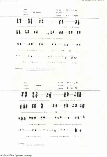
*24''.-Figure 1. Karyory^pe of an ALL-LI patient showing a) 46 XY,
t(4;lI)(q2l;q2j)
andb) 53 XXYfrot,t bone nnrrow culture
Karyotyping was done front G-banded chro,,toso,ile
t
:
translocation q:
long ann of the chronosonte G:
GiensaALL-LI
:
Acute lynrphoblasric leukenia type L-1,nnêe?./,
, 'ç21 ---22... y. !
Nart
lJ niur Àiâù01
No cM : :l:'7t'i'jt1) iiq. Ftâp :
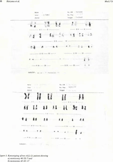
[image:5.595.83.548.61.639.2]Vol 3, No 4, October-December 1994 ALL Cytogenetic
Pattern
2O5ll{ilrl ùocM I 15/ft./92
inorial I 9-:.1-199A
rril
ff *,
r{
** *t
tâ.
r*
*
r
f
l,
.-,
n0,
*l
r
Ê
Jl
;t
r{
t*
al tô 1É
rr
'.
la
,;?1 - 22 <+
l\i êrê :
1.)n1$ . 3 tsà6
Àlêr?rl
t4â *ftt n n ,l 4 Ç
-- * t? -- ;q - '- -- É 21 '*- 22
--.-î\Ttaft? :
r,r
ln. èM l{o. Êr(}r 7 at'ûal
2ç'fttl91
)*t
-9?
Figure 2. Karyoq.pe of an ALL-Ll patient shov,ing
a) 46 XY,
(9;22)(q34;qIt.2I)
(Ph+) and b) 56 XY (poliploidl,)Karyoq'pittg was done front G-banded chronosone
I
:
translocation q:
long ann of the chronosone (Ph+1:
Philadelphia chromosone positiveG
:
Gientsa [image:6.595.114.521.67.664.2]206
Haryana et aI. MedJ
Univ Indontldiljù : 11o DM : l:;/'ii/'j'i
'Ja1$ : J'it'.Hrii: Nc ?r':P :
Llatttt : \arçgal '".'-:l:eti|
Figure 3. Karyotlytittg of two ALL-LI patient showirtg a) nonosony 46 XX-7 and
b) nonosony 45
XY-I7
karyo4, pittg was done.froilt G-banded chroiltoso,,rc G
:
GiensaALL-LL
:
Acure lyntphobkLstic leukenia \'peL-l
41 , _. un I
t*Kâ&&fr&ffi
in&
.: r; i)
*& *t #*
** &
&*
tx t1a 2*
*n
çï
*{
4,
€
àF19 2B ç* -." ZZ r.. y
al.çtiJTi? :
ljt !:i4 :
[image:7.595.64.517.59.704.2]Vol 3, No 4, October-Decetttber 1994
CONCLUSION.
Our results
suggestedthat
multiple
abnormalities
andmonosomies
indicate poorer prognosis
compare tohy-perploidy, poliploidy
andnormal
karyotype.
Patients
with
translocation
or deletion
cf
whole
chromosome
should be
categorized as
high
risk
patients regarldless
theHb,
V/BC
andplatelets
count.Acknowledgement
We
thankedProf
T.Tokuhisa, Kobe
University
Medi-cal
School
for his
valuable correction
andcritics.
We
also
thank
the assistanceof Wiwied,
Suwarsih, Maya
and
Ani for
thetechnical
works;
Sudaryonofor typing
the manuscript; the Director
of
Telogorejo
Hospital
and the Director
of
the
Live
Science
Laboratory
of
Gadjah
Mada University
for
providing the facilities
needed
for
this
work.
To
Prof.
Dr. A.G.
Sumantri,
Faculty
of Medicine
Diponegoro
University,
who
hasbeen
very supportive
in
managing
the
collaboration
between
GMU
andDiponegoro
University,
wewish to
express
our
sincere thanks.
We
also express
our
gratitude
toDP4M for
rhe granrNo. 33g/p4M/DppM/
L-331llBBIl1992;
and
grant
from UGMl34l5t
M/09/01,
June
1993.RBFERENCES
1. Cossrnan J. Approaching the molecular genetic era of diag_ nostic pathology.
In:
Molecular geneticsin
Cancer Diag_nosis. Elsevier, New York, 1990.
2. Solomon E, Barrow I, Goddard AD. Chromosom aberration
and cancer. Science 1991;54:1 153-9.
3. Rooney DE, Czepulkowski BH. Tissue culture methods in Human cytogenetics. In: Hurnan cytogenetics.
A
practicalapproach. Washington : IRL press, 1992:
l-36.
ALL C)'togenetic
Pattern
2O74. Medical Record Sardjito Hospital, 1994.
5. Sutaryo. Incidence and management therapy of leukemia in Sardjito Hospital. Scientific Meeting
of
Oncology Group, 1993.6. Soenjoyo. Incidence leukemia
in
Semarang. Kopapdi,Semarang, 1990.
7. Catovsky
D,
Mutates E, BuccheriV,
ShettyV,
HaslipI,
Yoshida N, et al.
A
classification of acute leukemia for the 1990s. Ann Hematoll99l;62:
16-21.8. Bain BJ. Immunological, cytogenetic and other markers. In: Leukemia Diagnosis
A
guideto
theFAB
classification. London: Gower Medical Publishing, 1990.9. Le Bean. Cytogenetic analysis of Hematological malignancy
disease. ALT Cytogenet
l99l;
39: 441.10.
Third International
Workshop
on
Chromosomein
Leukemia. 1980.
11. Benn PA, Perle
MA.
Chromosome staining and banding techniques. In: Human cytogenetics.A
practical approach. Vy'ashington: IRL Press, 1992; 57 -83.12. Secker-Walker
LM,
Chessels JM, StewarEL,
SwansburyGJ, Richards S, Lawler SD. Chromosomes and other prog-nostic features in acute lymphoblastic leukemia; a long term
follow up Brit J Haematol l9B9;12:336.
13. Watt JL. Stephen GS. Lymphocyte culture for chromosome analysis:
In:
Hurnan cytogenetics,A
practical approach.Washington: IRL Press, 1987.
14. Raimondi SC, Behm FG, Robertson
pK, pui
C-H, RiveraGK, Murphy SB, et al. Cyrogenetics
of
childhood T-cellleukemia. Blood 1988; '12:1560.
15. Saglio G, Guerrasio A, Ponzetto C, Zaccaria A, Testoni R, Celso B, et al. Variability of the molecular corresponding to
the
presenceol
a
Philadephia chromosomein
human hematologic malignancies Blood 1988; 72: 1203.16. Kapelushnik J, Dube I, Wilson p, Grunberg M. Acute
lym-phoblastic Leukemia
with
(4;11) translocation after