Correlation between hypoxia inducible factor -1α and renin expression
in rats kidney induced by cobalt chloride
Ani R. Prijanti,1 Raafqi Ranasasmita,2 Yurika Sandra,3 Septelia I. Wanandi1
1 Department of Biochemistry & Molecular Biology, Faculty of Medicine, Universitas Indonesia, Jakarta, Indonesia 2 Magister Program, Faculty of Medicine, Universitas Indonesia, Jakarta, Indonesia
3 Department of Biochemistry, Faculty of Medicine, University of YARSI, Jakarta, Indonesia
Abstrak
Latar belakang: Kobalt klorida dapat digunakan sebagai senyawa yang dapat menimbulkan kondisi mimikri hipoksia tanpa kadar rendah oksigen di dalam tubuh, dan menstabilkan hypoxia inducible factor-1α. Kami memutuskan untuk mengobservasi apakah terdapat regulasi ekspresi renin oleh HIF-1α. Dengan demikian kami menyelenggarakan beberapa penelitian untuk memastikan kemungkinan dan memulai dengan penelitian induksi tikus secara intraperitoneal kobalt klorida (CoCl2) untuk membangkitkan kondisi mimikri hipoksia dan mendapatkan konsentrasi dan pola ekspresi HIF-1α
dan mRNA.
Metode: Dua puluh empat ekor tikus dibagi menjadi 4 grup: kontrol, 2, 8, dan 24 jam inkubasi pasca injeksi intraperitoneal 30 mg/kg berat badan CoCl2. Setelah tikus dikorbankan, organ ginjal digunakan untuk pemeriksaan parameter berat ginjal,
kadar RNA, kadar protein HIF-1α (ELISA) dan mRNA renin (RT-PCR).
Hasil: Hasil menunjukkan bahwa terdapat perbedaan rasio berat ginjal/berat badan tikus, namun secara statistik tidak bermakna (p > 0,05). Secara statistik tidak terdapat perbedaan bermakna kadar protein HIF-1α antar kelompok (p > 0,05). Ekspresi relatif mRNA renin meningkat tajam (30 x kontrol), mulai pada 8 jam inkubasi pasca induksi intraperitoneal CoCl2
dan terus meningkat sampai inkubasi 24 jam (2465 x kontrol). Korelasi antara protein HIF-1α dan ekpresi relatif mRNA renin menggunakan analisis Pearson menunjukkan positif kuat (R = 0,91) (p = 0,09).
Kesimpulan: Terdapat kemungkinan yang besar bahwa gen renin diregulasi oleh HIF-1α. (Med J Indones. 2012;21:128-32)
Abstract
Background: Cobalt chloride can be used as an agent to stabilize hypoxia inducible factor-1α (HIF-1α) and to imitate hypoxia without low levels of oxygen inside the body. We intended to investigate if there was any regulation of renin expression by HIF-1α. Therefore, we conducted several studies to clarify this possibility starting with the induction of hypoxic mimicry in rats by intra-peritoneal (IP) injection of cobalt chloride (CoCl2) to obtain the levels and pattern of
HIF-1α and renin mRNA and protein expression.
Methods: Twenty-four rats were randomly divided into four groups, control group and incubation groups 2, 8, and 24 hours after intra-peritoneal injection of 30 mg CoCl2 per kg BW. After the rats were sacriiced, kidneys were excised, weighed and
kidney weight compared to BW. Tissue parameters were measured such as RNA concentration, HIF-1α protein by ELISA, and renin mRNA by RT-PCR.
Results: Differences between the groups in the ratios of kidney weight to BW and in the concentrations of HIF-1α protein were statistically not signiicant (p > 0.05). Relative expression of renin mRNA increased markedly starting 8 hours after CoCl2 IP injection (30 times over controls) and further rising until 24 hours (2465 times over controls). Correlation between
HIF-1α and renin mRNA by Pearson analysis was strongly positive, but not signiicant (R = 0.91; p = 0.09).
Conclusion: Renin gene regulation in renal hypoxic mimicry strongly correlates with HIF-1α. (Med J Indones. 2012;21:128-32)
Keywords: Cobalt chloride (CoCl2), hypoxia inducible factor-1α (HIF-1α), renin
Correspondence email to: [email protected]
activity and increase HIF-1α.2 Prolyl-hydroxylase
activity needs the presence of substrates and co-substrates such as oxygen, 2-oxoglutarate, and vitamin C to catalyze thereaction, which produces hydoxylated HIF-1α, succinate and CO2.
5 Besides lack of oxygen,
2-oxoglutarate analogues and CoCl2 can inhibit
prolyl-hydroxylase.6 Intraperitoneal-CoCl2 injection to rats
stabilizes HIF-1α and induces hypoxic mimicry.6
Kidney infections lead to the disappearance of blood vessels, which causes ischemia and hypertension.1,7,8
Since ischemia can trigger renin secretion,1,7-10 we
considered that HIF-1α may regulate renin expression. Hypoxia inducible factor (HIF) is a master transcription
factor that regulates transcription of several genes to maintain energy homeostasis.1 In normoxic condition,
HIF-1α is degraded through ubiqitin-proteasomes.1,2
Degradation of HIF-1 begins with the hydroxylation of prolines 402 and 564 in an oxygen-dependent-degradation domain (ODDD) catalyzed by prolyl-hydroxylase (PHD).2,3 Hydroxyprolines 402 and 564
in HIF-1α are recognized by the tumor suppressor vonHippel-Lindau protein (pVHL), which acts as an E3 ubiquitin ligase. After ubiqitinylation, HIF-1α is
degraded proteasomally.4 In hypoxic condition, lack of
Hence, we investigated renin regulation by HIF-1α through intra-peritoneal injection of cobalt chloride (CoCl2) into the rats to induce hypoxic mimicry and
obtain the levels and pattern of HIF-1α and renin mRNA and protein expression.
METHODS
Animals
This experimental animal study was conducted in the Department of Biochemistry and Molecular Biology, Faculty of Medicine, Universitas Indonesia, in 2011 and aimed to observe the levels and pattern of HIF-1α and renin during renal hypoxic mimicry. Twenty-four male Sprague-Dawley rats with 150 – 200 gram of body weight from BALITVET Bogor were divided randomly into four groups of 6 rats each: control group and three CoCl2 treated groups. The treatment was performed by intra-peritoneal injection of CoCl2 (30
mg/kg BW). Incubation times after induction were 2, 8 and 24 hours before the rats were sacriiced. After the rats were sacriiced, kidneys were removed and kept in a deep-freezer at -84 ºC until used. Each kidney was homogenized with a micro-pestle and RNA was isolated using “TriPure DNA RNA protein isolation” kit (Roche 11 667 165 001, Indonesia). From tissue homogenates we obtained the concentration of HIF-1α protein. The RNA isolates were used in Real Time RT-PCR to measure the relative expression of HIF-1α and renin. HIF-1α protein was measured by Western blot. HIF-1α and renin mRNA relative expression were measured by “iScriptTM One Step RT-PCR with SYBR®
Green” kit) (BioRad # 170-8892, USA).
Sample preparation
Preparation of kidney homogenate
100 mg of kidney tissue were put into 1.7 mL microtubes, then 0.5 mL TriPure Isolation Reagent was added. The tissue was homogenized at 15-25ºC using a micro-pestle. Another aliquote of 0.5 mL TriPure Isolation Reagent was added to the homogenate, then centrifuged at 12000 g for 10 minutes. The supernatant was used for measurements.
RNA isolation from kidney tissue
Total RNA was extracted with TriPure (DNA-RNA-protein) Isolation Reagent (Roche 11 667 165 001, Indonesia).
Separation: Kidney homogenates were incubated at 15-25ºC for 5 minutes to ensure that nucleoprotein
complexes completely dissociated. Then, 0.2 mL of chloroform (CHCl3) were added, shaken vigorously for 15 seconds and incubated again at 15-25ºC for 2-15 minutes. Centrifugation at 12000 g and 2-8ºC for 15 minutes separated 3 phases. The clear aqueous phase was on top and contained RNA. Interphase and organic phase at the bottom were colored red and contained DNA and proteins.
Isolation of RNA: The clear phase at the top was moved to another microtube. The residual red phases were stored at -20°C. To the clear phase 0.5 mL isopropanol was added, the microtube closed and 15 times shaken up and down. Subsequently, the samples were incubated for 10 minutes at 25ºC to precipitate RNA, centrifuged at 12,000 g at 2ºC for 10 minutes and the supernatant was discarded. To clean the RNA, one mL of 75% EtOH was added to the precipitate, vortexed and precipitated again by centrifugation at 7500 g and 2ºC for 5 minutes. The supernatant was discarded. Subsequently, the precipitate was half-dried to become a semi-dry RNA pellet and re-suspended in 50 µL of free water containing DEPC (DEPC-treated RNAse-free water). The precipitate was dissolved several times by pipette the solution, and then incubated at 55-60ºC for 15 minutes.
Measurement of parameters
Primer design of HIF-1α, renin, and β-actin genes
We used Primer-3 program to design the primers of HIF-1 α and renin genes. The respective sequences were obtained from the NCBI Gene Bank with the code (NC_005105.2) for rat HIF-1α and code (NC_005112) for renin. First, a Bioinformatics in silico search through the NCBI web site found the sequences of renin promoter, TATA box, and the structural renin gene. From this information, we made the design of the forward and reverse primer sequences of the promoter containing the HRE with the Primer-3 program. The primers were used for measurement of mRNA relative expression.
cDNA ampliication by Real Time - PCR
We used iScript One-Step RT-PCR Kit with SYBR Green (BioRad), primers of HIF-1α and renin, microtubes, RT-PCR tubes, microcentrifuge, single and multichannel micropipettes, and RT-PCR with the CFX program (MiniOpticon BioRad®) as follow:
seconds each at 95ºC, 30 seconds at 59ºC, and 30 seconds at 72ºC. Melting curve analysis: 1 minute at 95ºC, 1 minute at 55ºC, 10 seconds at 55ºC, totally 80 cycles, increasing 0.5ºC for each cycle. By using RT-PCR the amount of cDNA copies can be determined as quantitative gene expression of HIF-1α or renin. We used aquabidest as negative controls to exclude false positive results. As an external standard gene we used β-actin gene. Through RT-PCR we can obtain efficiency and cycle threshold (Ct) values. Expression of HIF-1α and renin genes was counted by relative quantification and relative concentration of mRNA was calculated using the Livax formula.
Measurement of HIF-1α by ELISA
This technique was run using SurveyorTM Intracellular
Human/Mouse Total HIF-1α Immunoassay (R&D,SUV1935.). A series of standard solutions was made: 31.25 pg/mL; 62.5 pg/mL; 125 pg/mL; 250 pg/mL; 500 pg/mL; 1000 pg/mL. Reagent diluent 2 was used as blank. One hundred microlitre of blank, standards, and sample homogenates of the kidneys were put into each well of an ELISA micro-plate, incubated with shaking at room temperature for 2 hours. After incubation, the wells were washed 3 times with 250 µL washing buffer. Each well was added with 100 µL total HIF-1α detection antibody at a concentration of 50 ng/mL. Wells were incubated for 2 hours with shaking. After washing wells were added with 100 µL streptavidine-HRP 1/200, and incubated for 20 minutes with shaking. After washing, each well was added with 100 µLmixture of reagent color A and B at 1:1 ratio. The micro-plate was kept in the dark for 20 minutes, 50 µL of stop solution was added and during the next 45 minutes, the absorbance was read with the ELISA reader at wavelength of 450 nm.
RESULTS
The average ratios between kidney and body weights are shown in table 1.
The ratios between kidney and body weights were calculated. There is an increase at 2 hours of incubation, slightly stronger in right than in left kidneys. At 8 hours of incubation weight ratios decrease again to about controls normal with no difference between left and right kidneys. At 24 hours of incubation, the kidney-body weight ratios decrease below controls, equally left and right.
HIF-1α protein
HIF-1α protein concentration increases in the 2 hours incubation group and reaches its peak at 8 hours of incubation (Figure 1). After 24 hours protein concentration is back to control values. Statistically, the differences between groups were not signiicant (p > 0.05).
Rat groups Right kidney ± SD (%)
Left kidney ± SD (%) Control 0.36 ± 0.04 0.35 ± 0.04
2 hours 0.39 ± 0.02 0.37 ± 0.03
8 hours 0.37 ± 0.05 0.37 ± 0.03 24 hours 0.34 ± 0.04 0.34 ± 0.04 Table 1. Average kidney-body weight ratios in rats with CoCl2
induced hypoxic mimicry
HIF 2.00 2.10 2.20 2.30 2.40 2.50 2.60 2.70
pg
HI
F
1
/g
protein
Contr
1a 2.26
0 0 0 0 0 0 0 0
Conc
kidn
rol 2
6 2
centratio
neys afte
hours 2.44
on of HIF
er CoCl
28 hours 2.61
F 1 in r
injectio
24 hours 2.27
rat
on
Figure 1.Concentration of HIF-1α protein in rat kidneys (pg/g tissue protein) after CoCl2 injection
Renin and HIF-1α mRNA
HIF-1α primers were 5’-CTG CCT CTG AAA CTC CAA AGC CAC T-3’ (forward) and 5’CTC ACT GGG ACT GTT AGG CTC AGG T-3’ (reverse). Renin primers were 5’-CTT TGT ACCGACTTG GGT CA-3’ (forward) and 5’-ATT TAG TCT CGT CCC GGA CA-3’ (reverse) with a PCR product of 262 bp. Primers for the β-actin reference gene were 5’-ACC ACA GCT GAG AGG GAA ATC-G-3’ (forward) and 5’-AGA GGT CTT TACGGATGT CAA CG-3’ (reverse) with a PCR product of 277 bp.
Relative expression of HIF-1α mRNA
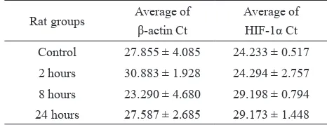
Averages of β-actin Ct and HIF-1α Ct of rat kidneys induced by CoCl2 are shown in table 2.
Rat groups Average of β-actin Ct
Average of HIF-1α Ct Control 27.855 ± 4.085 24.233 ± 0.517
2 hours 30.883 ± 1.928 24.294 ± 2.757
8 hours 23.290 ± 4.680 29.198 ± 0.794 24 hours 27.587 ± 2.685 29.173 ± 1.448 Table 2. Average of HIF-1α and β-actin Ct in rat kidney tissue
Relative expression of HIF-1α increased at 2 hours of incubation after CoCl2 intra-peritoneal injection (7.8 times over controls). In the group of 8 hours incubation after CoCl2 injection HIF-1α relative expression was decreased markedly, 1/1000 of controls (Figure 2). In the 24 hours incubation group, HIF-1α mRNA increased slightly vs. 8 hours, but remained still far below the control group (27/1000).
Renin
Renin mRNA relative expression starts to increase markedly (30 times over controls) at 8 hours after IP CoCl2 injection and further increase until 24 hours (2465 times over controls; igure 3).
Ratio
Relative expression of HIF-1α mRNA) in rat kidneys induced by CoCl2 injection
Figure 2.
Figure 3.Relative expression of renin mRNA (in logarithmic scale) in rat kidneys induced by CoCl2 injection
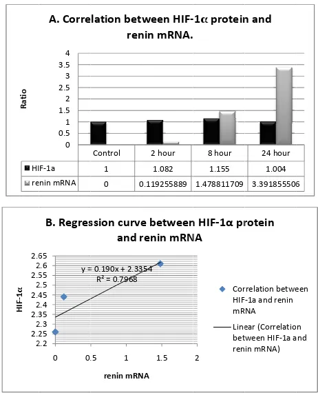
Correlation between HIF-1α protein and renin mRNA by Pearson analysis was strongly positive (R = 0.91) (p = 0.09) (Figure 4).
desferroxamine.9-11 Oxygen is one of the PHD substrates
and if enough oxygen is present, PHD hydroxylates prolines of HIF-1α. Subsequently, hydroxylated HIF-1α will be degraded. Conversely, low oxygen levels in hypoxia inhibit PHD and stabilize HIF-1α. Another regulation of HIF-1α is on the level of its biosynthesis.9 HIF-1α
synthesis can be triggered by activation of p44/42MAPK pathway, a signal transduction pathway through tyrosin kinase and activation of phosphoinositol threephosphate kinase (PI3PK) and mitogen-activating protein kinase (MAPK). MAPK pathway increases the activity of HIF-1α transcription. Morerover, some cytokines such as IL-1β and TNF-α can stimulate the binding of HIF-1α to DNA. The reason why there were no signiicant different in expression of HIF-1α protein in oxidative stress and hypoxia is that the process of translation is depressed (suppressed) for energy eficiency.9,11,12
HI
tion isstrongly positive (Pearsson correlation 0.91; p = 0.09); B: Regression curve of HIF-1α protein and renin mRNA
In this research, we saw that renin gene was regulated by HIF-1α as we had seen in our previous research.10 It was already known that HIF-1α is an acute response to hypoxic condition or to hypoxic mimicry induced by CoCl2, besides renin is the
between HIF-1α protein and HIF-1α mRNA relative expression by Pearson (SPSS 17) was 0.134, which is means the correlation was weakly positive. In other side, correlation between HIF-1α protein and renin mRNA in control, 2 hour and 8 hour groups by Pearson analysis was strongly positive (R = 0.91) (p = 0.09) (Figure 4).
Renin is a component of the renin-angiotensin-aldosteron-system (RAAS). The RAA system plays a role to maintain homeostasis of body luids and blood pressure. Renin cleaves angiotensinogen to be angiotensin I. Angiotensin I (ANG I) then is cleaved to angiotensin II (ANG II) by angiotensin converting enzyme (ACE). Angiotensin II binds to its receptor at targeted cells and leads to biological actions or effects such as vasoconstriction, increase of vascular volume by increasing sodium and water retention in renal tubules. Until now, 4 types of ANG II receptors have been revealed: AT1R, AT2R, AT3R, and AT4R. AT1R
is the main receptor of ANG II. AT2R is only found
in fetuses and neonates. The characteristics of AT3R and AT4R are not yet known.13 Recently, Matsuura
et al14 proved that CoCl2 induction suppressed AT1R expression and thus, biological effects of ANG II. It was considered that mimicry of hypoxia using CoCl2 did not generate hypertension different from hypoxia that causes renin elevation and hypertension. Hypoxia acts as a key role in cardiovascular defects such as systemic or pulmonary hypertension.14-17
Kramer et al15 found that acute hypoxia leads to
renin mRNA expression and renin activity in the plasma (plasma renin activity/ PRA). The increase of renin expression and PRA leads to hypertension. Kramer suggested that the increase of renin mRNA expression and PRA in acute hypoxia were caused by the increase of catecholamine concentration in circulation.15 Our studies found that renin was one of several genes, which are regulated by HIF-1α (in progress of publishing).10 The elevation of
HIF-1α increases renin expression. Therefore, in hypoxia or hypoxic mimicry, besides elevation of catecholamines renin elevation is regulated by the increase of HIF-1α protein.
In this research, we found that in CoCl2 intra-peritoneal
induction of rats, there is a strong correlation between the level of HIF-1α protein and renin mRNA in the kidney tissues. This result is supporting our previous study that renin expression is regulated by HIF-1α. From this research, we conclude that CoCl2 causes an increase
of HIF-1α protein and renin mRNA relative expression.
Acknowledgment
The authors are thankful to DRPM UI, which has funded this research through Hibah Riset Unggulan UI 2011.
REFERENCES
1. Nangaku M, Eckardt KU. Hypoxia and the HIF system in kidney disease. J Mol Med. 2007;85:1325–30.
2. Semenza GL. HIF-1 mediates the Warburg effect in clear cell renal carcinoma. J Bioenerg Biomembr. 2007;39:231-4. 3. Webb JD, Coleman ML, Pugh CW. Hypoxia,
hypoxia-inducible factors (HIF), HIF hydroxylases and oxygen sensing. Cell Mol Life Sci. 2009;66:3539-54.
4. Fedele AO, Whitelaw ML, Peet DJ. Regulation of gene expression by the hypoxia-inducible factors. Mol Interv. 2002;2(4):229-43.
5. Haase VH. Hypoxia-inducible factors in the kidney. Am J Physiol Renal Physiol. 2006;291:F271-81.
6. Yildirim O. The effect of vitamin C and cobalt supplementation on antioxidant status in healthy and diabetic rats. African Journal of Biotechnology. 2009;8(19):5053-8.
7. Nangaku M. Chronic hypoxia and tubulointerstitial injury: a inal common pathway to end-stage renal failure. J Am Soc Nephrol. 2006;17:17-25.
8. Edckardt K-U, Bernardt WM, Weidermann A, Warnecke C, Rosenberg C, Weisener MS, et al. Role of hypoxia in pathogenesis of renal disease. Kidney Inter. 2005;68:S46-51. 9. Lee JW, Bae SH, Kim SH, Kim KW. Hypoxia inducible
factor-1α (HIF-1α): its protein stability and biological function. Exp Mol Med. 2004;36:1-12.
10. Prijanti AR. Peran hypoxia inducible factor-1α (HIF-1α) dalam pengaturan ekspresi renin [Dissertasion]. Fakultas Kedokteran Universitas Indonesia; 2010. Indonesian. 11. Semenza GL, Shimoda LA, Prabhakar NR. Regulation of
gene expression by HIF-1α. Chichester: Wiley & Sons; 2005. p. 2-14.
12. Stroka DM, Burkhardt T, Desbaillets I, Wenger RH, Neil DAH, Bauer C, et al. HIF-1 expressed in normoxic tissue and display an organ-speciic regulation under systemic hypoxia. FASEB J. 2001;15:2445-53.
13. Ichihara A, Sakoda M, Karuaci MA, Kaneshiro, Itoh H. Renin, prorenin and the kidney: a new chapter in old saga. J Nephrol. 2009;22:306-11.
14. Matsuura H, Ichiki T, Ikeda J, Takeda K, Miyazaki R, Hashimoto T, et al. Inhibition of prolyl hydroxylase domain-containing protein downregulates vascular angiotensin II type 1 receptor. Hypertension. 2011;58:386-93.
15. Kramer BK, Ritthaler T, Schweda F, Kees F, Schricker K, Holmer SR, et al. Effects of hypoxia on renin secretion and renal renin gene expression. Kidney Int. 1998;54(Suppl 67):S155-8.
16. Chung JW, Shin JE, Han KW, Ahn JH, Kim YJ, Park JW, et al. Up-regulation of hypoxia inducible factor-1 alpha by cobalt chloride prevents hearing loss in noise exposed mice. Environ Toxicol Pharmacol. 2011;31(1):153-9. 17. Vengellur A, LaPres JJ. The role of hypoxia inducible