EISSN: 2086-4094
Morphological Characteristics of Ectomycorrhizas on Merbau
[Intsia bijuga (Colebr.) O. Kuntze]
JULIUS DWI NUGROHO1‡∗∗∗∗∗, IRDIKA MANSUR1, AGUS PURWITO2, ENDANG SUHENDANG1
1Department of Silviculture, Faculty of Forestry, Bogor Agricultural University, Darmaga Campus, Bogor 16680, Indonesia
2Department of Agronomy, Faculty of Agriculture, Bogor Agricultural University, Darmaga Campus, Bogor 16680, Indonesia
Received October 29, 2009/Accepted March 31, 2010
Merbau [Intsia bijuga (Colebr.) O. Kuntze] is one of valuable timber tree in south-east Asia which has been known having ectomycorrhizae (EcM) though being ignored. Identification of the ectomycorrizae is prime important for being basis of further studies. This study investigated the EcM fungi associated with Merbau by using both sporocarp morphology and EcM morphotypes. Morphological characters of sporocarps and basidiocarps of the fungi and EcM morphotypes obtained from seedlings and trees from natural and plantation stands of merbau, as well as from nurseries were compared to the description of those resulted from baiting method. Only one species of ectomycorrhizal fungus was found associated with merbau [Intsia bijuga (Colebr.) Kuntze] which has not been described yet. The fungus formed mycorrhizae with monopodial pinnate branching. The fungus was identified belonging to the genus of Scleroderma. The fungus was more common to occur beneath merbau seedlings than trees. The sporocarps may be yielded under greenhouse condition and could be cultured in MMN agar media, thus it facilitates to the production of inoculums used for further studies.
Key words: Merbau, ectomycorrhizae, Scleroderma, morphological characters, morphotype
___________________________________________________________________________
DOI: 10.4308/hjb.17.2.68
_________________
‡Current address: Department of Silviculture, Faculty of
Forestry the State University of Papua, Manokwari 98314, Indonesia
∗ ∗ ∗ ∗
∗Corresponding author. Phone/Fax: +62-986-211364,
E-mail: [email protected]
INTRODUCTION
Merbau [Intsia bijuga (Colebr.) O. Kuntze] belongs to family of Fabaceae, subfamily of Caesalpinoideae which is geographically distributed from Madagascar towards Malaysia, Indonesia, Australia, and Polinesia. Main ecological regions of this species in Indonesia are Sumatera, Borneo, Celebes, Java, Moluccas, East Nusa Tenggara, and Papua. However, this species is naturally dominant in all tropical lowland forest of Papua and Papua Barat Province of Indonesia. The species considers as one of valuable timber tree species in South-East Asia and is the main targeted species for commercial production of sawn timber (Dress 1938; PROSEA 1994).
Merbau, as subfamily of Caesalpinoideae, is characteristically not a nodulated tree, and has been well known exclusively forming ectomycorrhizas (EcM). No evidence is being reported yet that this tree species has an association with endomycorrhizas (vesicular-arbuscular mycorrhizas) fungi (Smith & Read 2008). Mycorrhiza association takes importance roles in mechanisms of survival for plants in areas with poor soil conditions by enhancing the plant ability to take up nutrients especially phosphorus which are frequently present as limited factors for plant growth (Pedersen & Silvia 1996; Smith & Read 2008). Until now, the role of fungi involved in the symbiosis of EcM with merbau is unknown.
Although the ectomycorhizal association has long been recognized (Wattling et al. 2002; Smith & Read 2008), the comprehensive study of the ectomycorrhizae on merbau is having less scientific attention. On the other hand, identification of the ectomycorrhizal fungi is prime important for further studies. EcM fungi may be identified based on morphology of sporocarp (Brundett et al. 1996), morphotype of the EcM (Agerer 2006) and later molecular biology (Smith & Read 2008). However, although molecular biology is considered as more accurate method for identification yet, the classical method based on morphology and morphotype are still regarded as more applicable and practical methods to be used in the field. Recently, only Tedersoo et al. (2007) identified 15 species of EcM fungi associated with natural stands of merbau of the Seycheles by using DNA sequencing of mycorrhizal root tips, but their study was lack of morphological descriptions.
The objective of the present study was to investigate the EcM fungi associated with Merbau by using both sporocarp morphology and EcM morphotypes. The results obtained in our work increases the knowledge of morphology characters of the fungi and EcM which provides helpful basic information for further studies.
MATERIALS AND METHODS
in groups and associated with other vegetations such as Pometia pinnata J.R. Forster & J.G. Forster, Calophyllum inophyllum L. and Palaquium amboinense Burck. Soils are sandy soils in the beach forests and calcareous soils in lowland forest. There are also plantation and non-permanent nurseries of merbau in this area. The stands in plantations were 50 years old with planting distance of 4
x 5 m. Meanwhile, the nursery is placed at Bogor District West Java Province, geographically located in between 106o43’30"E and 06o33’15"S. Soil in this site is originated from alluvial deposits.
Field Survey and Baiting Method. Natural stands, plantations and nurseries of merbau in Manokwari District, West Papua Province and a nurseries in Bogor District, West Java were visited three times during the months of December 2006 to March 2007 for collecting sporocarps, merbau ectomycorrhizal root tips, merbau wildlings/ seedlings and soils. Each sporocarps found were photographed to describe its size and colour, and further dried in an oven at 50 oC for 24 h and stored at room temperature. Fresh sporocarps were also brought to the laboratory for isolation. Root samples were collected using a trowel and digging around the trees to a maximum depth of 20 cm, following the root system. The root samples were taken out with the soil to minimize damages to ectomycorrhizal tips, placed in plastic bags, subsequently transported to the laboratory and stored in refrigerator at the temperature of 4 oC until its examination.
Baiting method was performed to get sporocarps of EcM fungi associated with merbau especially the fungi which colonized during seedling stage, as well as EcMs formed by the association. Merbau wildlings/seedlings with the height of 20-50 cm (approximately 4-8 moths old) which were collected from the field were further planted in pots filled with soil media taken from natural stands of merbau. The baiting plants were maintained in greenhouse. The sporocarps and EcMs produced through this method were described and compared to the characters of those found in the field.
Morphology and Anatomy Characterization. The main features were described based on sporocarps and basidiospore morphologies using naked eyes and a compound microscope, oftenly requiring a 100x oil immersion objectives. Colours, measurements and shapes of all microscopic characteristics were obtained in 3% KOH, 70% Et.OH and Melzer’s reagent. Spores were also examined under scanning electronic microscope (SEM). All macroscopic and microscopic characteristics were used for determination and identification by comparing them to the references (Giovanni 1985; Rifai 1987; Largent & Baroni 1988; Sims et al. 1995; Brundrett et al. 1996; Keizer 1998; Chen 2006; Watling 2006; Sanon et al. 2009). Ectomycorrhizal root tip fragments were fixed in FAA (5% Formalin, 5% acetic acid, and 90% alcohol) for 24 hours. Cross sections of ectomycorrhizal were observed under a compound microscope for histology studies of morphotypes (following Agerer 2006).
Characterization of Scleroderma sp. Fresh sporocarps of Scleroderma sp. from the field were clean and washed in sterilized water containing 40% alcohol for 10 sec,
followed by sterilization in 0.2% HgCl2 for 10 sec. Slices of the sporocarp were planted in Modified Melin-Norkrans (MMN) agar media. Composition of MMN agar media followed the receipt of Kjøller and Bruns (2002) with little modification, i.e. 8.0 g glucose, 2.5 g malt extract, 1 g yeast extract, 50 mg CaCl2·2H2O, 25 mg NaCl, 150 mg MgSO4·7H2O, 250 mg (NH4)2HPO4, 500 mg KH2PO4, FeCl3 (1%, v/v) 2 ml, 1 mg thiamine-HCl, and agar 15 g, for 1000 ml. The growth of fungi in MMN agar media was recorded, including the diameter of the growing fungus colony for 2 months and, the description of the colour and the textures of mycellia.
RESULTS
Identification based on the morphological characters of sporocarps and basidiospores of the specimens collected from beneath the natural stands, plantations and nurseries of merbau, it was found only one species i.e. Scleroderma sp. forming ectomycorrizae with merbau. Strongly confirmation that only one species of fungus formed EcM with merbau was obtained by carefully observing the connection of the root systems of the plant with the mycelia system of the fungi, and the consistency of occurrences of the fungi in the fields. Furthermore, histological analysis of all EcM root samples showed similar characteristics of morphotype. The fungus sporocarps most commonly occurred on the root system of merbau seedlings than that on mature trees.
Through baiting method, the sporocarps were successfully produced on 8-12 months after planting the wildlings in the greenhouse. Furthermore, based on the characteristics of the sporocarps and the morphotype of the mycorrhizae, the fungus and the mycorrhizae found in the field were identical.
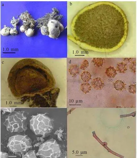
Scleroderma sp. is a member of Basidiomycota, fam. Sclerodermataceae. The species has basidioma: epigenous, globose, later irregular when mature, 0.66 + 0.17 cm in diam., with smooth surface, creamy white to brownish white. Peridium: rigid, simple, 0.2-1.0 mm thick, yellowish white. Sliced fresh of basidioma produced yellow exudates in Et.OH. Gleba: yellowish to light brown when young and dark brown when mature, without columella but having external basal pad. Clamp connection: present. Badiospores: pale brown in Melzer’s reagent and KOH, globose with reticulate ornamentation, 8 + 10 ì m in diam. (Figure 1).
Ectomycorrhizae formed very thick mantel (12 + 25 ì m), rough surface, creamy white in colour and monopodial pinnate or simple branching in the part of the young ectomycorrhizae, but becoming irregular pinnate in the base parts of the older ectomycorrhizae, sometimes ectomycorrhizae roots were interwoven very densely. The mantel structure was consisted of one layer of pseudoparenchymatous tissues. Moreover, hartig net was well developed within epidermal layer (Figure 2).
The growth of fungus in the media was 2.52 + 1.10 cm for 2 months. The colony of the fungus was yellowish white in colour and had soft texture and dense air mycelia. The bottom view of the colony apparently had yellowish brown, with the edge of the colony was white in colour. The isolates frequently produced yellowish brown
exudates (Figure 3). The growth of the cultured fungus was slower after two months and cease after three months in the culture.
DISCUSSION
Merbau does not have any symbiosis interaction with rhizobium and as well as with endomycorrhiza (vesicular-arbuscular mycorrhizas) fungi (Smith & Read 2008). Based on our histological observation of the root samples, there was no evidence that this tree species had endomycorrhiza (vesicular-arbuscular mycorrhizas) symbiont, thus the tree only formed Ectomycorrhiza (EcM).
Based on the morphological characteristics of sporocarps and badiospores, we found one species of EcM fungus associated with merbau, which was identified as a new species belonging to the genera of Scleroderma. There were no similar morphological characters of the fungus compared to the characters of Scleroderma species described by Rifai (1987), Sims et al. (1995), Chen (2006) and Sanon et al. (2009). By comparing the anatomical and morphological characters of the sporocarps, basidiospores and the EcM produced between baiting method and to those found in the fields, we confirmed that only one species of Scleroderma formed EcM association with merbau seedlings.
Based on the study, the characters observed from the culture were relatively difficult to be used for identification, since the characters were likely similar to those of other fungi such as isolate of S.dictyosporum Pat. However, it requires more taxonomic and culture experience to distinguish fungal isolates using identification keys based on cultural characters (Hutchinson 1991). Although there were similarities in cultural characters between Scleroderma sp. and S.dictyosporum Pat, but in fact the
a b
c d
e f
1.0 mm
1.0 mm
1.0 mm 10 µm
3.0 µm 5.0 µm
Figure 1. Scleroderma sp. (a) sporocarps globose to irregular forms; (b) Longintudinal section of sporocarp showing white yellowish peridium containing yellowish light brown spore mass; (c) mature sporocarp; with brown spore mass (d) Light microscopy view of basidiospores; (e) Scanning electron microscopy view of basidiospores, showing reticulate ornamentation; and (f) clamp connection.
Figure 2. Ectomycorrhizae of merbau. (a) Ectomycorrhizal root system of merbau with monopodial pinnate branching; (b) Scanning electron microscopy image of an ectomycorrhizal root tip of merbau with dense interwoven mycelium; (c) Cross-section of the ectomycorrhizal association of Scleroderma sp on merbau, showing thick mantel; (d) morphotype of mantel, showing pseudoparenchymatous tissue mantel type. M: matel, HN: hartig net, E: epidermal cells, C: cortex cell, En: Endodermal cells.
a b
1.0 mm 110 µm
c d
20 µm 20 µm
Figure 3. Cultural characters of Scleroderma sp. in Modified Melin-Norkrans (MMN) agar media. (a) Exudates production during isolation in MMN agar media; (b) the top view of the culture; (c) the bottom view of the culture.
sporocarps and basidiospores of these two fungi were distinctly different. Sporocarps of Scleroderma sp. were creamy white to brownish white in colour and never had a size of more than 1 cm in diameter. The sliced sporocarps excluded yellow exudates in Et.OH and in culture medium. All these characters can not be found in S.dictyosporum Pat which having sporocarps with brownish yellow to dark brown in colour (Sanon et al. 2009). Basidiospores of Scleroderma sp. have a size of 8-10 ì m, while those of S.dictyosporum Pat have a size of 10-13 ì m (Sims et al. 1995). Basidiospore of Scleroderma sp. showed flattened reticulate ornamentation comparing to that of S. dictyosporum which have prominent reticulate ornamentation (Rifai 1987; Kuo 2004; Sanon et al. 2009). However, the utilization of morphology features of EcM fungi solely based on basidiocarps for identification is not sufficient owing to variability of some characters (Wurzburger et al. 2001; Nouhra et al. 2005). The colour of the basidiocarp of Scleroderma sp. may be affected by environmental factors such as light intensity, exposure, and litter composition (Way et al. 1995). During rainy season the color of the basidiocarp tended being lighter, close to white instead of creamy white. The identification based on the anatomy and morphology of EcM is regarded as complementary of the first method though sometimes both methods may provide contradictory results (Wurzburger et al. 2001; Nouhra et al. 2005). EcM formed by different fungi on the same host plant, or by the same fungi on different host plant may be structurally distinct and differ (Dames et al. 1999), such as found in the case of mycorrhizas of white truffles of the genus Tuber (Giomaro et al. 2000). Moreover, study in Scleroderma species showed that it is difficult to distinguish between Scleroderma species based only on morphology of basidiocarps and basidiospores. This is because although they display different morphological characters, they might show identical genetic patterns (Sanon et al. 2009). However, the fungal and EcM description in this study is invaluable being used at least for initial identification in the field prior to molecular analysis.
Therefore, further work of taxonomic status of the species using DNA sequences is still needed to compare with other studies and to confirm whether the species is a new species. Correct identification of the species taxa of the fungus is prime essential for further studies (Brundrett et al. 1996), and as well as of the EcM (Dames et al. 1999). Tedersoo et al. (2007) previously used DNA sequences of ectomycorrhizal merbau root tips and revealed 15 fungi species, 2 species among them were Scleroderma, which forming EcM with merbau. If all fungi species associated with merbau were rightfully confirmed as ectomycorrhizal fungi, as previously observed by Tendersoo et al. (2007), further supported by the fact that merbau may occur in primary or old secondary forests on a wide variety of soils (PROSEA 1994), we may suggest that the finding of many different EcM fungi is highly possible.
Possibly, Scleroderma sp. takes its main role in the association of the early stage mycorrhizae than in the late stage mycorrhizae, since this fungus commonly occurred
beneath merbau seedlings. This also means that other EcM fungi could probably replace the previous species as merbau grows older. The dynamic of EcM fungal communities is a normal phenomenon in natural condition such as the different at EcM fungal community on different age of Quecus rubra L. (Gebhardt et al. 2007), and the declining of EcM fungi taxonomic richness as soil N availability increased (Lilleskov et al. 2002). In fact in merbau the fungal species may be overlap to those of other host plants (Tedersoo et al. 2007).
The fungus was easily isolated and cultured in MMN agar media culture in vitro although the growth was relatively slow. It was only 2.52 + 1.10 cm per two months. The success of the culture and the production of the sporocarp in the greenhouse through baiting method facilitates to the production of the mycelium aggregates and the spore mass for inoculums.
ACKNOWLEDGEMENT
This research was financially supported by a competitive scholarship of Ministry of Education, Indonesia 2007 and a research grant of doctoral student, Postgraduate Program, Bogor Agricultural University (IPB) 2009.
REFERENCES
Agerer R. 2006. Fungal relationships and structural identity of their ectomycorrhizae. Mycol Progress 5:67-107.
Brundrett M, Bougher N, Dell B, Grove T, Malajczuk N. 1996. Working with mycorrhizas in Forestry and Agriculture. Wembley, WA: Australian Centre for International Agriculture Research (ACIAR).
Chen YL. 2006. Optimization of Scleroderma spore inoculum for Eucalyptus nurseries in south China [Thesis]. Pert: Murdoch University.
Dames JF, Straker CJ, Scholes MC. 1999. Ecological and anatomical characterization of some Pinus patula ectomycorrhizas from Mpumalanga, South Africa. Mycorrhiza 9:9-24.
Dress EM. 1938. Kort oversicht der geslachten Intsia en Pahudia. Korte medeeling van het Boschbouwproefstation No. 67. Gebhardt S, Neubert K, Wöllecke J, Münzenberger B, Hüttl RF.
2007. Ectomycorrhiza communities of red oak (Quercus rubra
L.) of different age in the Lusatian lignite mining district, East Germany. Mycorrhiza 17:279-290.
Giomaro G, Zambonelli G, Sisti D, Cecchini M, Evangelista V, Stocchi V. 2000. Anatomycal and morphological characterization of mycorrhizas of five strains of Tuber borchii Vittad.
Mycorrhiza 10:107-114.
Giovanni P. 1985. MacDonald encyclopedia of mushrooms and toadstolls. London: Macdonald & Co (Publisher) Ltd. Hutchinson LJ. 1991. Description and identification of cultures
of ectomycorrhizal fungi found in North America. Mycotaxon
42:387-504.
Keizer GJ. 1998. The complete encyclopedia of mushrooms. Lisse, The Netherland: Rebo Publ.
Kjøller R, Bruns TD. 2002. Rhizopogon spore bank community within and among California pine forest. Mycologia 95:603-613.
Largent DL, Baroni TJ. 1988. How to identify mushrooms to genus I-VI. Eureka, C.A: Mad River Pr, Inc.
Nouhra ER, Horion TR, Cazares E, Castellano M. 2005. Morphological and molecular characterization of selected
Ramaria mycorrhizae. Mycorrhiza 15:55-59.
Pedersen CT, Sylvia DM. 1996. Mycorrhiza: ecological implications of plant interactions. In: Mukerdji KG (ed).
Concepts in mycorrhizal research. Netherlands: Kluwer Acad Publ.
[PROSEA] Plant Resources of South-East Asia. 1994. Plant Resources of South-East Asia 5. In: Soerianegara, Leummans RHMJ (eds). (1) Timber Trees: Major commercial timbers. Bogor: PROSEA.
Rifai MA. 1987. Malesian Scleroderma (Gasteromycetes). Trans Mycol Soc Japan 28:97.
Sanon KB, Bâ AM, Delaruelle C, Duponnois R, Martin F. 2009. Morphological and molecular analysis in Scleroderma species associated with some Caesalpinioid legumes, Dipeterocapaceae and Phyllantaceae trees in southern Burkina Faso. Mycorrhiza
19:571-584.
Sims KP, Watling R, Jeffries P. 1995. A revised key to the genus Scleroderma. Mycotaxon 56:403-420.
Smith SE, Read DJ. 2008. Mycorrhizal symbiosis. 3rd edition. New York: Acad Pr.
Tedersoo L, Suvi T, Beaver K, Kõljag U. 2007. Ectomycorrhizal fungi of the Seychelles: diversity patterns and host shifts from the native Vateriopsis seychellarum (Dipterocarpaceae) and
Intsia bijuga (Caesalpiniaceae) to the introduced Eucalyptus robusta (Myrtaceae) but not Pinus caribeae (Pinaceae).
Phytologist 175:321-333.
Watling R. 2006. The Sclerodermatoid fungi. Mycoscience 47:18-24.
Watling R, Lee SS, Turnbull E. 2002. The occurrence and distribution of putative ectomycorrhizal basidiomycetes in regenerating south-east asian rain forest. In: Watling R, Frankland AM, Isaac S, Robinson CH (eds). Tropical mycology, Volume I: Macromycetes. New York: CABI Publ. p 25-53. Way Y, Sinclair L, Hall IR, Cole ALJ. 1995. Boletus eduli Semen
Lato: a new record for New Zealand. NZ J Crop & Hort Sci
23:227-231.
![The significance of mycorrhiza in regeneration of merbau [Intsia bijuga (Colebr.) O. Kuntze] originating from Papua](data:image/gif;base64,R0lGODlhAQABAIAAAP///wAAACH5BAEAAAAALAAAAAABAAEAAAICRAEAOw==)