www.pjbt.org ISSN Online: 2312-7791
MUCOXIN ENHANCED TRANSCRIPTION AND PROTEIN EXPRESSION OF P53 IN BREAST CANCER CELL LINE T47D
Muhartono*, Ermin R**, Bayu PDJ*
*Department of Pathology, Medical Faculty of Lampung University. **Department of Physiology, Medical Faculty of UIN Maulana Malik Ibrahim, Indonesia
E. mail: [email protected]
Article received 13.11.2017, Revised 20.3.2018, Accepted 23.3.2018
ABSTRACT
Mucoxin is a type of acetogenin isolated from Rollinia mucosa leaves which is known to inhibit cell
proliferation and induce apoptosis. However, the mechanism of mucoxin in regulating and eliminating cancer cells was not clear. This study investigated the mucoxin effect on the transcriptional-translational and posttranslational processes of p53 gene in breast cancer cells line T47D. Breast cancer cells line T47D was divided into three groups referred to hours of assays, namely hour 24th, 48th, 72nd, where each group was given mucoxin with six
difference doses, namely 0.1 ng/mL, 0.5 ng/mL, 1 ng/mL, 5 ng/mL and 10 ng/mL with three replicates. Transcription of p53 gene was assayed by quantitative PCR (qPCR), whereas the expresssion of p53 protein assayed by immunocytochemistry. Mucoxin enhance p53 gene and protein in all treatment group. p53 gene transcription increased significantly in 48 h, while expression of p53 protein increased significantly in 72 h. Conclusion: Mucoxin increased transcription of p53 gene and expression of p53 protein on cell line T47D.
Keywords: Acetogenin, anticancer, cell line T47D, mucoxin, p53
INTRODUCTION
Breast cancer is one of the most frequent cancer in women [Koss and Melamed, 2006, Sumitha and Devi, 2016] and the second leading cause of death after cervical cancer [Rachmani et al., 2012]. Bre-ast cancer treatment can be done through surgery, chemotherapy, hormone therapy, radiation ther-apy, and immunological therapy [Kumar et al., 2005). Chemotherapy is one of the most common treatments. Unfortunately, chemotherapy has vari-ous side effects such as hair loss, nausea, vomit-ing, sleep disorders, diarrhea, skin redness and weight loss. Moreover, some cancer cells begin resistant to chemotherapy [Yuan et al., 2003]. The development of new anti-cancer drugs is a strategic choice in an effort to improve the sensi-tivity of pre-existing therapies. One of the prom-ising antitumor compound is acetogenin and its derivative compounds [Betancur et al., 1999, Yuan et al., 2003, Oberlies et al., 1997). One of the commercially type of acetogenin compound is mucoxin. Mucoxin is a non-classical acetogenin compound that was first isolated from the leaf extract of Rollinia mucosa by McLaughlin in (1996). This compound has a selective cytotoxic effect against pancreatic cancer cells (PACA-2) and breast cancer cells (MCF-7) (Yang et al., 2013). The results of our previous study showed, these compounds are able to induce apoptosis and inhibit the proliferation of breast cancer cell line T47D [Muhartono et al., 2016].
However, the molecular mechanisms of mucoxin in inducing apoptosis and inhibiting proliferation has not fully understood. One of the genes that play a role in the apoptosis process and prolife-ration is the p53 gene encoding the p53 protein. p53 is a tumor suppressor that acts as a trans-cription factor from genes involved in the cell cycle and apoptotic checkpoint phase in cancer cells. This causes p53 become gene target for new anticancer drugs. This study investigated the muc-oxin effect on transcriptional-translational and post translational process of p53 gene in breast cancer cell line T47D. The transcription-transla-tional process of p53 gene assayed by quantitative PCR (qPCR), which is shows the number of amp-lified gene products. Whereas the posttranslatio- nal process assayed by expresssion of p53 protein tested by immunocytochemistry. If mucoxin is proven to enhance p53 genes and protein, so this compound will become very potential to be used as a safe anticancer compounds.
MATERIALS AND METHODS
48th, 72nd hours, where each group was given mucoxin with six difference doses, namely 0.1 ng/mL; 0.5 ng/mL; 1 ng/mL; 5 ng/mL and 10 ng/mL with three replication of each.
Cell Culture: The cells were grown in Roswell Park Memorial Institue Medium (RPMI1640). The media supplemented with 10% Fetal Bovin Serum (FBS) GibcoTM (Thermo Fisher Scientific Cat. 26140-079) and 0.2 mL bovine insulin (Sig-ma Aldrich Cat. No. 15500 and CAS RN 11070-73-8) at 37oC in 5% CO2.. Thawing process per-formed in waterbath at 37oC for 2-4 min. Then, 5x10⁴ cells/cm2 was taken into T-flask and incuba-ted at 37o C in 5% CO2. When cells density reac-hed 80% confluent, trypsinization was done using 0.25% trypsin + 0.53 mM EDTA solution and then subcultured in new culture vessels and incu-bated at 37o C in 5% CO2. Afer two times passa-ging, the T47D cells ready to be treated.
Mucoxin Treatment: Mucoxin preparation was made by diluting the powder of mucoxin in 1 mL of 0.1% DMSO. The stock solution then diluted further in accordance with the needs of the six treatment concentration. After subcultured for two times, cells were diluted with RPMI and seeded in 24 well plate with a cell density of 5x10⁴ cell/cm2 in each well. Once the cell density reach 80% confluent, the cells treated with mucoxin of diff-erent concentration as follows: 0 ng/mL (K), 0,1 ng/mL (P1), 0,5 ng/mL (P2), 1 ng/mL (P3), 5 ng/ mL (P4) and 10 ng/mL (P5). After being treated, the cells were incubated in accordance with the lenght of hours that have been assigned to each group, i.e. 24, 48 dan 72 h.
Gene Transcription Assays: Expression of p53 gene in T47D cells were determined by quantita-tive PCR (qPCR) methods using RealMODTM Green Real Time PCR kit (Intron Biotecnology). RNA was extracted from breast cancer cell line T47D using easy Total RNA Extracion Kit (Intron Biotechnology). Primer for determining p53 exp-ression was forward primer 5’‒CTGAGGTTGG- CTCTGACTGTACCACCATCC‒3’ and reverse primer 5’‒CTCATTCAGCTCTCGGAAGCATT
-TGCGGTGGAC‒3’. β-actin gene was used as the
internal control (house keeping gene) while cont-rol sample was used as the calibrator gene. The qPCR data, analyzed using Light Cycler software from ROCHE.
Protein expression assays: After incubation per-iod, cells were washed using PBS, then fixed in 4% paraformaldehid. The primary anti body used for P53 assay were p53 polyclonal anti body (Rabbit IgG anti p53) from Bioss USA with dilut-ion of 1:100 in PBS with 1% FBS. The secondary anti body used were Ultratek HRP Anti-Polyval-ent (DAB) from SkyTex Laboratories. The results were visualized using Nikon Coolpix 4500. The expression of P53 protein in each treatment was calculated by summing the number of brownish product arising from the reaction between HRP and DAB in five fields over view and then divided by the number of replicates (n=3).
Statistical Analysis: Comparison of mean values between treatment presented as mean±SD and analyzed using ANOVA followed LSD test with a 95% confidence level.
RESULTS
Effects of Mucoxin on p53 Gene Transcription Effect of mucoxin treatment on p53 gene trans-cription are presented in Table 1. Based on that data, it is clear that mucoxin treatment enhanced p53 gene transcription comparing to control in all group (p<0,05). Furthermore, the highest trans-cription was exposed to mucoxin for 48 hours, at a mucoxin dose of 5 ng/mL (P4).
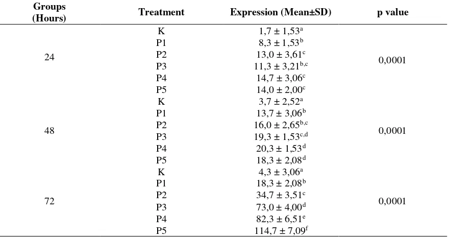
Effect of Mucoxin on p53 Protein Expression Effect of mucoxin treatmen on p53 protein expre-ssion in T47D cells are presented in Table 2. Comparing to control, mucoxin treatment affected p53 protein expression in all groups. The highest mucoxin effect was found in treated groups for 72 hours, with 10 ng/mL (P5) of mucoxin. Based on this data, it can be inferred that mucoxin is able to stabilize and activate the p53 protein by some post translation modification. The inference was supp-orted by highly significant difference (p<0,001) between mean values of p53 protein expression.
Table 1. Mean transcription of p53 gene in T47D cells treated with mucoxin
Groups
(Hours) Treatment
Transkription
(Mean±SD) p value
24
K 233,8 ± 7,45a
0,0001
P1 290,2 ± 6,40b
P2 315,1 ± 9,53c
P3 374,9 ± 11,38d
P4 432,6 ± 12,18e
P5 285,1 ± 14,37b
48
K 278,9 ± 11,10a
0,0001
P1 535,5 ± 14,12b
P2 863,4 ± 12,94c
P3 913,7 ± 5,60d
P4 1045,2 ± 19,58e
P5 746,1 ± 11,80f
72
K 235,9 ± 10,46a
0,0001
P1 482,9 ± 13,52b
P2 321,6 ± 5,17c
P3 443,5 ± 22,76d
P4 375,8 ± 20,13e
P5 118,2 ± 15,61f
Mean±SD values in the same hour group followed by the same superscript are not
different at α 0,05 by LSD test
Table 2. Mean expression of p53 protein in T47D cell treated with mucoxin Groups
(Hours) Treatment Expression (Mean±SD) p value
24
K 1,7 ± 1,53a
0,0001
P1 8,3 ± 1,53b
P2 13,0 ± 3,61c
P3 11,3 ± 3,21b,c
P4 14,7 ± 3,06c
P5 14,0 ± 2,00c
48
K 3,7 ± 2,52a
0,0001
P1 13,7 ± 3,06b
P2 16,0 ± 2,65b,c
P3 19,3 ± 1,53c,d
P4 20,3 ± 1,53d
P5 18,3 ± 2,08d
72
K 4,3 ± 3,06a
0,0001
P1 18,3 ± 2,08b
P2 34,7 ± 3,51c
P3 73,0 ± 4,00d
P4 82,3 ± 6,51e
P5 114,7 ± 7,09f
Mean±SD values in the same hour group followed by the same superscript are not different at α 0,05 by LSD test
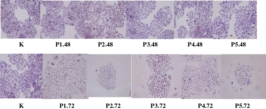
Fig. 1: Immunocytochemistry expression of p53 protein in breast cancer cells treated with mucoxin in different concentration and exposure time
DISCUSSION
The p53 is a tumor suppressor gene that plays an important role in maintaining genomic integr- ity.This gene is able to mediate cellular responses against cellular damage, through transcriptional regulation of genes involved in cell cycle proce-sses, DNA repair, apoptosis and aging [Adike-savan et al., 2014]. Various conditions that can trigger cell damage, such as DNA damage, hypo-xia and chemical induction, inducethe p53 activity [An et al., 1998]. p53 regulation activity, can be grouped into 2 mechanisms; transcriptional-trans-lational mechanism and post transtranscriptional-trans-lational mecha-nism. This study was conducted to determine the effect of mucoxin on both mechanisms.
The results of this study showed that mucoxin administration increased p53 gene transcription in accordance with the dose and duration of treat-ment (Table 1). These results are consistent with previous studies of several other acetogenin com-pounds, such as desacetyluvaricin and anonacin, which is showed that the acetogenin compound can increase p53 expression in some cancer cells [He et al., 2011, Yuan et al., 2003].
Increased transcription of the p53 gene by muc-oxin, is thought to occur through several mecha-nisms. Mucoxin appears to increase or inhibit one or several transcription factors that bind to the promoter portion of the p53 gene sequence, such as HOXA 5, NF‒κB, Myc/Max, C/EBPβ and RPB-Jk [Meyer and Targa, 2011; Reisman et al., 2012]. Mucoxin may also affect the p53's epige-netic mechanism. Mucoxin is thought to prevent methylation of the p53 gene promoter. Several studies have found a correlation between DNA hypermethylation in p53 gene promoters with low
mRNAs formed in hepatocellular cancer and leuk-emia [Hervouet et al., 2013, Schoofs et al., 2014]. Mucoxin was also suspected to affect the configu-ration settings of p53 gene chromatin. More open chromatin configuration, it is easier to activate p53 transcription [Nicoll et al., 2001].
In line with its transcription, mucoxin was also able to increase p53 protein expression in T47D breast cancer cells (Table 2). These results sho-wed that p53 protein synthesized is active and stable which is characterized by an increase in expression in accordance with the duration of treatment.
P53 protein is a very unstable and has a short half-life, so it does not affect the progress of cell cycle [Sabary et al., 2017] Mucoxin treatment seems that p53 protein undergoes posttranslational modi-fication, so this protein is stable. Posttranslational modification of p53, commonly occurs by cova-lent modification, in which p53 proteins will und-ergo phosphorylation at various points, resulting in a p53 ubiquitination mission, which results in a decrease in the degradation of this protein [Gu and Zhu, 2012].
In addition, posttranslational modifications also made p53 proteins being active [Gu and Zhu, 2012]. This occurs through changes in DNA bin-ding activity in specific sequences of p53 gene. p53 is a transcriptional activator of a particular gene based on its ability to bind to the sequence of the gene. Normally, this binding is inhibited by inhibition of C domain terminal. When there is exposure to stress, this inhibition will be elimi-nated, resulting in increased DNA binding. Conse-quently, there is an increased activity of the p53
K P1.48 P2.48 P3.48 P4.48 P5.48
protein [Sakaguchi et al., 1999]. In addition to C domainterminals, p53 protein activation capabili-ties can also be induced by changes in N transac-tivation domain terminal.
Another mechanism of p53 activation may also occur through changes in the subcellular location of the p53 protein. Normally, latent p53 protein will reside in the cytoplasmic, when exposure to stress, p53 protein will be accumulated in the cell nucleus [Shaulsky et al., 1990]. As a result there is accumulation of this protein in cells, proven by higher expression of this protein along with the increase of observation time.
The p53 gene encodes the p53 protein which is a tumor suppressor. As a tumor suppressor, p53 plays a very important role in preventing exce-ssive cell proliferation and maintaining genomic integrity [Yang et al., 2013]. p53 will be activated in response to the presence of stress signals origi-nating inside or outside the cell. The presence of a stress signal, will induce various upstream media-tors, such as 14ARF and Mdm2, make p53 stable and active [Christopher et al., 2006]. In this study, stress derived from mucoxin with various doses postulated would be a future chemotherapy agent to overcome current chemotherapy drug, which is not resistance or giving minimal side effects. The activated p53 protein will act as a regulatory pro-tein that triggers a variety of major biological responses to cell proliferation and the process of cell apoptosis [Mollereau and Ma, 2014].
Based on the fact that mucoxin significantly incre-ased transcription and expression of protein p53, where is the p53 are the main factor in the apo-ptosis dan proliferation of cancer cells, it can be concluded that mucoxin can be an aternative therapy for breast cancer.
Acknowledgement
Authors are grateful for supports from Faculty of Medicine, University of Lampung.
REFERENCES
Adikesavan, A.K., Sudipan, K., Patricia, P., Liguo, W., Shuang, L. and L. Wei, Activation of p53 Transcription Activity by SMRT: a Histone Deacetylase 3-Independent Function of a Transcription Corepressor. Molecular and Cellular Biology 34(7): 1246-61 (2014). An, W.G., Meera, K., Celeste, S., Emin, M.,
Mikhall, V.B. and M.N. Leonard, Stabiliza- tion of Wild Type p53 by Hypoxia-Induci- bleFactor1α.Nature 39(26):405-408 (1998) Betancur, G.L.A., Saez, J., Granados, H., Sala-
zar, A. and J . E . Ossa, Antitumor and Anti-viral Activity of Colombian Medicinal Plant
Extracts. Mem. Inst. Oswaldo Cruz. 94 (4): 531-535 (1999).
Christopher, L.B. and W. Gu, p53 Ubiquitination: Mdm2 and beyond. Mol. Cell 21(3): 307–315 (2006).
Gu, B. and W.G. Zhu, Surf the Post–translational Modification Network of p53 Regulation. Int. J. Biol. Sci. 8(5): 672–684 (2012).
He, H.B., Wu, X.L. and B. Yu, The Effect of Des- acetyluvaricin on The Expression of TL-R4 and
p 53 Protein in Hepg 2.2.15. Hepat. Mon. 11(5): 364-367 (2011).
Hervouet, E., Cheray, M. and F.M. Vallette, DNA Methylation and Apoptosis Resistence in Can-cer Cells. Cells 2: 545–573 (2013).
Koss, L.G. and M.R. Melamed, D iagnostic Cyto-logy and Its Hitopathologic Basis, 5th ed. Philadelphia Lippincott Williams & Wilkins Pp .1104-30 (2006)
Kumar, V., Abbas, A.K. and N. Fausto, Robbins and Cotran Pathologic Basis and Disease, 7th ed. Philadelpia Elsevier Sounders Pp. 1149 (2005).
Meyer,S.R.andR.F.Targa,Transcriptional and Epigenetic Regulation of p53 Tumor Supp-ressor Gene Epigenetis 6(9):106877 (2011). Muhartono, Asep, S., Sutyarso and M. Kanedi, Anti Proliferative and Apoptotic Effect of Mucoxin (Acetogenin) in T47D Breast Cancer Cells. Biomedical and Pharmacology Journal 9 (2): 491-498 (2016).
Mollereau, B. and D. Ma, The p53 Control of Apoptosis and Proliferation: Lesson from Dro-sophila. Apoptosis 19(10): 1421-9 (2014). Nicoll, G., Crichton, D.N., McDowell, H.E.,
Ker-nohan, N., Hupp, T.P. and A.M. Thompson, Expression of the Hypermethylated in Cancer Gene (HIC–1) is Associated With Good Out-come in Human Breast Cancer. Br. J. Cancer 85(12): 1878–1882 (2001).
Oberlies, N.H., Croy, V.L., Harrison, M.L. and J. R. McLaughin, The Annonaceous Acetogenin Bullatacin is Cytotoxic Against Multidrug Res-istantHumanMammaryAdenocarcinoma (MC-F-7/Adr)Cells.Cancer Letters 115:73-79 (1997) Rachmani, E.P.N., Suhesti, T.S., Widiastuti, R. and Aditiyono, The Breast of Anticancer from Leaf Extract of Annona muricata Againts Cells Line T47D. International Journal of Applied
Science and Technology 2(1): 157-164 (2012). Reisman, D., Paula, T., Amanda, P. And B.
Sabary, A.H., Mohammed, S.A.R. and A.W.T. W.T. Moshtak, Deection of P53 and Bax Gene Associated with Helicobacter pylorii Infection. Pak. J. Biotechnol. 14(3): 383-388 (2017). Sakaguchi, K., Herrera, J.E., Saito, S., Miki, T.,
Bustin, M., Vassilev, A., et al., DNA Damage Activates p53 Through a Phosphorylation– Acetylation Cascade. Genes Dev. 15(12): 1677–1679 (1998).
Schoofs, T., Berdel, W.E. and C.M. Tidow, Spot light on Epigenetics in Hematologic Malig-nancies. Origins of Aberrant DNA Methylation in Acute Myeloid Leukemia. Leukemia 28: 1– 14 (2014).
Shaulsky, G., Zeev, A.B. and V. Rotter, Sub-cellular Distribution of the p53 Protein During the Cell Cycle of Balb/c 3T3 Cells. Oncogene 5(11): 1707–11 (1990).
Sumitha, J., T. Devi, Breast Cancer Diagnosis Through Analysis of BRCA Gene Using Mac-hine Learning Algorithms. Pak. J. Biotechnol. 13(4): 231-235 (2016).
Yang, H.Y., Wen, Y.Y. and C.H. Chen, 14-3-3σ Positively Regulates p53 and Suppresses Tumor Growth. Mol. Cell Biol. 23(20): 7096-7107 (2013).