*Contact address:
Assistant Prof. Dr. Amin Derakhshanfar, Department of Pathobiology, Faculty of Veterinary Medicine, Shahid Bahonar University of Kerman, P.O. Box 7616914111, Kerman, Iran, Phone and Fax: +98 341 263079; E-mail: [email protected]
A study on avian cellulitis in broiler chickens
Amin Derakhshanfar∗∗∗∗∗
,,,,,
and Reza GhanbarpourDepartment of Pathobiology, Faculty of Veterinary Medicine, Shahid Bahonar University of Kerman, Kerman, Iran
DERAKHSHANFAR, A., R. GHANBARPOUR: A study on avian cellulitis in broiler chickens. Vet. arhiv 72, 277-284, 2002.
ABSTRACT
Avian cellulitis in broiler chickens, especially on the thighs and abdominal wall, has been observed more frequently in recent years. In the present study, over a one-year period 98 broiler carcasses with cellulitis were diagnosed at slaughterhouse. The lesions were characterized by thickening and brown discoloration of the skin. A fibrinopurulent exudate with some caseation was seen in the subcutaneous tissues. Microscopically, hyperkeratosis, thickening of the dermis, infiltration of mononuclear cells and heterophils, along with fibrinocaseous exudates, were present. In 90 of the 98 (91.8%) broiler bacteriological samples E. coli was isolated, and in 82 (91.1%) of these samples it was the only bacterial species found. Serotyping results revealed that E. coli isolates were distributed among 6 different serotypes. The most prevalent serotype was O78 (52.2%). In addition, Staphylococcus aureus and Actinomyces pyogenes were isolated from 12 and 2 cases, respectively. This study confirms the frequent association of E. coli with cellulitis lesions in broiler chickens, along with isolation of S. aureus and A. pyogenes. The latter have not been reported in the previous studies.
Key words:cellulitis, broiler chickens, Escherichia coli, Staphylococcus aureus, Actinomyces pyogenes
Introduction
Skin diseases are the main reason for condemnation of carcasses in
slaughtered broilers (BERGMANN et al., 1995). However, the number of
A. Derakhshanfar and R. Ghanbarpour: A study on avian cellulitis in broiler chickens
the slaughterhouses is increasing (MESSIER et al., 1993). Avian cellulitis, also
known as inflammatory process, infectious process, or IP, is a chronic skin disease which is characterized by sheets of caseated, heterophilic exudate in subcutaneous tissues. Lesions are located in the skin between the thigh
and midline (CALNEK et al., 1997). E. coli is that most often isolated from the
lesions, although other agents, such as Pasteurella multocida,
Pseudomonas aeruginosa, Enterobacter agglomerans, Proteus
vulgaris, and Streptococcus dysgalactiae have also been isolated (GOMIS
et al., 2001; PEIGHAMBARI et al., 1995a). Usually, the lesions can be detected
only after the feathers have been removed (MESSIER et al., 1993). No clinical
signs are visible in the living flock (GLÜNDER, 1990).
The object of the present study was to determine the role of various
serotypes of E. coli and possibly other bacteria in the development of
lesions of cellulitis, as seen in a broiler slaughterhouse in Kerman.
Materials and methods
Selection of carcasses. Over a one-year period (2000-2001) a total of
98 defeathered broiler carcasses with non-lacerated lesions of cellulitis were sampled. The lesions were located in the thigh and abdominal area and characterized by brown discolouration of the skin. The skin was thickened and scabby. A fibrinopurulent exudate with some caseation was seen in the subcutaneous tissues. Ten carcasses with no visible lesions were also selected as controls. The carcasses were shipped in ice to the Kerman Veterinary College and were immediately examined. A sterile cotton swab was used to remove moist material from the lesion in subcutaneous tissue, without any contamination from the outer surface.
Bacteriology. A sterile swab was used to collect fibrinopurulent material
for bacteriological examinations. All swabs were streaked onto MacConkey agar (Biolife Laboratories, Italy) and bovine blood agar (Biolife Laboratories,
Italy). Plates were incubated at 37 oC for 24 hours under aerobic condition.
E. coli isolates were identified by standard biochemical techniques (QUINN,
et al., 1994) and were maintained on tryptic soy agar (Biolife Laboratories,
Italy) slants until they were serotyped. For serotyping of E. coli isolates,
slide agglutination test. The other isolates (on blood agar) were identified
as described by QUINN et al. (1994).
Pathology. Samples from both diseased and healthy skins were collected
and fixed in 10% buffered formalin. After fixation the tissues were processed by standard methods and embedded in paraffin. Sections were cut at 5 ìm and stained by hematoxylin and eosin.
Results
Gross pathology. The colour of the skin around the inflamed area
changed to brown. Carcasses with skin lesions were in good body condition. Thickening of the diseased skins was obvious. The size of the lesions ranged from 7 to 12 cm in diameter. The lesions were localized to the skin of the thigh and the side of the abdominal wall (Fig.1). Yellowish plaques were observed in the subcutaneous tissues.
Fig. 1. Thickened and brown discolouration skin of a 7-week-old broiler chicken with cellulitis
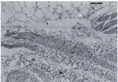
Histopathology. Microscopically, the lesions were characterized by
hyperkeratosis and thickening of the dermis with evidence of infiltration of mononuclear cells and heterophils. Necrotic cellular debris and fibrinocaseous exudates were present. In a few cases there was evidence of epidermis ulceration (Figs. 2, 3).
A. Derakhshanfar and R. Ghanbarpour: A study on avian cellulitis in broiler chickens
Fig. 2. Skin and subcutis of a 7-week-old broiler chicken with cellulitis. Note the presence of ulcer in the epidermis (short arrow) and infiltration of inflammatory cells
(arrow heads) in the subcutaneous tissue. H&E, scale bar = 500 µm.
Fig. 3. Subcutis of a 7-week-old broiler chicken. Note the marked accumulation of heterophils and mononuclear cells (arrow head) in the subcutaneous fatty tissue. H&E,
A. Derakhshanfar and R. Ghanbarpour: A study on avian cellulitis in broiler chickens microscopic agglutination acording to the trapping area with corresponding results
Table1. Results of serotyping cellulitis isolates of E. coli
O groups No of isolates O1 5 O2 13 O20 3 O36 2 O78 47 O115 1 Untypable 19 Total 90 Discussion
Avian cellulitis in broiler chickens is characterized by a diffuse inflammatory reaction in the subcutaneous tissue which results in the
complete or partial condemnation of the carcass at processing (SINGER et
al., 1999). E. coli is most often isolated from the lesions, although a variety
of other bacteria occasionally have been recovered (CALNEK et al., 1997;
GOMIS et al., 2001). Pathogenicity of E. coli cellulitis strains has been
determined in previous studies. Gross and microscopic findings showed that cellulitis isolates induced more severe lesions than airsacculitis and
Bacteriology. In 90 of the 98 (91.8%) broilers sampled, E. coli was
isolated from the lesion, and in 82 (91.1%) of these samples it was the only
bacterial species found. In one case E. coli and Actinomyces pyogenes
were present. In addition, in one case Actinomyces pyogenes was the
only microorganism which could be isolated. Staphylococcus aureus was
the only bacterium, which grew in 5 cases. Additionally, E. coli along with
Staphylococcus aureus observed in 7 cases. No bacteria were isolated
from 2 instances. Table 1 shows the results of serotyping cellulitis isolates
of E. coli. These isolates belong to 6 O groups, and 19 of them were
faecal isolates (PEIGHAMBARI et al., 1995b). In the present study a total of 6
different O serogroups of E. coli were identified, with O78 serogroup being
the most frequent. This serogroup is known to include virulent strains
associated with severe E. coli infection in poultry. It is also recognized as
one of the serogroups associated with enterotoxigenic E. coli strains that
can affect humans. This could have public health implications since it has
been demonstrated that E. coli strains colonizing the intestinal tract of
poultry can be transmitted to humans in close contact with the birds, and
these E. coli can be isolated from human faecal samples (MESSIER et al.,
1993). Severe cephalic swelling and facial cellulitis in turkeys associated
with fowl cholera was diagnosed. This unusual presentation of fowl cholera could be the result of local infection introduced by pecking trauma. Most turkeys with facial cellulitis had no internal lesions typical of fowl cholera,
whereas birds with lung or liver lesions had facial cellulitis (JEFFREY et al.,
1993). Some evidence exists indicating that certain clonal groups identified
by multilocus enzyme electrophoresis may be specific to cellulitis E. coli,
although in many cases the cellulitis E. coli were identified with other
pathogenic strains (NGELEKA et al., 1996). Other observations of
cellulitis-affected broiler carcasses support the hypothesis of cellulitis-type E. coli,
because the vast majority of broilers with cellulitis lesions are otherwise normal at necropsy. Scratches and injuries to the skin, increase in relative bird density, litter and chick quality and onset of sexual maturity are
predisposing factors for cellulitis (PEIGHAMBARI et al., 1995b; HARRIS et al.,
1978; JEFFREY et al., 1999).
In the present study, S. aureus was isolated from 12 cases.
Staphylococcus spp. is a normal inhabitant of skin and mucous membranes.
Some have the potential to be pathogenic and produce disease if allowed
entry through skin or mucous membranes (CALNEK et al., 1991). This organism
was isolated from skin infection in chickens in industrialized poultry units (KOHLER et al., 1980).
In this study, Actinomyces pyogenes was isolated from two cases.
Although serious outbreaks of osteomyelitis caused by this organism in
commercial turkeys have been reported (BARBOUR et al., 1991), none of the
previous studies reported this species from poultry skin diseases. However,
A. pyogenes should be considered an avian pathogen (CALNEK et al., 1991).
References
BARBOUR, E. K., M. K. BRINTON, A. CAPUTA, J. B. JOHNSON, P. E. POSS (1991): Characteristics of Actinomyces pyogenes involved in lameness of male turkeys in North-Central United States. In: Diseases of poultry (Calnek, B. W., H. John Barnes, C. W. Beard, L. R. McDougald, Y. M. Saif , Eds.), 10th ed, 1997, Mosby-Wolfe, p.
289.
BERGMANN, V., K. KOGLIN, A. VALENTIN (1995): Skin diseases as a reason for condemnation of broiler carcasses. Tierärztl. Prax. 23, 374-380.
CALNEK, B. W., H. JOHN BARNES, C. W. BEARD, W. M. REID, H. W. YODER (1991): Diseases of poultry. 9th ed., Iowa State University Press, pp. 294-295.
CALNEK, B. W., H. JOHN BARNES, C. W. BEARD, L. R. MCDOUGALD, Y. M. SAIF (1997): Diseases of poultry. 10th ed., Mosby-Wolfe, pp. 136-137.
GLÜNDER, G. (1990): Dermatitis in broilers caused by Escherichia coli: isolation of Escherichia coli from field cases, reproduction of the disease with Escherichia coli O78:K80 and conclusion under consideration of predisposing factors. Zentralbl. Veterinärmed. B 37, 383-391.
GOMIS, S. M., C. RIDDELL, A. A. POTTER, B. J. ALLAN (2001): Phenotypic and genotypic characterization of virulence factors of Escherichia coli isolated from broiler chickens with simultaneous occurrence of cellulitis and other colibacillosis lesions. Can. J. Vet. Res. 65, 1-6.
HARRIS, G. C., M. MUSBAH, J. N. BEASLEY, G. S. NELSAN (1978): The development of dermatitis (scabby-hip) on the hip and thigh of broiler chickens. Avian Dis. 22, 122-130.
JEFFREY, J. S., L. SHIVAPRASAD, C. J. CARDONA, B. R. CHARLTON (1993): Facial cellulitis associated with fowl cholera in commercial turkeys. Avian. Dis. 37, 1121-1129.
JEFFREY, J. S., R. P. CHIN, R. S. SINGER (1999): Assessing cellulitis pathogenicity of Escherichia coli isolated in broiler chickens assessed by an in vivo inoculation model. Avian Dis. 43, 491-496.
KOHLER, B., H. NATTERMANN, W. WITTE, F. FRIEDRICHS, E. KUNTER (1980): Staphylococcus aureus infection in chickens in industrialized poultry units. 2. Microbiological studies: Staphylococcus aureus and other pathogens. Arch. Exp. Veterinärmed. 34, 905-936.
MESSIER, S., S. QUESSY, Y. ROBINSON, L. A. DEVERIESE, J. HOMMEZ, J. M. FAIRBROTHER (1993): Focal dermatitis and cellulitis in broiler chickens: bacteriological and pathological findings. Avian Dis. 37, 839-844.
It seems that further investigations are needed to clarify the significance of this organism.
A. Derakhshanfar and R. Ghanbarpour: A study on avian cellulitis in broiler chickens
DERAKHSHANFAR, A., R. GHANBARPOUR: Istraživanje avijarnog celulitisa u tovnih piliæa. Vet. arhiv 72, 277-284, 2002.
SAŽETAK
Avijarni celulitis posebice na koži nogu i trbušne stijenke tovnih piliæa èesto je opisivan u posljednje vrijeme. U radu je tijekom jednogodišnjeg razdoblja celulitis dokazan u 98 tovnih piliæa na jednoj klaonici. Promjene su se oèitovale zadebljanjem i smeðkastom bojom kože. Utvrðen je fibrinopurulentni eksudat s kazeifikacijom u potkožnom tkivu. Mikroskopski je utvrðena hiperkeratoza, zadebljanje kože, infiltracija mononuklearnih stanica i heterofila zajedno s fibrinokazeoznim eksudatom. U 90 (91,8%) od 98 tovnih piliæa izdvojena je bakterija Escherichia coli, a u 82 (91,1%) bila je jedina izdvojena vrsta. Serotipizacijom je potvrðen nalaz šest razlièitih serovarova. Najèešæi serovar bio je O78 (52,2%). Bakterija Staphylococcus aureus bila je izdvojena iz 12 piliæa dok je bakterija Actinomyces pyogenes izdvojena iz 2 pileta. Ovim istraživanjem je potvrðena èesta povezanost bakterije Escherichia coli, Staphylococcus aureus i Actinomyces pyogenes sa znakovima avijarnog celulitisa, a po prvi put su dokazane i vrste S. aureus i A. pyogenes. To nije dokazano dosada.
Kljuène rijeèi: celulitis, tovni piliæi, Escherichia coli, Staphylococcus aureus, Actinomyces pyogenes
Received: 4 June 2002 Accepted: 30 October 2002 NGELEKA, M., J. K. KWAGA, D. G.WHITE, T. S.WHITTAM, C. RIDDELL, R.
GOODHOPE, A. A. POTTER, B. ALLAN (1996): Escherichia coli cellulitis in broiler chickens: clonal relationships among strains and analysis of virulence-associated factors of isolates from diseased birds. Infec. Immun. 64, 3118-3126.
PEIGHAMBARI, S. M., J. P. VAILLANCOURT, R. A. WILSON, C. L. GYLES (1995a): Characteristics of Escherichia coli isolates from avian cellulitis. Avian Dis. 39, 116-124.
PEIGHAMBARI, S. M., R. J. JULIAN, J. P. VAILLANCOURT, C. L. GYLES (1995b): Escherichia coli cellulitis: experimental infections in broiler chickens. Avian Dis. 39, 125-134.
QUINN, P. J., M. E. CARTER, B. K. MARKEY, G. R. CARTER (1994): Clinical veterinary microbiology. 1st edn., Wolfe Publishing, pp. 209-242.
SINGER, R. S., J. S. JEFFREY, T. E. CARPENTER, C. L. COOKE, R. P. CHIN, E. R. ATWILL, D. C. HIRSH (1999): Spatial heterogeneity of Escherichia coli DNA fingerprints isolated from cellulitis lesions in chickens. Avian Dis. 43, 756-762.