*Corresponding author: E-mail: [email protected]
Available online at htp://medpet.journal.ipb.ac.id/
The Potential Use of Secondary Metabolites in
Moringa oleifera
as an Antioxidant Source
A. Fitria,*, T. Toharmatb, D. A. Astutib, & H. Tamurac
aStudy Program of Nutrition and Feed Science, Faculty of Animal Science, Graduate School, Bogor Agricultural University
bDepartment of Nutrition and Feed Technology, Faculty of Animal Science, Bogor Agricultural University Jalan Agatis, Kampus IPB Darmaga Bogor 16680, Indonesia
cDepartment of Applied Biological Science, Faculty of Agriculture, Kagawa University Miki-cho, Kagawa 761-0795, Japan
(Received 26-06-2015; Reviewed 07-08-2015; Accepted 17-09-2015)
ABSTRACT
This present study determined antioxidant activity, lipid peroxidation, total phenolic, total la -vonoids and phytochemicals in moringa leaves and moringa stem. Analysis used in this study was 1, 1-diphenyl-2-picrylhydrazyl (DPPH) method for antioxidant activity, thiobarbituric acid reactive substances (TBARS) method for lipid peroxidation, Folin-Ciocalteu method for total phenolic, total lavonoid and UFLC (Ultrafast Liquid Chromatography) for identiication and quantiication of phe -nolic compounds. The results showed that moringa leaves had higher ability to scavenge free radical, total phenolic, and total lavonoid than moringa stem (P<0.001). Malondialdehyde production, the end product of lipid peroxidation, in moringa leaves was lower than moringa stem (P<0.001). Ferulic acid was the major active compound in both moringa leaves and moringa stem. This present study indicated that moringa leaves and moringa stem could be used as feed additive which had a good potential to prevent oxidative stress in animals.
Key words: antioxidant, lipid peroxidation, moringa leaves, moringa stem, phytochemicals
ABSTRAK
Penelitian ini dilakukan untuk mengidentiikasi aktivitas antioksidan, peroksidasi lemak, to -tal fenolik, to-tal lavonoid, dan senyawa fenolik yang terdapat pada daun kelor dan batang kelor. Analisis yang digunakan dalam penelitian ini adalah analisis aktivitas antioksidan (metode DPPH atau 1, 1-diphenyl-2-picrylhydrazyl), analisis peroksidasi lemak (metode TBARS), analisis total feno-lik (metode Folin-Ciocalteu), analisis total lavonoid, identiikasi dan kuantiikasi senyawa fenolik
(Ultrafast Liquid Chromatography). Hasil menunjukkan bahwa daun kelor memiliki kemampuan
dalam meredam radikal bebas (DPPH) lebih tinggi daripada batang kelor (P<0,001). Produksi malon-dialdehida pada proses peroksidasi lemak pada sampel daun kelor lebih rendah daripada batang kelor (P<0,001). Kandungan total fenolik dan lavonoid pada daun kelor lebih tinggi dibandingkan batang kelor. Asam ferulat merupakan senyawa fenolik yang paling banyak di daun dan batang kelor. Penelitian ini mengindikasikan bahwa daun kelor dan batang kelor dapat digunakan sebagai pakan suplementasi yang berpotensi dalam mencegah terjadinya oksidatif stress di ternak.
Kata kunci: antioksidan, peroksidasi lemak, daun kelor, batang kelor, itokimia
INTRODUCTION
A tropical country like Indonesia has temperatures
ranging from 23 to 33o C and humidity of 45%-97%
(BMKG, 2013). In the tropics, with a combination of high ambient temperatures and humidity, livestock are prone
to heat stress. Heat stress can cause a lowered produc
-tion and high mortalities leading to economic losses at farm level. Oxidative stress occurred because the production of free radicals is higher than the antioxidant defense system in the body. Free radicals can damage cell walls and impair the function of organs that play crucial roles in the body’s metabolic system (Yoshikawa & Naito, 2002). Oxidative stress in animals can inhibit growth rate, decrease appetite, decrease nutrient di
decrease the production quality, and increase animals
mortality (Sugito et al., 2007; Rajani et al., 2011; Hashemi
et al., 2012).
The antioxidants could counteract free radicals thus it could prevent oxidative damage in the cells. There are two principle mechanisms of antioxidant action. The irst is the primary antioxidant donates an electron to the free radicals and the second is removal of the secondary antioxidant by quenching chain-initiating catalyst (Lobo et al., 2010). Some studies have already proven that using antioxidant for animals could give a positive impact on health and production (Rajani et al., 2011; Hashemi et al., 2012). Antioxidant compounds can be found in the plant that it has phytochemical sub
-stances such as α-tocopherol, β-carotene, ascorbic acid, lavonoids, carotenoids, anthocyanins, phenolic com
-pounds, zinc and selenium (Moyo et al., 2012; Atowadi et al. 2010). In addition, the natural antioxidant sources is the most recommended to be used for animals feeding.
Moringa oleifera (moringa) is widely cultivated in many locations in the tropics. Moringa leaves contain protein, β-carotene, vitamins A, B, C and E, minerals, steroids, alkaloids, quercetin and kaempferol. Several
studies have proven that moringa leaves have several
functions as antioxidant, anticancer, anti-atherosclerotis, anti-inlammatory, antitumor, to regulate thyroid status, improve growth performance in broiler chickens, im -prove the immune system (Chumark et al., 2008; Iqbal &
Bhanger 2006; Nkukwana et al., 2014; Verma et al., 2009; Rao et al., 2001; Sreelatha et al., 2011). Moringa leaves and fruits have high nutrients contents and advanta
-geous for health, it is not only used for human food but also for animal feed. In order not to compete with human being, some parts of moringa plant can be used as animal feed such as stem and middle to old leaf. Thus the objectives of this study are to evaluate antioxidant activity in moringa leaves and moringa stem that can be used for animals feed.
MATERIALS AND METHODS
Plant Materials
Moringa forage was collected from Bekasi, West Java, Indonesia. The samples were obtained in February 2014. Moringa forage consisted of leaves and twigs that were at least the third branch and smaller. The moringa forage was separated into leaves and stems. The stems were twigs or small branches that were not more than 1 cm in diameter. All samples were dried at 50oC for 24 h
and were ground to a ine powder.
Plant Extract Preparation
In a polypropylene centrifuge tube (50 mL) contain
-ing 2 g of powdered samples, 10 mL of distilled water and 15 mL of acetonitrile were added. This extraction method called QuEChERS (quick, easy, cheap, efective, rugged and safe) method that is normally used for de
-termining of pesticides residues in agricultural product. However, with a litle modiication this method can also be used to analyze the chemical compound in foods
(Sato et al., 2015). The solution was homogenized for 1
min at 1000 rpm, followed by the addition of sodium chloride (1 g), trisodium citrate dehydrate (1 g), diso
-dium hydrogen citrate sesquihydrate (0.5 g), and anhy
-drous magnesium sulfate (4 g) and then shaked it for 1 min.Then, the mixture was centrifuged at 3000 rpm for 5 min. The acetonitrile extract was evaporated and dried with a vacuum pump. The extracts were stored at -20oC.
The amounts of extracts obtained from moringa leaves and moringa stem were 4.6% and 2.1%, respectively.
Analysis of DPPH Free Radical Scavenging Activity
The efect of extracts and standard solution (Trolox) on the DPPH (1, 1-diphenyl-2-picrylhydrazyl) were determined by using method described by Zhu et al. (2014). Samples were diluted with methanol and acetic acid bufer (1:1). A volume of 0.25 mL of extracts or the standard in diferent concentrations (200, 100, 50, 10, 5, 1 μg/mL) was mixed with 0.25 mL of acetic acid bufer (0.10 M), 0.25 mL of methanol and 0.25 mL of DPPH (0.4 mM in methanol). The reaction mixtures were vigorous
-ly mixed and incubated for 30 min at room temperature in the dark. The absorbance of mixtures was measured by spectrophotometer at 517 nm. The SC50 (scavenging capacity in 50%) value was determined by GraphPad PRISM 6 from the output of scavenging activity values. Each sample was done in triplicates. The SC50 value was expressed as μg/mL.
Lipid Peroxidation Analysis (TBARS Assay)
Lipid peroxidation inhibition was measured by using a thiobarbituric acid (TBA) method (Tamura & Yamagami, 1994). A 100 μL of each sample solution (10 mg/mL) and linoleic acid (5 mg) was mixed with 4.8 mL of 0.2% SDS Tris-HCl bufer. Then, 100 μL of 20 mM ferrous sulfate aqueous solution was added and the mixture was incubated for 16 h at 37oC. The production
of TBARS, mainly malondialdehyde, was measured in the following way. One mL of the reaction solution above was mixed with 3 mL of 0.05 N HCl and 1 mL of 0.05 M TBA-50% acetic acid and then incubated for 30
min at 100oC. After cooling to room temperature, 4 mL
of n-butanol was added and the mixtures were shaken
vigorously and added 200 μL of EtOH. The mixtures were centrifuged (10 min, 2500 rpm) and the absorbance
of the n-butanol layer was measured at 535 nm. To make
a standard curve, 1, 1, 3, 3-tetraethoxypropane standard solution (0, 2.5, 5, 10, 20, 30, 40, 50, 60, 70, 80 nmol/mL) were measured. Lipid peroxidation was expressed in nmol of MDA per 1 mg of linoleic acid (nmol MDA/mg linoleic acid).
Total Phenolic Content (TPC) Analysis
Total phenolic content was estimated by following the procedure of Folin-Ciocalteu by Asada & Tamura (2012) with slight modiication. A 20 μL of extracts (1 mg in 1 mL methanol) was mixed with 200 μL of 50% phenol reagent, 200 μL of 10% sodium carbonate aque
was stored for 1 h in the dark place at room tempera
-ture. Then the absorbance was read at 760 nm with spec
-trophotometer by 5 mm length of a quarz cell using an UV−vis spectrophotometer (JASCO V-520-SR). A calibration curve was prepared with standard gallic acid (0, 200, 400, 600, 800, and 1000 μg/mL) and the results were expressed as mg gallic acid equivalent (GAE) per g of extract (mg GAE/g dry extract).
Total Flavonoid Content (TFC) Analysis
Total lavonoid content was measured following
the method of Poudel et al. (2008), using quercetin as a
standard. A volume of 0.25 mL of the sample (5 mg in 1 mL 95% ethanol) or standard solution of quecertin (1-0.0625 mg/mL) was pipeted in a test tube and mixed with 0.25 mL 0.1% HCl in 95% ethanol (v/v) and 4.55 mL 2% HCl (v/v). The solution was incubated for 15 min and the absorbance was read at 360 nm with a spectro
-photometer. Total lavonoid content was expressed as mg quercetin equivalent per g of extract (mg QE/g dry extract).
Ultrafast Liquid Chromatography (UFLC) Analysis
The UFLC analysis of all samples was carried out on a Shimadzu UFLC system equipped with LC-20AD pump and SPD-M20A detector. The separation was performed on a Mightysil RP-18 GP column (3.0 mm i. d x 100 mm). The mobile phases were 10% CH3CN-0.5% TFA as eluent A and 100% CH3CN-0.5% TFA as eluent
B. The low rate was ixed at 0.5 mL/min and the column temperature was set at 40 oC. A gradient program was
performed as follows: 0 min, 100% (A); 1 min, 100% (A);
All data were expressed as mean ± standard devia
-tion (SD) in triplicate at least. For comparisons between
samples, data were analyzed by ANOVA and Duncan test (SPSS, version 16.0). A probability of 5% or less was accepted as statistically signiicant.
RESULTS AND DISCUSSION
Total Phenolic Content and Total Flavonoid Content
Phenolic compounds are secondary metabolites in fruits or plants. These compounds are derivatives of the pentose phosphate, shikimate, and phenylpropanoid pathways in plants (Randhir et al., 2004). Phenolics, including lavonols, lavones, phenolic acids, proan
-thocyanidins and tannins, are reported as the major contributors to the biological properties like antioxidant activities of moringa leaves (Astuti et al., 2011; Vongsak et al., 2013). extracted with acetone (120.33 mg tannin equivalent (TE)/g dry extract) was higher than extracted with aque
-ous (40.27 mg TE)/g dry extract). The TPC of moringa leaves was 105 mg GAE/g in aqueous extracts (Singh et al. 2009), 103 mg GAE/g in methanol extracts, 97.2 mg GAE/ g in ethanol extracts (Sultana et al., 2014) and 123.3 mg GAE/g in 80% methanol extracts (Siddhuraju & Becker, 2003).
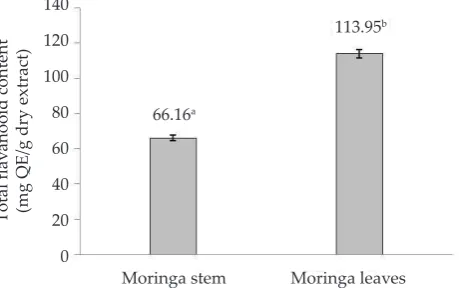
Quecertin and kaempferol are the major lavonoid compounds reported in this plant (Singh et al., 2009; Atowadi et al., 2010; Sultana & Anwar, 2008). In this paper, quecertin was used as a standard chemical to measure total lavonoid. Quecertin is one of lavonoid compound in lavonols group and is abundant in fruit and vegetables. Quecertin was reported had a great an
-tioxidant and antiallergic activity (Sato et al., 2015; Singh et al., 2009). All samples tested showed signiicantly dif
-ferences to the total lavonoid value (P<0.001). Moringa leaves (113.95 mg QE/g dry extract) had the highest lavonoid content, followed by moringa stem (66.16 mg QE/g dry extract) (Figure 2). Moyo et al. (2012) reported lavonoid content in moringa leaves extracted with
Figure 1. Total phenolic content in moringa leaves and moringa leaves (n= 4). Means with diferent superscript are sig
-niicantly diferent (P<0.001).
Figure 2. Total lavonoid content in moringa leaves and mor
-inga stem (n= 4). Means with diferent superscript are signiicantly diferent (P<0.001).
Total phenolic content (mg GAE/g dry extract)
acetone (295.01 mg QE/g dry extract) was higher than extracted with aqueous (45.1 mg QE/g dry extract).
These diferences could be due to several factors such as type of cultivation, climate, fruit variety, geo
-graphic origin, ripeness and extraction method (Deng et al. 2010; Vasco et al., 2008). All samples tested could be considered as a good source of phenolic compounds and antioxidants. Phenolic compounds as free radical scavengers could be due to their redox properties, pres
-ence of conjugated ring structures and carboxylic group which have been reported to inhibit lipid peroxidation (Oyedemi et al., 2010).
Moyo et al. (2012) reported that supplementa
-tion of 200 g powder of moringa leaves (equal to 0.5 g quecertin/g extract) increased antioxidant enzymes in goats such as glutathione, superoxide dismutase and catalase. The supplementation of moringa leaves and moringa stems could also be safely used as a source of antioxidant in goats feed and the others animals
(Sultana et al., 2014).
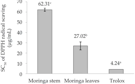
DPPH Radical Scavenging Activity
The method of DPPH radical scavenging activity to evaluate the antioxidant activity is an established pro
-cedure and is widely used to estimate the antioxidant activities of food. This method is easy to handle, low cost, reasonably and fast method to evaluate radical scavenging activity (Sharma & Bhat, 2009). This assay has also been used to establish antioxidant activity of herbal extract and phytochemicals (Moyo et al., 2012).
The result of DPPH radical scavenging activity is interpreted with SC50 values. All samples had an activ
-ity to scavenge free radicals (Figure 3). The SC50 values
of DPPH of moringa leaves (27.02 μg/mL) had lower than moringa stem (62.31 μg/mL), but trolox had the lowest values SC50 of DPPH (P<0.001). The lower the
SC50 value, the beter the antioxidant activity. The SC50
of DPPH in moringa leaves extracted with 70% ethanol was 62.94 μg/mL (Vongsak et al., 2013). The diferences
of SC50 values of DPPH scavenging radical activity was
possibly caused by diferent solvent used, cultivation, extraction method and varieties of plant. The high activ
-ity of antioxidant in moringa leaves was caused by the
high value of phenolic content and lavonoid content in this sample. Phenolic compounds in the sample have a linear correlation with antioxidant activity.
Lipid Peroxidation
Lipid peroxidation is one of the markers of oxida
-tive stress. Lipid peroxidation is always occurred in polyunsaturated faty acids such as linoleic acid, linolenic acid and arachidonic acid (cell membrane component) which are oxidized in various pathological conditions (Yoshikawa & Naito, 2002). Malondialdehyde (MDA) and 4-hydroxy-2-hexenal (HHE) are the end products from lipid peroxidation process. These products have cytotoxic, mutagenic, and neurotoxic properties and can promote cancer development in the gastrointestinal (GI) tract and liver (Del Rio et al., 2005; Long & Picklo, 2010).
The efects of moringa leaves and moringa stem on lipid peroxidation were summarized in Figure 4. In this study, there was a signiicant diferent in the efect of moringa leaves and moringa stem on lipid peroxida
-tion (P<0.001). In lipid peroxida-tion, the lower value of MDA indicates the stronger activity of inhibited lipid peroxidation. The moringa leaves (4.9 nmol MDA/mg of linoleic acid) had the strongest activity for inhibiting of lipid peroxidation compared to moringa stem (7.77 nmol MDA/mg of linoleic acid).
The percentages of lipid peroxidation inhibition in moringa leaves and moringa stems were 85.88 and 77.63%, respectively. Moringa leaves extracts that were used as goats feed inhibited lipid peroxidation by 81.33%. This value was higher than used sunlower (38.76%) and grass hay (1.99%) (Moyo et al., 2012).
Supplementation of moringa leaf meal up to 5% of dry mater intake in broiler chickens could improve faty acid proile and reduce lipid peroxidation in meat (Nkukwana et al., 2014). In addition, feeding 30% of moringa in sheep ration increased glucose and triglyc
-erides, increased albumin, globulin and IgG, and also decreased cholesterol (Astuti et al., 2011). It proved that inhibition of lipid peroxidation in moringa can improve product quality and health status in animals.
Figure 3. The SC50 of DPPH free radicals in moringa leaves and moringa stem (n= 3). Means with diferent super
-scripts are signiicantly diferent (P<0.001).
Figure 4. The lipid peroxidation in moringa leaves (n= 3), mor
-inga stem (n= 3) and control (n=4). Means with difer
Ultrafast Liquid Chromatography (UFLC) Analysis
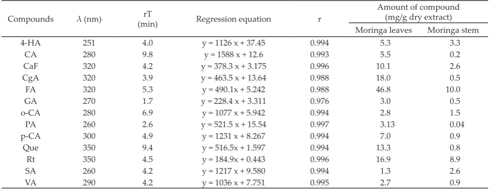
In the present study, identiication of chemical compounds in moringa leaves and stems was done by using UFLC. Some reference standards that were used
in this study are representative of three main group of
phenolics compound such hydroxybenzoic acids (gallic acid, protocatechuic acid, syringic acid, vanilic acid and 4-hidroxybenzoic acid), hydroxycinnamic acids (cafeic acid, ferulic acid, p-coumaric acid, o-coumaric acid, cin
-namic acid and chlorogenic acid), and lavonoids (rutin and quercetin).
Figure 5 (A) shows the chromatogram of standard compounds monitored at diferent wavelengths, se
-lected on the basis of maxima absorbance and maximum of peak area (Arimboor et al., 2008; Zu et al., 2006). The chromatogram of some standards was overlapped such as VA, SA and CgA, 4-HA, possibly because these stan
-dards were eluted closely. However, using absorption maxima we were able to compare them. For VA and SA, their chromatogram showed that SA had highest absor
-bance at 269 nm and smaller at 290 nm, this wavelength would be the detection wavelength for VA. So, the de
-tection wavelength for 4-HA is at 260 nm and CgA is at 338 nm.
Table 1 showed the phenolic compounds in morin
-ga leaves and morin-ga stem (Figure 5 B and 5 C). Ferulic acid was the most abundant phenolic compounds in moringa leaves (46.8 mg/g dry extract) and moringa stem (10.0 mg/g dry extract). Chlorogenic acid, rutin, quecertin, cafeic acid was predominantly phenolic compounds in moringa leaves. Then in the moringa stem, rutin was the second most of phenolic compounds in this study. It is possible that ferulic acid, chlorogenic acid, rutin, quecertin, cafeic acid provided strong scav
-enging free radicals and inhibition lipid peroxidation.
Figure 5. The chromatograms of standard reference of phenolic compounds (A), moringa leaves (B) and moringa stem (C) in the 270 and 340 nm. CA, cafeic acid; GA, gallic acid; PA, protocatechuic acid; 4-HA, 4-hidroxybenzoic acid; SA, syringic acid; Rt, rutin; p-CA, p-coumaric acid; FA, ferulic acid; o-CA, o-coumaric acid; Que, quercetin; CA, cinnamic acid; VA, vanilic acid; CgA, chlorogenic acid.
A
B
CONCLUSION
Moringa leaves and moringa stem had the ability to scavenge free radicals and to inhibit lipid peroxidation. Total phenolic and lavonoids content in moringa leaves were higher than moringa stem. Ferulic acid and rutin were found in both samples. Although the moringa stem had lower activity than moringa leaves, but both of them could be used as feed supplement (100 g of mor
-inga leaves powder or 360 g of mor-inga stem powder) to improve health status in animals.
ACKNOWLEDGEMENT
This study was done in Department of Applied Biological Science, Faculty of Agriculture, Kagawa University, Japan. This study was also supported by Japan Student Services Organization (JASSO) Scholarship.
REFERENCES
BMKG. Badan Metereologi, Klimatologi dan Geoisika. 2013. Prakiraan cuaca Indonesia. htp://www.bmkg.go.id/ BMKG_Pusat/Meteorologi/Prakiraan_Cuaca_Indonesia. bmkg [13 Februari 2014]
Arimboor R, K. S. Kumar, & C. Arumughan. 2008. Simultane
-ous estimation of phenolic acids in sea buckthorn (
Hip-pophae rhamnoides) using RP-HPLC with DAD. J. Pharm.
& Biomed. Anal. 47: 31–38. htp://dx.doi.org/10.1016/j. jpba.2007.11.045
Asada, T. & H. Tamura. 2012. Isolation of bilberry anthocyani
-din 3-glycosides bearing ortho-dihydroxyl groups on the b ring by forming an aluminum complex and their antioxi
-dant activity. J. Agric. Food Chem. 60: 10634-10640. htp:// dx.doi.org/10.1021/jf302476n
Astuti, D. A., A. S. Baba, & I. W. T. Wibawan. 2011. Rumen fer
-mentation, blood metabolites, and performance of sheep fed tropical browse plants. Med. Pet. 34: 201-206. htp:// dx.doi.org/10.5398/medpet.2011.34.3.201
Atowadi, S. E., J. C. Atowadi, G. A. Idakwo, B. Pfundstein, R. Haubner, G. Wurtele, H. Bartsch, & R. W. Owen. 2010. Evaluation of the polyphenol content and antioxidant properties of methanol extracts of the leaves, stem, and root barks of Moringa oleifera Lam. J. of Medicinal Food. 13: 710-716. htp://dx.doi.org/10.1089/jmf.2009.0057
Chumark P., P. Khunawat, Y. Sanvarinda, S. Phornchirasilp, N. P. Morales, L. Phivthong-ngam, P. Ratanachamnong, S. Srisawat, & K. S. Pongrapeeporn. 2008. The in vitro and
ex vivo antioxidant properties, hypolipidaemic and anti
-atherosclerotic activities of water extract of Moringa oleif-era Lam. leaves. J. Ethnopharm. 116: 439-446. htp://dx.doi. org/10.1016/j.jep.2007.12.010
Del Rio, D., A. J. Stewart, & N. Pellegrini. 2005. A review of recent studies on malondialdehyde as toxic molecule and biological marker of oxidative stress. Nutr., Metab. Car
-diovasc. Dis. 15: 316−328. htp://dx.doi.org/10.1016/j.num
-ecd.2005.05.003
Deng, S., B. J. West, & C. J. Jensen. 2010. A quantitative com
-parison of phytochemcial components in global noni fruits and their commercial products. Food Chem. 122: 267–270. htp://dx.doi.org/10.1016/j.foodchem.2010.01.031
Hashemi, S.R., I. Zulkili, H. Davoodi, Z. Zunita, & M. Ebra -himi. 2012. Growth performance, intestinal microlora, plasma faty acid proile in broiler chickens fed herbal
plant (Euphorbia hirta) and mixed of acidiiers. Anim. Feed Sci. & Tech. 178: 167-174. htp://dx.doi.org/10.1016/j.ani
-feedsci.2012.09.006
Iqbal, S., & M. I. Bhanger. 2006. Efect of season and produc
-tion loca-tion on antioxidant activity of Moringa oleifera leaves grown in Pakistan. J. Food Comp. & Anal. 19: 544– 55. htp://dx.doi.org/10.1016/j.jfca.2005.05.001
Lobo, V., A. Patil, A. Phatak, & N. Chandra. 2010. Free radi
-cals, antioxidant and functional foods: Impact on hu
-man health. Pharmacogn. Rev. 4: 118-126. htp://dx.doi. org/10.4103/0973-7847.70902
Long, E. K., & M. J. Picklo. 2010. Trans-4-hydroxy-2-hexenal, a product of n-3 faty acid peroxidation: make some room HNE. Free Radical Biol. Med. 49: 1−8. htp://dx.doi. org/10.1016/j.freeradbiomed.2010.03.015
Moyo, B., S. Oyedemi, P. J. Masika, & V. Muchenje. 2012. Polyphenolic content and antioxidant properties of
Mo-ringa oleifera leaf extracts and enzymatic activity of liver
Compounds λ (nm) (min)rT Regression equation r
Amount of compound (mg/g dry extract)
Moringa leaves Moringa stem
4-HA 251 4.0 y = 1126 x + 37.45 0.994 5.3 3.3
CA 280 9.8 y = 1588 x + 12.6 0.993 5.5 0.2
CaF 320 4.2 y = 378.3 x + 3.175 0.996 10.1 2.6
CgA 320 3.9 y = 463.5 x + 13.64 0.988 18.0 0.5
FA 320 5.3 y = 490.1x + 5.242 0.988 46.8 10.0
GA 270 1.7 y = 228.4 x + 3.311 0.976 3.0 0.5
o-CA 280 6.9 y = 1077 x + 5.942 0.994 2.8 1.5
PA 260 2.6 y = 521.5 x + 15.54 0.997 3.13 0.04
p-CA 300 4.9 y = 1231 x + 8.267 0.994 7.0 0.9
Que 350 9.4 y = 516.5x + 1.597 0.994 13.3 0.8
Rt 350 4.5 y = 184.9x + 0.443 0.996 16.9 8.9
SA 260 4.2 y = 1217 x + 9.580 0.994 1.3 2.6
VA 290 4.2 y = 1036 x + 7.751 0.995 2.7 0.9
Table 1. Wavelength detection (λ), retention time (rT), regression equation, and quantiication of phenolic compounds in moringa
leaves and moringa stems
from goats supplemented with Moringa oleifera leaves/ sunlower seed cake. Meat Sci. 91: 441-447. htp://dx.doi. org/10.1016/j.meatsci.2012.02.029
Nkukwana, T. T., V. Muchenje, E. Pieterse, P. J. Masika, T. P. Mabusela, L. C. Hofman, & K. Dzama. 2014. Efect
of Moringa oleifera leaf meal on growth performance, ap
-parent digestibility, digestive organ size and carcass yield in broiler chickens. Livest. Sci. 161: 139–146. htp://dx.doi. org/10.1016/j.livsci.2014.01.001
Oyedemi, S.O., G. Bradley, & A. J. Afolayan. 2010. In-vitro and
-vivo antioxidant activities of aqueous extract of Strychnos
henningsii Gilg. Afr. J. Pharm. Pharmacol. 4: 70-78.
Poudel, P.R., H. Tamura, I. Kataoka, & R. Mochioka. 2008. Phenolic compounds and antioxidant activities of peels and seeds of ive wild grapes and two hybrids native to Japan. J. Food Comp. & Anal. 21: 622-625. htp://dx.doi. org/10.1016/j.jfca.2008.07.003
Rajani, J., M. A. K. Torshizi, & S. Rahimi. 2011. Control of as
-cites mortality and improved performance and meat shelf-life in broiler using feed adjuncts with presumed antioxi
-dant activity. Anim. Feed Sci. & Tech. 170: 239-245. htp:// dx.doi.org/10.1016/j.anifeedsci.2011.09.001
Randhir, R., Y. T. Lin, & K. Shety. 2004. Phenolics, their anti
-oxidant and antimicrobial activity in dark germinated fen
-ugreek sprouts in response to peptide and phytochemical elicitors. Asia Pac. J. Clin. Nutr. 2004. 13: 295-307.
Rao, A. V., P. U. Devi, & R. Kamath. 2001. In vitro radioprotec
-tive efect of Moringa oleifera leaves. Indian J. Exp. Biol. 39: 858–863.
Sato, A., T. Zhang, L. Yonekura, & H. Tamura. 2015. Antialler
-gic activities of eleven onions (Allium cepa) were atributed to quercetin 4’-glucoside using QuEChERS method and Pearson’s correlation coeicient. J. Functional Foods. 14: 581–589. htp://dx.doi.org/10.1016/j.jf.2015.02.029
Sharma, O. P. & T. K. Bhat. 2009. Analytical Methods; DPPH antioxidant assay revisited. Food Chem. 113: 1202-1205. htp://dx.doi.org/10.1016/j.foodchem.2008.08.008
Siddhuraju, P. & K. Becker. 2003. Antioxidant properties of various solvent extracts of total phenolic constituents from three diferent agroclimatic origins of drumstick tree (
Mo-ringa oleifera lam.) leaves. J. Agric. Food Chem. 51:
2144-2155. htp://dx.doi.org/10.1021/jf020444+
Singh, B. N., B. R. Singh, R. L. Singh, D. Prakash, R. Dhakarey, G. Upadhyay, & H. B. Singh. 2009. Oxidative DNA dam
-age protective activity, antioxidant and anti-quorum sens -ing potentials of Moringa oleifera. Food & Chem. Toxic. 47: 1109–1116. htp://dx.doi.org/10.1016/j.fct.2009.01.034
Sreelatha, S., A. Jeyachitra, & P. R. Padma. 2011. Antiprolif
-eration and induction of apoptosis by Moringa oleifera leaf
extract on human cancer cells. Food Chem. Toxic. 49: 1270-1275. htp://dx.doi.org/10.1016/j.fct.2011.03.006
Sugito, W. Manalu, D. A. Astuti, E. Handharyani, & Chairul. 2007. Morfometrik usus dan performa ayam broiler yang diberi cekaman panas dan ekstrak n-heksana kulit batang “jaloh” (Salix tetrasperma Roxb). Med. Pet. 30: 198–206. Sultana, B., & F. Anwar. 2008. Flavonols (kaempeferol, querce
-tin, myricetin) contents of selected fruits, vegetables and medicinal plants. Food Chem. 108: 879-884. htp://dx.doi. org/10.1016/j.foodchem.2007.11.053
Sultana, N., A. R. Alimon, K. S. Haque, A. Q. Sazili, H. Yaakub, & S. M. J. Hossain. 2014.The efect of cuting interval on yield and nutrient composition of diferent plant fractions
of Moringa oleifera tree. JFAE 12: 599-604.
Tamura, H., & A. Yamagami. 1994. Antioxidative activity of monoacylated anthocyanins isolated from muscat bailey a grape. J. Agric. Food Chem. 42: 1612-1615. htp://dx.doi. org/10.1021/jf00044a005
Vasco, C., J. Ruales, & A. Kamal-Eldin. 2008. Total phenolic compounds and antioxidant capacities of major fruits from Ecuador. Food Chem. 111: 816–823. htp://dx.doi. org/10.1016/j.foodchem.2008.04.054
Verma, A. R., M. Vijayakumar, C. S. Mathela, & C. V. Rao. 2009. In vitro and in vivo antioxidant properties of difer
-ent fractions of Moringa oleifera leaves. Food Chem. Toxic. 47: 21196–21201. htp://dx.doi.org/10.1016/j.fct.2009.06.005 Vongsak, B, P. Sithisarn, S. Mangmool, S. Thongpraditchote,
Y. Wongkrajang, & W. Gritsanapan. 2013. Maximizing total phenolics, total lavonoids contents and antioxidant activity of Moringa oleifera leaf extract by the appropriate extraction method. Ind. Crops Prod. 44: 566-571. htp:// dx.doi.org/10.1016/j.indcrop.2012.09.021
Yoshikawa T., & Y. Naito. 2002. What is oxidative stress? JMAJ. 45: 271–276.
Zhu, F., T. Asada, A. Sato, Y. Koi, H. Nishiwaki, & H. Tamura. 2014. Rosmarinic acid extract for antioxidant, antiallergic, and α-glucosidase inhibitory activities, isolated by su
-pramolecular technique and solvent extraction from pe
-rilla leaves. J. Agric. Food Chem. 62: 885-892. htp://dx.doi. org/10.1021/jf404318j
Zu, Y., C. Li, Y. Fu, & C. Zhao. 2006. Simultaneous determina
-tion of catechin, rutin, quercetin kaempferol and isorham