See discussions, stats, and author profiles for this publication at: https://www.researchgate.net/publication/313291243
Cytological analysis of ginseng carpel
development
Article in Protoplasma · February 2017 DOI: 10.1007/s00709-017-1081-4
CITATIONS
0
READS
142
9 authors, including:
Some of the authors of this publication are also working on these related projects:
Soil Microbial Community analysis
View project
Functional characterization of pollen development genes in rice
View project
Yu-Jin KimKyung Hee University
106PUBLICATIONS 943CITATIONS
SEE PROFILE
Johan Sukweenadhi
Universitas Surabaya
29PUBLICATIONS 93CITATIONS
SEE PROFILE
Deok-Chun Yang
Kyung Hee University
579PUBLICATIONS 4,432CITATIONS
SEE PROFILE
Dabing Zhang
Shanghai Jiao Tong University
187PUBLICATIONS 5,257CITATIONS
SEE PROFILE
All content following this page was uploaded by Jeniffer Silva on 23 October 2017.
1 23
Protoplasma
An International Journal of Cell Biology
ISSN 0033-183X
Protoplasma
DOI 10.1007/s00709-017-1081-4
Cytological analysis of ginseng carpel
development
1 23
ORIGINAL ARTICLE
Cytological analysis of ginseng carpel development
Jeniffer Silva1&Yu-Jin Kim1,2 &Dexin Xiao2&Johan Sukweenadhi1&Tingting Hu2&
Woo-Saeng Kwon1&Jianping Hu2,3&Deok-Chun Yang1&Dabing Zhang2
Received: 17 August 2016 / Accepted: 25 January 2017
#Springer-Verlag Wien 2017
Abstract Panax ginsengMeyer, commonly known as ginseng, is considered one of the most important herbs with pharmaceu-tical values due to the presence of ginsenosides and is cultivated for its highly valued root for medicinal purposes. Recently, it has been recognized that ginseng fruit contains high contents of triterpene such as ginsenoside Re as pharmaceutical compounds. However, it is unclear how carpel, the female reproductive tissue of flowers, is formed during the three-year-old growth before
fruit is formed in ginseng plants. Here, we reportP. ginseng
carpel development at the cytological level, starting from the initial stage of ovule development to seed development. The
carpel ofP. ginsengis composed of two free stigmas, two free
styles, and one epigynous bilocular ovary containing one ovule in each locule. Based on our cytological study, we propose that
the female reproductive development inP. ginsengcan be
classified into seven stages: early phase of ovule development, megasporogenesis, megagametogenesis, pre-fertilization, fertili-zation, post-fertilifertili-zation, and seed development. We also describe the correlation of the female and male gametophyte development and compare morphological differences in carpel development between ginseng and other higher plants. One unique feature for ginseng seed development is that it takes 40 days for the embryo to develop to the early torpedo stage and that the embryo is small relative to the seed size, which could be a feature of taxonomic importance. This study will provide an integral tool for the study
of the reproductive development and breeding ofP. ginseng.
Keywords Panax ginseng. Ontogeny . Ovule . Ultrastructure . Stages of carpel development
Introduction
Sexual reproduction is a major process in the life cycle of flowering plants, as it is essential for seed production to generate
a new population of offspring (Robert et al.2015; Zhang et al.
2015). This process depends on highly specialized reproductive
organs, stamen (male) and carpel (female), which are protected by flower buds until anthesis. Both reproductive organs have tightly coordinated mechanisms that are indispensable for effec-tive fertilization and production of viable seeds (Vivian-Smith
et al.2001; Robert et al.2015). Through the fusion of haploid
gametes, fertilization initiates the development of a new diploid
organism (Dresselhaus et al.2016). Following fertilization, the
ovule develops into a seed while the surrounding carpel differ-entiates into a fruit to complete a successful reproduction
(Coombe1976; Vivian-Smith et al.2001). The major seed
com-ponents, embryo and endosperm, are formed after the fusion of two sperm cells with two dimorphic female gametes, the egg and
the central cell, respectively (Dresselhaus et al.2016). Successful
Handling Editor: Liwen Jiang
Department of Oriental Medicine Biotechnology and Graduate School of Biotechnology, College of Life Science, Kyung Hee University, Yongin 446-701, South Korea
2
Joint International Research Laboratory of Metabolic and
Developmental Sciences, Shanghai Jiao Tong University–University of Adelaide Joint Centre for Agriculture and Health, School of Life Sciences and Biotechnology, Shanghai Jiao Tong University, Shanghai 20040, China
3 Department of Energy Plant Research Laboratory, Michigan State
University, East Lansing, MI 48824, USA Protoplasma
female reproductive development has to undergo several critical events, starting with the specification of the megasporocyte, which subsequently produces a functional megaspore (megasporogenesis) that afterwards forms the embryo sac (megagametogenesis). Through embryogenesis, the embryo sac
develops into an embryo (Reiser and Fischer1993).
Angiosperm carpels normally display the polygonum type of embryo sac, ovule with the anatropous curvature, and the nuclear endosperm. Although carpel development in angio-sperms share common characteristics, flower, carpel, and ovule in different species show a variety of developmental features, which have been investigated in a wide number of plant species
such asArabidopsis(Arabidopsis thaliana) (Schneitz et al.
1995), Iris tenaxDouglas (Wilson2001),Triteleia sp. (Berg
2003),Vochysiaceae (Litt and Stevenson2003),Araliaceae
(Costello and Motley 2004; Sokoloff et al.2007; Oskolski
et al.2010),Panaxsp. (Yu and Kim1992; Venugopal et al.
2013; Qi et al. 2015), Cytisus striatusand C. multiflorus
(Rodríguez-Riaño et al. 2006),Hydatellaceae (Rudall et al.
2008),Silene latifolia(Koizumi et al.2009),Drosera x obovata
(Rodondi et al.2009),Psychotria carthagenensisandRudgea
macrophylla(Figueiredo et al.2013), rice (Oryza sativa) (Kubo
et al.2013; Wu et al.2016a,b),TaraxacumWigg. (Musiałet al.
2013),Gaussia attenuata(Castaño et al.2014),Coffea arabica
(de Oliveira et al. 2014), and Encephalartos natalensis
(Woodenberg et al.2014). These studies clearly illustrate that
as a vital sexual reproductive organ, the ovule is involved in many complex systematic interactions during flower develop-ment. As the ovule is contained in the carpel, it is needed to understand the structure and development of the carpel as a whole. Many significant events in the life cycle of a plant, such as the production of the female gametophyte with the egg cell via meiosis, guidance and attraction of the pollen tube (male
gametes) at the micropyle—i.e., small opening formed from the
integuments through which the pollen tube enters to fuse the with the egg cell, canalization of male gametes toward the egg cell via the nucellus and female gametophyte, protection of the female gametophyte-containing nucellus, double fertilization, and the development of the new sporocyte, are all completed
within the carpel (Angenent and Colombo1996; Wang and
Ren2008; Endress2011). The anatomy and morphology of
carpel in different plant species are divergent, showing specific characteristics in the number of stigmas, position of the ovary, number of locules and ovules per locule, ovule size, ovule curvature, number and thickness of integuments, nucellus thickness, type of embryo sac, placentation, endosperm, and embryogenesis.
The genusPanaxL. belongs to the order Umbelliferales in
the Araliaceae family and is commonly used for medicinal
purpose. Among the 17 species in this genus,Panax ginseng
Meyer is the most widely used because of its high
pharmaco-logical efficacy (Kim et al.2015a). Propagation of ginseng
plants is dependent on seeds produced via self-fertilization.
At the third growth year, they initiate flowering and generate seed and undergo active reproductive development by forming
30–50 flowers in an umbel inflorescence after the fourth year.
Although studies onPanaxL. about post-fertilization to seed
development (Yu and Kim 1992; Venugopal et al.2013; Qi
et al.2015) and anther development (Kim et al. 2015b) have
conducted, it is still insufficient on understanding about carpel development. Elucidating and characterizing the carpel devel-opmental events at a cytological level will be helpful to develop inter- and intra-specific hybrids, which displayed hybrid vigor
on metabolite and plant yield (Kim et al.2016), and identify
genes at exact stages for hybridization barrier. In the present
research, we studied carpel development inP. ginseng. First, we
used light microscopic observations of transverse sections to analyze the complete ontogeny during carpel development
and cellular changes during P. ginseng carpel development.
We were able to divide the course of ginseng carpel develop-mental into seven stages, providing a comprehensive and de-tailed descriptive analysis at the ultrastructural level. In addi-tion, we correlated female gametophyte development with an-ther development that was described in our previous report.
Given the economic and medical importance ofP. ginseng, in
this study the timing of the occurrence of morphological events during reproductive development was investigated, aiming to improve the efficiency of ginseng production and breeding. We are providing a key asset in ginseng reproductive development that will allow interpreting defects and/or events derived from reproductive development, provide a basis knowledge for mo-lecular analysis of genes involved in carpel and stamen, and it will represent an essential tool to select specific reproductive developmental stages for ginseng breeding.
Materials and methods
Plant material
To study carpel development, inflorescences of 5-year-old
P. ginsengwere collected from the ginseng field of Kyung Hee
University (South Korea) from April to July (April 17, 23, May 1, 7, 12, 19, 25, June 1, 8, 15, 23, 29, and July 7) of 2015.
Light microscopy of semi-thin sections
Ginseng inflorescences at different stages were fixed using formalin-acetic acid alcohol (FAA, 50% ethanol, 5% glacial acetic acid, 3.7% formaldehyde). The fixed samples were dehydrated in a graded ethanol series (70%, 80%, 90%, and 100%) for 30 min per step and then embedded in Kulzer’s Technovit 7100 cold polymerizing resin (Heraeus Kulzer GmbH Philipp-Reis-Straße 8/13, D-61273 Wehrheim/Ts) by preinfiltration, infiltration, and
embedding at 45 °C (Igersheim and Cichocki1996; Beeckman
and Viane2000; Zhang et al.2013). Embedded samples were
J. Silva et al.
sectioned at 3–4μm thickness using an Ultratome III
ultramicro-tome (LKB) and stained with 0.25% toluidine blue O (Chroma Gesellshaft Shaud) at 42 °C. Bright-field photographs of the ovary sections were taken using a Nikon Eclipse 80i microscope and a Nikon DXM1200 digital camera.
Scanning electron microscopy
For scanning electron microscopy (SEM) examination, inflores-cences were collected, fixed, and washed using the same proce-dure for semi-thin section except the dehydration proceproce-dure, which used 3 min treatment of 20, 30, 40, 50, 60, 70, 80, 90, and 100% ethanol, respectively. After dehydration, samples were thoroughly dried at critical point (Leica EM CPD300). A Leica EM SCD050 ion sputter was used for aurum coating at 5-nm thickness. The aurum-coated samples were observed with a Hitachi S3400N scanning electron microscope.
Image analysis
Images were analyzed with the ImageJ version 1.49 m software (National Institutes of Health, USA) and the Fiji plugin. From ten samples at each developmental stage, more than five selected images of each stage samples were used for morphometric analysis on flower bud, stigma, ovules, and fruit.
Results
Ginseng carpel development during plant morphogenesis
P. ginsengis a slow-growing perennial herb that usually
pro-duces flowers in the third year of growth, when the stem de-velops three compound leaves, each with five leaflets, verticil-late—i.e., having a circular arrangement, at the apex of the stem
(Kim et al.2015a,2016). The inflorescence ofP. ginseng
con-sists of a single, terminal umbel—i.e., a rounded flower cluster
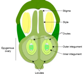
in which the individual stalks arise from about the same point, with small flowers. Ginseng carpel is composed of three parts: two free stigmas, two free styles, and one ovary (monocarpus). The ovary is epigynous—i.e., located below the petals and sepals, receptacular—i.e., the ovary is embedded in the floral
receptacle, and bicarpellar—i.e., the ovary has two locules and
a single ovule in each locule (Fig.1). These are consistent with
general characteristics of the Araliaceae family and the closely
related family, Umbelliferae (Costello and Motley2004). The
mature ovule is anatropous—i.e., the ovule is completely inverted so that the micropyle is situated next to the funicle,
bitegmic—i.e., the ovule has two integuments and
tenuinucellate—i.e., ovule with thin nucellus (Fig.1). The
sin-gle ovary develops into a simple flesh fruit (berry) and splits to release the seeds when ripe into two one-seeded mericarps
(Sachs and Bennett2011; Singh2010).
We used light microscopy of semi-thin sections to reveal
the cytological aspects ofP. ginsengcarpel development prior
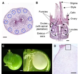
Fig. 1 Structure of aP. ginseng
flower.P. ginsengflower is monocarpus and has an epigynous and bilocular carpellate ovary with a single ovule in each locule, and anatropous, bitegmic, and tenuinucellate ovules. Cartoon is not drawn to scale
to seed formation.P. ginsengcarpel exhibits distinctive
fea-tures in different stages of plant morphology (Fig.2). On the
basis of visualized cellular events, we divided the carpel
de-velopment ofP. ginsenginto seven stages (Fig.3, Table1).
Stage 1—early phase of ovule development
When ginseng plant exhibits a parasol kind of shape with
unfolded leaves attached to the peduncle (Kim et al.2015b;
Fig.2a), the inflorescence has∼8–15 flower buds (Kim et al.
2015b; Fig.2h), each at 0.9686 ± 0.04 mm in length (Fig.4a), and stigmas that are each 0.3316 ± 0.01 mm in length
(Fig.2o). Ovule primordium was initiated at the center of
the ovary, from the placental tissues of carpel margins
(Fig.4b). Afterwards, each ovule primordium grows in
oppo-site directions and finally settling at 90° from the center
(Fig.4c). We denominated this stage as stage 1-I, at 1 to 5 days
after sprouting (DAS). Later, at 5 to 10 DAS, the ovule
pri-mordium elongates, becoming finger-like (Fig.4d), and then
undergoes the first meiotic division (Fig.4e), which we named
stage 1-II. Within the anther, the megasporocyte undergoes the
first meiotic division (Fig.4f, g).
Stage 2—megasporogenesis
During stage 2-I, at 10 to 14 DAS, the unfolded leaves detach
from the peduncle (Fig. 2b), the inflorescence holds more
flower buds (Fig. 2i) that are 1.4220 ± 0.02 mm in length
(Fig.5a) and the stigma 0.5000 ± 0.01 mm in length. Within
the ovary, a differentiated commissure and two locules are
observed (Fig. 2p). The megaspore mother cell enlarges
(Fig.5b), while the integuments have initiated as small
protu-berances (Fig.5c). The inner integument is composed of two
cell layers while the outer integument is composed of one cell
layer (Fig.5d). Concurrently, the anther is undergoing the first
meiotic division (Fig. 5(d1)). Afterwards, both integuments
elongate and the megaspore mother cell undergoes the second
meiotic division (Fig.5e). During this stage, the anther is also
under the formation of tetrads by meiotic division (Fig.5(e1)).
During stage 2-II, at 14–24 DAS, the outer integument
sur-rounds both the inner integument and nucellus, and the
nucel-lus is more prominent and becomes tenuinucellar (Fig.5e).
Stage 3—megagametogenesis
At 24 to 31 DAS, when ginseng leaves are partially expanded
and the inflorescence has ∼20–25 flower buds (b; Fig.2c, j),
differentiated funicles have developed within the ovary, and the
stigma length is 0.5598 ± 0.01 mm (Fig.2q). During this stage
(stage 3), the four-nucleus embryo sac appears and the outer integument completely encloses both the inner integument and
nucellus (Fig.5f). Ultimately, these four nuclei divide and
pro-duce an eight-nucleus embryo sac, with four nuclei in the chala-zal pole—i.e., the area below the antipodal cells, and four in the
micropylar pole—i.e., the area at where the integuments form an
opening for the pollen tube entry. Through this process, the em-bryo sac enlarges and the eight nuclei undergo reorganization and cellularization. Concurrently, the anther releases young
micro-spores from the tetrads (Fig.5(f1)).
Stage 4—pre-fertilization
At 31 to 40 DAS, when ginseng leaves are fully expanded and the peduncle have reached its maximum height (Kim et al. 2015b; Fig. 2d), theBsnowman shape^ flower buds (Kim
et al.2015b; Fig.2k) have stigmas of 1.0524 ± 0.11 mm in
length and ovules of 0.4722 ± 0.01 mm in length (Figs.2r and
6a), considered to be a common size in angiosperms ovules at
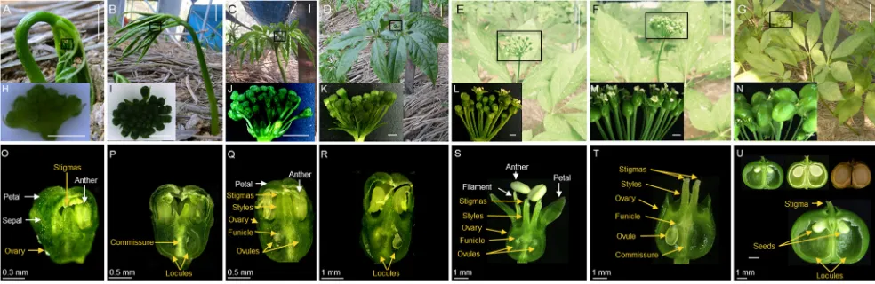
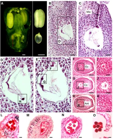
Fig. 2 Carpel development during plant growth inP. ginseng.aInitial stage, 1–5 DAS.bDeveloping stage, 10–14 DAS.c–eMaturing stage at 24 to 31 DAS (c), 31 to 40 DAS (d), and 40 to 45 DAS (e).f–gMature to senescence stage at 45 to 50 DAS (f) and 50 to 90 DAS (g). h–n
Inflorescence morphology at different plant developmental stages before fruit formation.o–uMorphogenesis of flower at different plant developmental stages before fruit formation.Scale bars= 1 cm (a–c), 5 cm (d–g), 2 mm (h–n).DASdays after sprouting
J. Silva et al.
the time of fertilization (Endress2011). Stage 4-I is defined as the stage when the ovule begins the formation of a vacuole
and the antipodals (Fig.6b, d). During stage 4-II, the
antipo-dals degenerate, while the central cell, synergids, and egg cell
appear (Fig.6e). The last two mitotic divisions give rise to the
cellularization process, where cells differentiate into different
lineages: egg cell, central cell, and antipodals (Fig.6f–k). In
stage 4-III, the micropyle opens (Fig.6c) to allow the entry of
the pollen tube.
In the course of stages 4-I to 4-III, the microspores undergo vacuolation, first and second mitotic divisions, and the anther
matures (Figs.3and6l–o). At the end of stage 4-III, the anther
starts to dehisce (Fig.6i) and the flower starts to open (Fig.6a).
Stage 5—fertilization
At 40 to 45 DAS, flowers in the inflorescence range from
closed flower buds to fertilized flowers (Kim et al.2015b;
Figs.2e, l, and 7a). The fertilized flowers exhibit withered
and pale yellow anthers, the filament has reached its
maxi-mum length (Kim et al.2015b; Fig.2s), and the stigma length
is 2.2473 ± 0.05 mm (Fig.7b), at which point pollination takes
place. During this stage, the fusion of sperm cells with the egg cell (to give rise to the zygote) and central cell (to give rise to the endosperm) takes place. We defined this stage as stage 5-I, when locules start to enlarge to make space for the fertilized
ovules to grow (Fig.2s).
Stage 6—post-fertilization
At 45 to 50 DAS, the inflorescence contains fertilized flowers
(Fig.2f, m) that each have a stigma of 2.1465 ± 0.09 mm in
length, enlarged ovules that are 1.387 ± 0.02 mm wide and
0.8853 ± 0.02 mm long (Fig. 2t). Eventually, the sepals,
petals, and stamens wither and fall off (Fig.7d). Stage 6-I
was defined as when the synergids disappear and the egg cells
break down (Fig.7e, f). At stage 6-II, the two polar nuclei fuse
with the second sperm cell to generate the endosperm (Fig.7e,
l) with visible haustoria at the micropylar pole.
Stage 7—seed development
During 50 to 90 DAS, the inflorescence is full of green fruits
that ultimately become yellow when matured (Fig.2g, n). The
size of the fruit ranges from 5.1140 ± 0.06 mm in length and 7 . 0 4 1 6 ± 0 . 0 3 m m i n w i d t h , a n d t h e s t i g m a i s
2.0092 ± 0.05 mm long (Fig.2u). In these fruits, the anthers
and petals have completely disappeared, the locules have reached its maximum size, to around 2.7430 ± 0.01 mm in width and 3.5177 ± 0.11 mm in length, to enclose the devel-oping seed, and the ovary subsequently hardens to form the
seed coat (Fig.8a). Stage 7-I was defined as when after
fertil-ization the egg is transformed into the zygote of about 0.072 mm in length. Asymmetric cell division leads to an apical-basal axis. As the endosperm cellularizes, the cells be-gin to take on differential fates with internal endosperm cells
that accumulate nutrient storage reserves (Fig.8b, h). During
stage 7-II, the zygote begins longitudinal division to a length
of about 0.116 mm (Fig.8c, i), and the endosperm is
persis-tent. At stage 7-III, two rounds of longitudinal and one round of transverse divisions of the apical daughter cell produce the
octant-stage embryo (Fig. 8d, j) that is thin and elongated
(∼0.143 mm), where the length of the embryo proper is
0.056 mm and the suspensor is 0.087 mm. At stage 7-IV, tangential division of each of the cells in the embryo proper produces inner cells and epidermis (protoderm) cells, main-taining the thin and elongated shape and the same size as the octant stage embryo, but the embryo properly increases to 0.104 mm in length and the suspensor becomes reduced to 0.039 mm in length, an indication that the embryo is at the
dermatogen stage (Fig.8e, k). At stage 7-V, after protodermal
divisions in the region of the future cotyledons, the cotyledon initials form, and the embryo develops into the triangular stage
(Fig.8f, l). At stage 7-VI, cotyledon initials are present, and
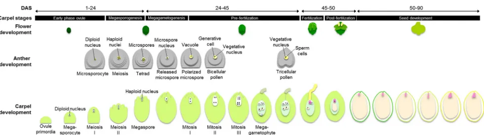
the central cells begin to elongate and divide to form the pro-vascular tissue. The embryo proper, with 0.170 mm in length and a suspensor of 0.078 mm in length, is enclosed by a cellularized endosperm, as the uppermost cells of the suspen-sor start to differentiate to form the hypophysis. The first two Fig. 3 Diagram ofP. ginsengreproductive development and the stages of carpel development. Male developmental schemes were adapted from Kim et al.
Table 1 Major cytological events during ginseng carpel development
1. Early phase of ovule development
1-I Primordium initiation 0.70–1.10 1∼5 ∼5 1 i ∼1
4-I Formation of a vacuole, antipodals appear
6-II Endosperm initiation 45∼50 M
7. Seed development
7-II Zygote elongation 50∼90 M
appendages of the shoot are initiated in an opposite phyllotax-is from the dphyllotax-istal pole of the embryo, which phyllotax-is at the early torpedo stage with 0.191 mm in length and a suspensor of
0.057 mm in length (Fig.8g, m). After the final fruit size is
reached to about 5.00 to 6.25 mm, the fruit begins desiccation and eventually dehisces.
Discussion
In this study, we classified ginseng carpel developmental
stages (Table 1), summarizing its development from the
early phase of ovule development when the primordium arises to seed development when the embryo develops into the early torpedo stage. This classification helps to better understand the timing of the morphological events that take place in the carpel. This knowledge will also provide a basis for proper analysis of defects in ovule development and expression of genes and proteins in-volved in carpel development. The staging criteria are derived from the development of the megaspore, megaga-metophyte, and embryogenesis, because these events pro-vide reliable indicators that appear in a consecutive
man-ner. Moreover, Fig. 3 shows the synchronized
develop-ment of the reproductive organs, ovule and anther, until pre-fertilization, in order to provide an integral view of ginseng plant reproduction.
Stages of carpel development
Stage 1 comprises the early phase of ovule development until the primordia are enlarged in the pistil. Stage 2 covers mega-sporogenesis, starting with the enlargement of the megaspore mother cell and finishes with the outer integument surround-ing both the inner integument and the nucellus. Stage 3 in-cludes megagametogenesis, from the formation of the four-nuclear embryo sac to the development of the outer in-tegument to enclose both the inner inin-tegument and the nucel-lus. Stage 4 involves pre-fertilization, starting with the forma-tion of a vacuole and the emergence of the antipodals and concluding with the degeneration of the antipodals and emer-gence of the central cell, synergids, and egg cell. Stage 5 constitutes fertilization. Stage 6 denotes post-fertilization de-velopment, starting with fertilization and finishing with the initiation of endosperm. Stage 7 encompasses seed develop-ment from the zygote stage to the developdevelop-ment of the early torpedo stage of the embryo. These stages cover the complete ontogeny of carpel development, an effort that was only
re-ported for ovule development inArabidopsis(Schneitz et al.
1995) and seed development in Panax quinquefolius
Fig. 4 Section analysis of early ovule development inP. ginseng,aFlower bud at the initial developmental stage.Dashed lineindicates the position in the ovary where the transverse sections were made,bovule primordium initiation,cseparation of each ovule primordium in two opposite directions,
delongation of ovule primordium into finger-like, andefirst meiotic division in the ovule primordium.fandgshow the concurrent events observed in the anther as differentiation of anther layers (f) and microspore mother cell formation (g).Scale bars= 100μm (b–e), 20μm (f,g)
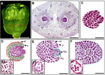
Fig. 5 Megasporogenesis and megagametogenesis inP. ginseng.aFlower bud at the developing stage. Thered dashed lineindicates the position in the ovary where transverse sections were made.bMegaspore mother cells of each locule;cinitiation of the integuments that appear as small protuberances;
dinitiation of the integument elongation, showing that the two-cell-layered inner integument is slightly longer than the one-cell-layered outer integument;
esecond meiotic division of the megaspore mother cell;fthe outer integument completely encloses both the inner integument and nucellus. The concurrent events observed in the anther were (d1) first meiotic division, (e1) tetrad formation, and (f1) young microspore formation.Scale bars= 200μm (b), 500μm (c–f), 20μm (d1,e1,f1).iiinner integument,Nu
nucellus,oiouter integument,Pcparietal cell
J. Silva et al.
Characteristics of ginseng carpel
Synchrony of female and male gametophytic development was observed in this study to begin when ovule primordium undergoes elongation, at which point the anther undergoes the formation of the microsporocyte, and finishes at the end of pre-fertilization when cellularization is completed. The micro-pyle opens, and the anther releases mature pollen grains
(Fig. 3). The significance of observing the synchrony in
timing of female and male gametes derives from proving that it will not lead to failed fertilization. If some fertilization bar-riers are found, we can infer that they are originated from failures in the development of one of the gametophytes.
One interesting feature of the young ovule ofP. ginsengis
the presence of vascular bundles in a concentric circle adjacent
to the ovary wall (Fig. 9a). This distribution pattern of the
vascular bundles differs from those reported in other higher plants. For instance, they appear in an heterocarpellary
posi-tion inSeemannaralia gerrardii(Oskolski et al.2010), form
an external circle inE. natalensis(Woodenberg et al. 2014),
form an external and a central complex inGaussia attetuata
(Castaño et al.2014), and form an arc inVochysiaceae(Litt
and Stevenson2003). Anatomical studies of floral vasculature
support the idea that in receptacular epigynous flowers, the vascular bundles provide traces to the floral appendages that run upward through the length of the ovary wall and then Fig. 6 Section analysis of
pre-fertilization inP. ginseng.a
Flower bud at the maturing stage. Thered dashed lineindicates the position in the ovary where longitudinal sections were made.
bFormation of the vacuole;c
opening of the micropyle;d
formation of the antipodals;e
degeneration of antipodals and formation of central cell and egg cell.f–hCellularization.i–k
enlargement of cellularization ofi
egg cell,jcentral cell, andk
antipodals. The concurrent events observed in the anther arel
microspore vacuolation,m
mitotic division,nmaturing pollen, andorelease of mature pollen grains.Scale bars =50μm
(b–c), 20μm (d–e), 100μm (f– h), 20μm (i–k), 20μm (l–o).Ant
antipodals,Cccentral cell,Ecegg cell,Micmicropyle
Fig. 7 Section analysis of fertilization and post-fertilization inP. ginseng.aOvary at the end of the maturing stage.bStigma showing remaining pollen grains attached. c Scanning electron microscopic observation of the outer surface of stigma.d
Withered stamens and fallen sepals and petals. Thered dashed lineindicates the position in the ovary where longitudinal sections were made.eDegeneration of synergids, breaking down of egg cells, and initiation of endosperm. Also shown are haustoria at the micropylar end and cellular endosperm at the edges.f
Enlargement of (e) showing the zygote and endosperm.Scale bars =0.5 cm (a,b), 150μm (c), 1 cm (d), 100μm (e), 50μm (f). Hshaustoria,Cecellular endosperm
Fig. 8 Section analysis of seed development in P. ginseng.aFruits showing completely wilted stamens and disappeared petals, ovary that has started to harden to form the seed coat, and the locules that have reached its maximum size to enclose the developing seed. Thedashed lineindicates the zygote, suspensor, and embryo proper.b–gEmbryos during consecutive developmental stages of zygote (b), elongated zygote
(c), octant stage (d), dermagoten stage (e), triangular stage (f), and early torpedo stage (g).h–mEnlargement of the boxes region inb–gshowing the embryo developmental stages.Scale barsindicate 2 mm (a), 500μm
(b–g), 50μm (h–m).Aaxis,Ccotyledon,Cecellular endosperm,EP
embryo proper,Nenuclear endosperm,Ssuspensor
J. Silva et al.
descend to supply nutrition to the ovule (Douglas 1957;
Kaplan1967; Costello and Motley2004). Interestingly
gin-seng accumulates triterpene metabolite, and ginsenosides in
vascular bundles of roots and stem (Kim et al.2015b),
espe-cially ginsenoside Re in the ginseng fruit. This leads to the speculation that the presence of vascular bundles in the flower bud might have a function in the transport of water and me-tabolites such as ginsenosides, which may contribute to the expansion of developing ovules prior to fertilization. Further research needs to be carried out to confirm the involvement of vascular bundles in ginsenoside transport.
Megasporogenesis inP. ginsengproceeds with the
forma-tion of a parietal cell (Fig.5e), which is a typical feature of
polypetalous dicotyledons with bitegmic ovules. It is the divi-sion of the archesporial initially into primary parietal cells and primary sporogenous cells that results in the formation of the tenuinucellate ovule, which is consistent with the
characteris-tic of Araliaceae (Johri et al.1992).
The initiation of integuments is an essential event in ovule morphogenesis, because the tissues that ultimately will form the micropyle, nucellus, chalaza and funicle are also defined
(Schneitz et al.1995; Schneitz1999; Endress2011). The inner
integument initiates slightly earlier than the outer integument
(Fig.5d). The outer integument originates from a subepidermal
cell close to the dividing epidermal cell; cell division extends to neighboring subepidermal cells and the covering epidermals. The integuments continue their development and eventually
enclose the nucellus completely. Although both the inner and outer integuments mostly contain two cell layers in angiosperms
(Endress2011), this feature differs between monocots and dicots.
In dicots the outer integument mostly has two cell layers; in contrast, in monocots, it can contain two to ten cell layers, as
reported forI. tenax(Wilson2001),Triteleiasp. (Berg2003), and
G. attenuata(Castaño et al.2014). The comparison ofP. ginseng
and other dicots shows that inP. ginseng, the outer integument
only has one layer of cells, which is different from dicots such as
Arabidopsis(Schneitz et al.1995),Cytisussp. (Rodríguez-Riaño
et al.2006), andDrosera x obovata(Rodondi et al.2009),
com-prising a unique feature ofP. ginseng. Moreover, we observed
that the curvature of the ovule is subtle inP. ginsengand not as
conspicuous as in other higher plants (Endress2011) such as
Triteleia sp. (Berg2003),Cytisus sp. (Rodríguez-Riaño et al.
2006), I. tenax (Wilson 2001), and Drosera x obovata
(Rodondi et al.2009). In angiosperms, the outer integument is
often thinner in orthotropous ovules than in anatropous ovules
(Endress2011), suggesting thatP. ginsengmight have evolved
from plants with orthotropous ovules. Knowing the ovule curva-ture type is of great importance since its function is to bring the micropyle close to the funicle to enable a direct passage of the pollen tube, thereafter, the integuments become the seed coat to protect the embryo, assist in seed dissemination, and regulate
seed germination (Kelley and Gasser2009). In addition, the
ovule has apical placentation (Fig.9b), which is characteristic
of P. ginsengbecause the most common placentation of the
Fig. 9 Histological and ultrastructural analysis of
P. ginsengcarpel.aCross section showing vascular bundles (circles) in the flower buds at stage 1.bLongitudinal section showing anatropous ovules that are slightly curved.cFruit showing one aborted ovule after fertilization.dLongitudinal section showing the early torpedo stage of an embryo within the developing seed.Scale bars
indicate 100μm (a), 200μm (b),
1 mm (c), and 500μm (d)
anatropous ovules is basal (Wilson2001; Endress2011; Musiał
et al.2013; Figueiredo et al.2013). The features of the
integu-ments show howP. ginsengcarpel has evolved to develop
ad-vanced ovules that help the mobility of the pollen tube to be unprolonged and secure the movement during fertilization.
Another noteworthy observation is that in the transition from megasporogenesis to megagametogenesis, the ovule exhibits a
twofold enlargement from 110 to 220μm in 10 days (Fig.5c–f).
A likely explanation of this enlargement is the subsequent growth of the embryo sac to prepare to form and encase the cell lineages (antipodals, central cell, synergids, and egg cell). The other ob-servation is that at the very beginning of stage 7, aborted ovules were observed. Although it is not commonly seen in seeds that
P. ginsengproduces, it is known to occur in Araliaceae (Johri
et al.1992) and P. quinquefolius(Schluter and Punja 2000),
suggesting that post-fertilization events are likely responsible for abortion. Our observations provide evidence to assume that abortion may be originated from defects during pre-fertilization stage where the anther development undergoes the microspore vacuolation, mitotic division, maturing pollen, and pollen release. Or it may be a failure during cellularization in the carpel. These are the two main aspects for fertilization to occur, in cell lineages in the embryo sac and two sperm cells in the mature pollen grain.
Characteristics of ginseng embryogenesis
In this study, we have found three distinctive features of
P. ginsengembryogenesis. First is the length of time that a
com-plete embryo development requires. UnlikeArabidopsis, in
which embryogenesis is completed typically within 20 days
post-fertilization (Le et al.2010),P. ginsengneeds 40 days to
reach the early torpedo stage. To achieve germination, the em-bryo needs up to 90 additional days of stratification (Baranov
1966; Li1995; Kim et al.2015a), leading to the question why it
needs longer time to develop. Based on the Baskin and Baskin
(1998) classification theory of seed dormancy,P. ginsengseeds
belong to the morphophysiological dormancy class, where seeds have underdeveloped embryos and require treatments to break the dormancy. As a result, to promote anatomical development,
stratification inP. ginsengis usually done in humidified sand for
3 months at 5 °C to complete embryo development and to
achieve germination (Kim et al.2015a).
The second feature ofP. ginsengis the small size of the
embryo relative to the seed size, leading to the question whether the size is related to the nutrition needed by the
de-veloping embryo. This characteristic is also observed inZea
mays, for which the endosperm persists through seed
devel-opment and provides storage reserves during seedling growth
(Kiesselbach 1949; Bai and Settles 2015). In contrast,
Arabidopsisembryo consumes the endosperm reserves as it
develops, degenerating most of the endosperm by seed
matu-rity (Bai and Settles2015). Small embryo relative to the size
of the seed is likely to be an ancestral characteristic in
angiosperms (Forbis et al. 2002) that has been kept in
P. ginseng. Forbis et al. (2002) reported that five species of
Araliaceae, including Acanthopanax sessiliflorus, Aralia
hispida,Oplopanax horridium,P. quinquefolius, andHedera
helix, have a small embryo, with a 0.38 ratio between the size
of the embryo and seed. Consistently, the seed ofP. ginsengis
filled mostly by endosperm with the embryo making up a small part of the seed volume. A likely explanation for the small embryo size in the albuminous seed lies in the unique life of Araliaceae family.
The third feature ofP. ginsengis that at the early torpedo
stage, the endosperm exhibits two types, nuclear and cellular,
and the embryo grows in the nuclear endosperm (Figs.8and
9d), as what has been reported to occur inPanax wangianus
(Venugopal et al. 2013). The embryogenesis type of
P. ginsengis chenopodiad, where the embryo is generated
from both basal and terminal cells. We observed some
differ-ences in embryogenesis of P. ginseng from study on
P. quinquefoliusby Qi et al. (2015). First is the size and shape
of the embryo. At octant and dermatogen stages, the suspensor
and embryo proper ofP. ginsengembryo are both thinner and
more elongated than that ofP. quinquefolius, with the
suspen-sor size difference responsible for the remarkable embryo size difference between the two species is at the octant stage. At
the dermatogen stage, the embryo proper in P. ginseng is
wider than inP. quinquefolius. The second difference is the
suspensor. At the triangular and early torpedo stages,
P. ginseng embryo retains the suspensor, while in
P. quinquefoliusit is already degenerated. However, both
spe-cies show an embryo of about 0.200 mm at the final stage of seed development. The suspensor is morphologically diverse throughout the plant kingdom (Kawashima and Golberg
2010) and according to our observations, it can also differ
betweenPanaxspecies. The role of the suspensor in embryo
development is essential, since it places the embryo proper into the endosperm cavity and connects the embryo proper to surrounding maternal and endosperm tissues, serving as a conduit for nutrients and growth regulators required for
em-bryonic development (Kawashima and Golberg 2010).
According to our observations, the suspensor completes ear-lier its contribution for the embryo development and
un-dergoes programmed cell death inP. quinquefoliusin
compar-ison withP. ginseng, although the mechanisms responsible for
this early shift remain to be determined.
Through this cytological analysis ofP. ginsengcarpel
devel-opment, we have been able to propose a 7-stage process for the development based on reliable indicators that appear in a consec-utive manner throughout carpel development. Correlation of the development of female and male gametophytes and identifica-tion of unique characteristics of ginseng carpel/embryo develop-ment are also summarized. Our study may offer a valuable tool for the study of reproductive developmental in Araliaceae family and provide knowledge basis for ginseng breeding.
J. Silva et al.
Acknowledgements This research was supported by a grant from the Basic Science Research Program through the National Research Foundation (NRF), Ministry of Education, Republic of Korea, to YJ Kim (2016R1A6A3A11931858), an iPET grant from the Korea Institute of Planning and Evaluation for Technology in Food, Agriculture, Forestry and Fisheries, Republic of Korea, to DC Yang (112142-05-4-SB010), and a grant from the Ministry of Science and Technology, People’s Republic of China, to JP Hu (2015DFG32560).
Compliance with ethical standards
Conflict of interest The authors declare that they have no conflict of interest.
References
Angenent GC, Colombo L (1996) Molecular control of ovule develop-ment. Trends Plant Sci 1(7):228–232
Bai F, Settles AM (2015) Imprinting in plants as a mechanism to generate seed phenotypic diversity. Front Plant Sci 5(780). doi:10.3389/fpls. 2014.00780
Baranov A (1966) Recent advances in our knowledge of the morphology, cultivation and uses of ginseng (Panax ginsengC. A. Meyer). Econ Bot 20(4):403–406
Baskin CC, Baskin JM (1998) Seeds: ecology, biogeography, and evolu-tion of dormancy and germinaevolu-tion. Academic Press, San Diego Beeckman T, Viane R (2000) Embedding thin plant specimens for
ori-ented sectioning. Biotech Histochem 75(1):23–26
Berg RY (2003) Development of ovule, embryo sac, and endosperm in
Triteleia(Themidaceae) relative to taxonomy. Am J Bot 90(6):937–948 Castaño F, Stauffer F, Marquinez X, Crèvevoeur M, Collin M, Pintaud JC, Tregear J (2014) Floral structure and development in the mon-oecious palmGaussia attenuata(Arecaeae; Arecoideae). Ann Bot 114(7):1483–1495
Coombe BG (1976) The development of fleshy fruits. Ann Rev Plant Physiol 27(1976):507–528
Costello A, Motley TJ (2004) The development of the superior ovary in
Tetraplasandra(Araliaceae). Am J Bot 91(5):644–655
de Oliveira RR, Cesarino I, Mazzafera P, Dornelas MC (2014) Flower development inCoffea arabicaL.: new insights into MADS-box genes. Plant Reprod 27(2):79–94
Douglas GE (1957) The inferior ovary, II. Bot Rev 23:1–41
Dresselhaus T, Sprunck S, Wessel M (2016) Fertilization mechanisms in flowering plants. Curr Biol 26(3):125–139
Endress PK (2011) Angiosperm ovules: diversity, development, evolu-tion. Ann Bot 107(9):1465–1489
Figueiredo RC, Masullo FA, Cardoso Vieira R, De Toni KLG (2013) Development of carpels and ovules inPsychotria carthagenensis
(Psychotrieae) and Rudgea macrophylla (Palicoureeae) (Rubioideae, Rubiaceae). South African J Bot 84:110–114 Forbis TA, Floyd SK, De Queiroz A (2002) The evolution of embryo size
in angiosperms and other seed plants: implications for the evolution of seed dormancy. Evolution 56(11):2112–2125
Igersheim A, Cichocki O (1996) A simple method for microtome sectioning of prehistoric charcoal specimens, embedded in 2- hydroxyethyl meth-acrylate (HEMA). Rev Palaeobot Palynol 92:389–393
Johri BM, Ambegaokar KB, Srivastava PS (1992) Comparative embryology of angiosperms, vol 1/2. Springer-Verlag, Berlin Heidelberg, Berlin Kaplan DR (1967) Floral morphology, organogenesis and interpretation
of the inferior ovary inDowningia bacigalupii. Am J Bot 54(10): 1274–1290
Kawashima T, Golberg RB (2010) The suspensor: not just suspending the embryo. Trends Plant Sci 15(1):23–30
Kelley DR, Gasser CS (2009) Ovule development: genetic trends and evolutionary considerations. Sex Plant Reprod 2(4):229–234 Kiesselbach TA (1949) The structure and reproduction of corn. Research
bulletin 161 Lincoln: Agricultural Experiment Station, University of Nebraska College of Agriculture, Nebraska
Kim YJ, Zhang D, Yang DC (2015a) Biosynthesis and biotechnological production of ginsenosides. Biotechnol Adv 33(6):717–735 Kim YJ, Jang MG, Zhu L, Silva J, Zhu X, Sukweenadhi J, Kwon WS,
Yang DC, Zhang D (2015b) Cytological characterization of anther development inPanax ginsengMeyer. Protoplasma. doi:10.1007/ s00709-015-0869-3
Kim YJ, Silva J, Zhang D, Shi J, Joo SC, Jang MG, Kwon WS, Yang DC (2016) Development of interspecies hybrids to increase ginseng bio-mass and ginsenoside yield. Plant Cell Rep 35(4):779–790 Koizumi A, Yamanaka K, Kawano S (2009) Carpel development in a
floral mutant of dioeciousSilene latifoliaproducing asexual and female-like flowers. J Plant Physiol 166(16):1832–1838
Kubo T, Fujita M, Takahashi H, Nakazono M, Tsutsumi N, Kurata N (2013) Transcriptome analysis of developing ovules in rice isolated by laser microdissection. Plant Cell Physiol 54(5):750–765 Le BH, Cheng C, Bui AQ, Wagmaister JA, Henry KF, Pelletier J, Kwong
L, Belmonte M, Kirkbride R, Horvath S, Drews GN, Fischer RL, Okamuro JK, Harada JJ, Goldberg RB (2010) Global analysis of gene activity duringArabidopsisseed development and identifica-tion of seed-specific transcripidentifica-tion factors. Proc Natl Acad Sci U S A 107(18):8063–8070
Li TSC (1995) Asian and American ginseng–a review. Hortechnol 5(1): 27–34
Litt A, Stevenson DW (2003) Floral development and morphology of
Vochysiaceae. I. The structure of the gynoecium. Am J Bot 90(11):1533–1547
MusiałK, Płachno BJ,Świątek P, Marciniuk J (2013) Anatomy of ovary and ovule in dandelions (Taraxacum, Asteraceae). Protoplasma 250(3):715–722
Oskolski AA, Sokoloff DD, Van Wyk BE (2010) False paracarpy in
Seemannaralia(Araliaceae): from bilocular ovary to unilocular fruit. Ann Bot 106(1):29–36
Qi J, Sun P, Liao D, Sun T, Zhu J, Li X (2015) Transcriptomic analysis of American ginseng seeds during the dormancy release process by RNA-Seq. PLoS One 10(3). doi:10.1371/journal.pone.0118558
Reiser L, Fischer RL (1993) The ovule and the embryo sac. Plant Cell 5(10):1291–1301
Robert HS, Crhak Khaitova L, Mroue S, Benková E (2015) The impor-tance of localized auxin production for morphogenesis of reproduc-tive organs and embryos in Arabidopsis. J Exp Bot 66(16):5029–
5042
Rodondi G, Beretta M, Andreis C (2009) Ovule and pollen development in the natural hybridDrosera x obovataMert. & Koch (Droseraceae) and its parents. Flora 204(9):685–691
Rodríguez-Riaño T, Valtueña FJ, Ortega-Olivencia A (2006) Megasporogenesis, megagametogenesis and ontogeny of the aril in
Cytisus striatusandC. multiflorus(Leguminosae: Papilionoideae). Ann Bot 98(4):777–791
Rudall PJ, Remizowa MV, Beer AS, Bradshaw E, Stevenson DW, Macfarlane TD, Tuckett RE, Yadav SR, Sokoloff DD (2008) Comparative ovule and megagametophyte development in Hydatellaceae and water lilies reveal a mosaic of features among the earliest angiosperms. Ann Bot 101(7):941–956
Sachs J, Bennett AW (2011) A text-book of botany: morphological and physiological. Cambridge University Press, Cambridge
Schluter C, Punja ZK (2000) Floral biology and seed production in cul-tivated North American ginseng (Panax quinquefolius). J Amer Hort Sci 125(5):567–575
Schneitz K (1999) The molecular and genetic control of ovule develop-ment. Curr Opin Plant Biol 2(1):13–17
Schneitz K, Huiskamp M, Pruitt RE (1995) Wild-type ovule development inArabidopsis thaliana: a light microscope study of cleared whole-mount tissue. Plant J 7(5):731–749
Singh G (2010) Plant systematics an integrated approach. CRC Press, Delhi Sokoloff DD, Oskolski AA, Remizowa MV, Nuraliev MS (2007) Flower structure and development inTupidanthus calyptratus(Araliaceae): an extreme case of polymery among asterids. Pl Syst Evol 268(1): 209–234
Venugopal N, Ahuja P, Lalchhanhimi (2013) A unique type of endosperm inPanax wangianusS. C. Sun. J Plant Develop 20(2013):45–50 Vivian-Smith A, Luo M, Chaudhury A, Koltunow A (2001) Fruit
devel-opment is actively restricted in the absence of fertilization in Arabidopsis. Development 128(12):2321–2331
Wang ZF, Ren Y (2008) Ovule morphogenesis in Ranunculaceae and its systematic significance. Ann Bot 101(3):447–462
Wilson CA (2001) Floral stages, ovule development, and ovule and fruit success inIris tenax, focusin on var.Gormanii, a taxon with low seed set. Am J Bot 88(12):2221–2231
Woodenberg WR, Berjak P, Pammenter NW, Farrant JM (2014) Development of cycad ovules and seeds. 2. Histological and
ultrastructural aspects of ontogeny of the embryo inEncephalartos natalensis. Protoplasma 251(4):797–816
Wu X, Liu J, Li D, Liu CM (2016a) Rice caryopsis development I: Dynamic changes in different cell layers. J Integr Plant Biol 58(9): 772–785
Wu X, Liu J, Li D, Liu CM (2016b) Rice caryopsis development II: Dynamic changes in the endosperm. J Integr Plant Biol 58(9):786–
798
Yu SC, Kim WK (1992) Structural changes and histochemical study of endosperm onPanax ginsengC. A. Meyer during embryo develop-ment. Korean J Ginseng Sci 16(1):37–43
Zhang Y, Liang W, Shi J, Xu J, Zhang D (2013) MYB56 encoding a R2R3 MYB transcription factor regulates seed size inArabidopsis thaliana. J Integr Plant Biol 55(11):1166–1178
Zhang J, Tang W, Huang Y, Niu X, Zhao Y, Han Y, Liu Y (2015) Down-regulation of a LBD-like gene, OsIG1, leads to occur-rence of unusual double ovules and developmental abnormali-ties of various floral organs and megagametophyte in rice. J Exp Bot 66(1):99–112
J. Silva et al.