Original Articles
Comparing the Effectivities of Chitosan Citrate and Chitosan
Acetate in Eradicating
Enterococcus faecalis
Biofilm
Uppalavanna Witedja1, Tien Suwartini1, Anastasia E Prahasti1, Armelia Sari Widyarman2 1Department of Conservative Dentistry, Faculty of Dentistry, Trisakti University – Indonesia
2Department of Microbiology, Division of Oral Biology, Faculty of Dentistry, Trisakti University – Indonesia
‘Corresponding Author: Armelia Sari Widyarman, Faculty of Dentistry, Trisakti University – Indonesia. Email: [email protected]
Received date:June 19, 2017.Accepted date:November 15, 2017.Published date:January 25, 2018.
Copyright:©2018 Witedja U, Suwartini T, Prahasti AE, Widyarman AS. This is an open access article distributed under the terms of the Creative Commons Attribution License, which permits unrestricted use, distribution, and reproduction in any medium provided the original author and sources are credited.
ABSTRACT
Background: Adequate biomechanical preparations, antibacterial irrigants, and intracanal medications to promote the elimination of bacteria and their products are required to succeed root canal treatment. Enterococcus faecalis with its
biofilm is known as an important etiological agent in endodontic treatment failures. Chitosan, as a natural product, has an antibacterial activity and is considered less toxic to the periapical tissue than other irrigants. However, the use of this natural product needs to be examined to determine its effectiveness as a root canal irrigant in endodontic treatment; this can be done by comparing it with the most common endodontic irrigant (NaOCl 5.25%) as a positive control.Objective:
The objective of the study was to compare the effectiveness between 1–3% chitosan acetate (CA) and 1–3% chitosan citrate (CC) againstE. faecalisbiofilm formation after treatment for 15, 30, and 60 minutes. Methods:The study was conducted using 12 groups, including 1–3% CA, 1–3% CC, and control groups.E. faecalis biofilms in 96-well plates were exposed to each sample for 15, 30, or 60 minutes. Subsequently, the biofilms were stained with crystal violet solution, and the optical density value was measured using a microtiter plate reader at a wavelength of 600 nm.Results:
CA and CC were effective in eradicating E. faecalis biofilm. However, the levels of effectiveness of CC and CA
depended on the concentration and application time. Three-way analysis of variance (ANOVA) showed a significant difference between the irrigants (p <0.05) and three application times (p <0.05). The CA was effective in eradicating
biofilm after 15 minutes of application, whereas the CC was more effective after 30 and 60 minutes of application.
Conclusion:CC and CA are both effective in eradicatingE. faecalisbiofilm.
Keywords : biofilm, chitosan acetate, chitosan citrate, Enterococcus faecalis, irrigation
Background
Endodontic treatment focuses on reducing the microbial load in the root canal to enable healing and prevent bacteria from re-entering the root canal in the
wall, which indicates that the cleaning process of the root canal cannot be achieved via preparation alone.2,3 To achieve a successful endodontic treatment, in addition to preparation, effective antibacterial irrigants are required to eliminate bacteria in the root canal.4,5
Recent studies have suggested that the presence of
Enterococcus faecalis in the root canal can increase the
risk of endodontic infections during or after the root canal treatment.6,7,8 Studies on NaOCl and chlorhexidine as antimicrobial irrigants have shown that both irrigants are effective to be used as antibacterial agents.9,10However, NaOCl is potentially toxic to human osteoblast cells; such toxicity can affect tissue regeneration, thereby causing a delay in the healing process.11.12Therefore, the search for ideal root canal irrigants continues with the development of new materials and methods. One material that has been investigated in many studies is chitosan, which has been considered for many dental applications. As the second most abundant polysaccharide after cellulose, it is widely distributed in nature, especially in Indonesia.13Chitosan can be easily generated from prawn and crab shells.14,15
In a previous study on chitosan dissolved in citric acid, it was found that 0.1–0.6% chitosan citrate (CC) solution showed antibacterial activity, significantly better cleansing of the smear layer, and a lack of dentin erosion compared with 10% citric acid. A previous study using electronic micrographs showed that chitosan weakens and even damages Gram-positive bacterial cell walls and disrupts the cytoplasm membrane in Gram-negative bacteria.16
Previous research on the effect of chitosan on the attachment and formation ofStreptococcus mutansbiofilm
has suggested that chitosan can inhibit the attachment and formation of biofilm, and it is able to remove existing biofilm.17Based on these findings in the literature, the aim of the present study was to evaluate the effectiveness of chitosan acetate (CA) and CC as antibacterial agents and their inhibition ofE. faecalisbiofilm formationin vitro.
Material and Methods
Bacterial Culture
The bacterial cultures were obtained by following the standard protocols at MiCORE Laboratory, Faculty of
Dentistry, Trisakti University, Indonesia. Bacterial strains of E. faecalis ATCC 29212 were maintained in stock cultures frozen at –80°C in brain heart infusion (BHI) broth containing glycerol (20% v/v). For biofilm assays,
E. faecalis colonies were inoculated in BHI broth and incubated at 37°C under anaerobic conditions using a gas pack jar system for 24 hours. Bacterial cells were then collected by centrifugation and suspended in fresh sterile BHI broth.
Sample Solution Preparation
Chitosan acetate (CA) solution was prepared with different concentrations ranging from 1 to 3% by mixing 1–3 g of chitosan powder into 100 mL of 1% acetic acid. The pH level of the CA solution was adjusted to 3.5 with NaOH. This solution was stirred for 24 hours with a magnetic stirrer, using materials from Biotech Surindo.18 The CC solution was prepared with different concentrations ranging from 1 to 3% by mixing 1–3 g of chitosan powder into 100 mL of 10% citric acid.19The pH level of the CC solution was adjusted to 3.2 with NaOH. This solution was stirred for 24 hours with a magnetic stirrer, using materials from Biotech Surindo.18
Minimum Inhibitory Concentration (MIC)
A broth dilution test was performed to determine the minimum inhibitory concentration (MIC) of the CC and CA solutions. Three milliliters of BHI broth were inserted into 12 glass tubes, and then 100 μL of bacterial cell suspension (1 × 10⁸CFU/mL) was added to each tube, followed by 100 μL solution of CC and CA at various concentrations (1%, 1.5%, 2%, 2.5%, and 3%) and 5.25% NaOCl as the positive control. A solution containing only BHI and chitosan was also made for a comparison of the optical density values. The plate was incubated at 37°C for 24 hours. The MIC was determined by observing the optical density value obtained using a microtiter plate reader.12
Biofilm Assay
to form biofilm. Subsequently, 200-μL samples of each irrigation solution (1%, 1.5%, 2%, 2.5%, 3% CA; 1%, 1.5%, 2%, 2.5%, 3% CC; and 5.25% NaOCl) were added to theE. faecalisbiofilm-containing well plate. The plate was incubated for 15, 30, or 60 minutes. Afterward, the biofilms in each well were stained with 0.1% violet crystalline solution for 15 minutes and rinsed with phosphate-buffered saline (PBS); following this, 90% ethanol was added. The effectiveness of each irrigation solution in terms of disrupting theE. faecalisbiofilm was analyzed by observing the optical density values of the crystal violet dyes absorbed by the biofilms.20 The absorbencies of the remaining biofilms after being treated by CA, CC, and NaOCl were assessed at 600 nm using a microtiter plate reader. Five wells for each group were used in this study.
Statistical Analysis
Differences between experimental groups were analyzed using three-way analysis of variance (ANOVA). A p-value of less than 0.05 was considered statistically significant. Shapiro Wilk test was used to test for normality previously. Statistical calculations were performed with SPSS Statistics for Windows software version 20 (IBM, USA).
Results
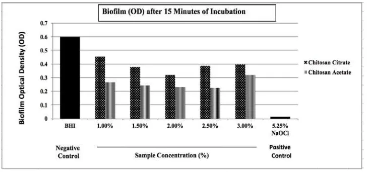
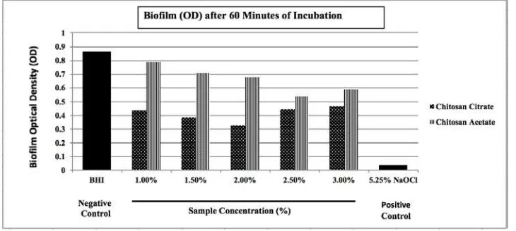
The MIC test results showed that the minimum concentration of CA and CC to inhibit E. faecalis when grown in the planktonic state was 1%. The data normality test, which was performed using the Shapiro–Wilk test for each group, showed a normal data on the optical density values of the CC (1%, 1.5%, 2%, 2.5%, 3%), CA (1%, 1.5%, 2%, 2.5%, 3%), and 5.25% NaOCl groups after 15, 30, and 60 minutes of application time (p>0.05). The biofilm assay result showed that, for the 15-minute application, the highest optical density values were evident in the group of negative controls, followed by 1% CC, 3% CC, 2.5% CC, 1.5% CC, 2% CC, 3% CA, 1% CA, 1.5% CA, 2% CA, and 2.5% CA; 5.25% NaOCl had the lowest value (Fig. 1). For the 30-minute application, the optical density values, ranging from highest to lowest, were obtained from the group of negative controls, 1% CA, 1.5% CA, 2% CA, 3% CA, 3% CC, 2.5% CC, 1% CC, 1.5% CC, 2.5% CA, 2% CA, and 5.25% NaOCl (Fig. 2). For the 60-minute application, the optical density values, ranging from highest to lowest, were obtained from the group of negative controls, 1% CA, 1.5% CA, 2% CA, 3% CA, 2.5% CA, 3% CC, 2.5% CC, 1% CC, 1.5% CC, 2% CC, and 5.25% NaOCl (Fig. 3).
Figure 2. Optical density measurement of Enterococcus faecalis biofilm eradication using a microtiter plate reader after 30 minutes of incubation.
Figure 3. Optical density measurement of Enterococcus faecalisbiofilm eradication using a microtiter plate reader after 60 minutes of incubation.
Discussion
The results of three-way ANOVA on the crystal violet absorption assay of E. faecalis biofilms, after treatment with CC, CA, and 5.25% NaOCl, showed that both the CC and CA solutions at all given concentrations for application time of 15, 30, and 60 minutes generated significant differences between irrigation time (F = 6.50;
p <0.05) and each type of irrigant (F = 2.92; p <0.05). The effectiveness of 1.5–3% CC was lower than that of 1–3% CA for 15 minutes; 1.5–2% CC and 1–3% CA for
30 minutes; 1–2% CC for 60 minutes; and 5.25% NaOCl for 15, 30, and 60 minutes. There was also a difference between the effectiveness levels of CC and CA in eradicating the E. faecalis biofilm, depending on the
Multiple comparison tests were performed using Tukey’s HSD to investigate significant differences among each type of irrigant and irrigation time. The posterior multiple comparison test showed that there
were some differences in the effectiveness of each irrigant, depending on the concentration and irrigation time. The following irrigants, with the indicated concentrations and irrigation times, suggested the same level of antimicrobial activity as 5.25% NaOCl for eradicating the E. faecalis biofilm: 1.5–3% CA for 15 minutes, 1–2% CC for 30 minutes, 1.5–2% CC for 60 minutes, 1–3% CA for 15 minutes, and 2.5% CA for 30 minutes.
A previous study concluded that there was no significant difference betweenCAand NaOCl solution in eradicatingE. faecalisbiofilm.21However, in the current
study, each irrigant had different effective concentrations against bacterial biofilms, in contrast to the previous study. This may have been caused by the different sources and molecular weights of the chitosan between the two studies. In this study, the chitosan powder was extracted from crab shells, and it had a higher molecular weight of 900 kDa (>150 kDa).In this study, acetic acid and citric acid were used to prepare the chitosan solutions, as they have been commonly used as chitosan solvents. In addition, 10% citric acid is often used as a root canal irrigant, as it is ideal for cleansing the smear layer and exhibits antimicrobial activity.22,23The common concentration of citric acid is 10%, with pH 3.2. However, in this study, we used citric acid with a higher concentration and lower pH than 3.2 to dissolve the chitosan; still, the chitosan powder was not completely dissolved. The chitosan powder was successfully dissolved in 1% acetic acid at pH 3.5. Therefore, we suggest that acetic acid is more effective than citric acid as a solvent for chitosan powder.
The optical density values obtained at an application of 15 minutes were significantly better than those at 30 and 60 minutes. The least effective irrigant was in the group of 1% CA for an application time of 60 minutes, whereas the most effective one was found in the NaOCl group as the positive control. However, no significant differences were found in the effectiveness levels of NaOCl, 1–2% CC for 60 minutes, 2.5% CA for 30 minutes, 2% CC for 15 and 30 minutes, and 1–3% CA for
15 minutes. The most effective application time of 5.25% NaOCl was observed to be 15 minutes, whereas applications for 30 and 60 minutes showed less effective antimicrobial activity due to the instability of the NaOCl solutions and the loss of active chlorine compounds during the incubation period.24The current study also suggested that CC was more stable than CA, as observed in its effectiveness in eradicating theE. faecalisbiofilms.
Conclusions
Chitosan acetate (CA) and chitosan citrate (CC) are effective in eradicatingE. faecalisbiofilms. The levels of effectiveness of CC and CA differ depending on the concentration and application time. CA is more effective in eradicating biofilm after 15 minutes of application, whereas CC is more effective after 30 and 60 minutes of application. Both solutions may be used as alternative irrigants in root canal treatment. However, further research is needed to evaluate the interaction between the chitosan solutions to evaluate their cleaning efficacy in vivo.
Acknowledgement
The authors would like to thank the Faculty of Dentistry, Trisakti University and the Microbiology Center of Research and Education (MiCORE) laboratory for their invaluable support. The authors also thank Stella Pranoto, S.Si and Aradhea Monica Drestia, S.Si, for their laboratory assistances.
Conflict of Interest
The authors declare that there are no conflicts of interest.
References
2013;39(2):249-53. DOI: 10.1016/j.joen.2012.11.008
2. Mohammadi Z, Giardino L, Mombeinipour A.
Antibacterial substantivity of a new antibiotic-based endodontic irrigation solution. Aust Endod J.
2012;38(1):26-30. DOI:
10.1111/j.1747-4477.2010.00263.x
3. Mohammadi Z. Jafarzadeh H, Shalavi S. Antimicrobial efficacy of chlorhexidine as a root canal irrigant: A literature review. J Oral Sci. 2014;56(2):99-103.
4. De Andrade FB, Arias MPC, Maliza AGA, Duarte MAH, Graeff MSZ, Amoroso - Silva PA,et al. A new improved protocol for in vitro intratubular dentinal bacterial contamination for antimicrobial endodontic tests: Standardization and validation by confocal laser sanning microscopy. J Appl Oral Sci. 2015; 23(6):591-8. DOI: 10.1590/1678-775720140261
5. Yeung W, Raldi DP, Cunha RS, Mello I. Assessment of smear layer removal protocols in curved root canal. Aust Endod J. 2014;40(2):66-71. DOI: 10.1111/aej.12038 6. Stuart CH, Schwartz SA, Beeson TJ, Owatz CB.
Enterococcus faecalis: its role in root canal treatment failure and current concepts in retreatment. J Endod. 2006; 32(2):93-8. DOI: 10.1016/j.joen.2005.10.049
7. Wang QQ, Zhang CF, Chu CH, Zhu XF. Prevalence of Enterococcus faecalis in saliva and filled root canals of teeth associated with apical periodontitis. Int J Oral Sci. 2012; 4(1): 19–23. DOI: 10.1038/ijos.2012.17
8. Demiryürek EÖ, Kalyoncuoğlu E, Duran E, Çoban AY,
ÇaycıYT. Efficacy of different instrumentation techniques on reducing Enterococcus faecalis infection in experimentally infected root canals. J Dent Sci. 2014; 9(1): 23-28. DOI: 10.1016/j.jds.2012.03.024
9. Del Carpio-Perochena AE, Bramante CM, Duarte MAH, Cavenago BC, Villas-Boas MH, Graeff MS, et al.Biofilm dissolution and cleaning ability of different irrigant solutions on intraorally infected dentin. J Endod. 2011;37(8):1134-8. DOI: 10.1016/j.joen.2011.04.013 10. Gomes BPFA, Vianna ME, Zaia AA, Almeida JFA,
Souza-Filho FJ, Ferraz CCR. Chlorhexidine in endodontics. Braz Dent J. 2013;24(2):89-102. DOI: 10.1590/0103-6440201302188
11. Isabela NR, Siqueira JF Jr. Comparison of the in vivo antimicrobial effectiveness of sodium hypochlorite and chlorhexidine used as root canal irrigants: A molecular microbiology study. J Endod. 2011;37(2):143-50. DOI:10.1016/j.joen.2010.11.006
12. Anurhada B, Indira R, Lalitha MK, Sriram T. A new irrigant againstE. faecalisin root canal disinfection. Biosci
Biotech Res Asia. 2014;11(1):121-7. DOI:
dx.doi.org/10.13005/bbra/1242
13. Arif AR, Ischaidar, Natsir H, Dali S. Isolasi kitin dari limbah udang putih (penaeus merguiensis) secara enzimatis. Paper presented at : Seminar Nasional Kimia. 2013. Prosiding Seminar Nasional Kimia 2013-Universitas Islam Negeri Alauddin Makasar: 2013 May 4th; Makasar Indonesia; 2013.10-6.
14. Kumari S, Kumar Annamareddy SH, Abanti S, Kumar
Rath P. Physicochemical properties and characterization of
chitosan synthesized from fish scales, crab and shrimp shells. Int J Biol Macromol. 2017;104(Pt.B):1697-1705. DOI: 10.1016/j.ijbiomac.2017.04.119
15. Younes I, Rinaudo M. Chitin and chitosan preparation from marine sources. Structure, properties and applications. Mar Drugs. 2015;13(3):1133–74. DOI: 10.3390/md13031133
16. De Carvalho MMSG, Stamford TCM, Dos Santos EP, Tenorio P, Sampaio F. Chitosan as an oral antimicrobial agent. In Mendez-Vilas A. editor. Science against microbial pathogens: communicating current research and technological advance. Formatex; 2011. 542-51.
17. Costa EM, Silva S, Tavaria FK, Pintado MM. Study of the effects of chitosan upon Streptococcus mutans adherence and biofilm formation. Anaerobe. 2013 Apr;20:27-31. DOI: 10.1016/j.anaerobe.2013.02.002
18. Vidya N, Sreedhara KS, Sharath CSM. Comparison of antimicrobial activity of two chelating agents chitosan and etidronate against Enterococcus faecalis using agar diffusion test. Int J Appl Dent Sci. 2015;1(4):75-8. 19. Gusiyska A, Vassileva R, Dyulgerova E, Ilieva R,
Mironova J, Gyulbenkiyan E. Scanning electron microscopy studies of root canal dentin irrigated with a chitosan-citrate solution: A preliminary report. Int J Sci Res. 2014;5:539-42.
20. Widyarman AS, Widjaja SB, Idrus E. Strawberry extract’s effects on Enterococcus faecalis and Porphyromonas gingivalisbiofilms in vitro. Sci Dent J. 2017;01:1-5. DOI: https://doi.org/10.26912/sdj
21. Jaiswal N, Sinha DJ, Singh UP, Singh K, Jandial UA, Goel S. Evaluation of antibacterial efficacy of chitosan, chlorhexidine, propolis and sodium hypochlorite on Enterococcus faecalis biofilm: An in vitro study. J Clin Exp Dent. 2017; 9(9):e1066-74. DOI: 10.4317/jced.53777 22. Suzuki S, Masuda Y, Morisaki H, Yamada Y, Kuwata H,
Miyazaki T. The study of chitosan citrate solution as a root canal irrigant: A preliminary report. J Oral Hyg Health. 2014;2(4):142. DOI: 10.4172/2332-0702.1000142
against Enterococcus faecalis biofilms. Int Endod J. 2015;48:1188–93. DOI: 10.1111/iej.12424. Epub 2015 Jan 8