E D I T O R I A L B O A R D
Editor in Chief Richard Robinson [email protected] Tucson, Arizona
Advisory Editors
Peter Bruns, Howard Hughes Medical Institute
Rex Chisholm, Northwestern University Medical School Mark A. Davis, Department of Biology, Macalester College Thomas A. Frost, Trout Lake Station, University of Wisconsin Kenneth S. Saladin, Department of Biology, Georgia College and State
University
Editorial Reviewer
Ricki Lewis, State University of New York at Albany
Students from the following schools participated as consultants: Pocatello High School, Pocatello, Idaho
Eric Rude, Teacher
Swiftwater High School, Swiftwater, Pennsylvania Howard Piltz, Teacher
Douglas Middle School, Box Elder, South Dakota Kelly Lane, Teacher
Medford Area Middle School, Medford, Wisconsin Jeanine Staab, Teacher
E D I T O R I A L A N D P R O D U C T I O N S T A F F Linda Hubbard, Editorial Director
Diane Sawinski, Christine Slovey, Senior Editors
Shawn Beall, Bernard Grunow, Michelle Harper, Kate Millson, Carol Nagel, Contributing Editors
Kristin May, Nicole Watkins, Editorial Interns Michelle DiMercurio, Senior Art Director Rhonda Williams, Buyer
Robyn V. Young, Senior Image Editor
Julie Juengling, Lori Hines, Permissions Assistants Deanna Raso, Photo Researcher
Macmillan Reference USA Elly Dickason, Publisher
biolog
y
V O L U M E
3
I – Po
Copyright © 2002 by Macmillan Reference USA
All rights reserved. No part of this book may be reproduced or transmitted in any form or by any means, electronic or mechanical, including photo-copying, recording, or by any information storage and retrieval system, with-out permission in writing from the Publisher.
Macmillan Reference USA Gale Group 300 Park Avenue South 27500 Drake Rd.
New York, NY 10010 Farmington Hills, 48331-3535
Printed in the United States of America 1 2 3 4 5 6 7 8 9 10
Library of Congress Catalog-in-Publication Data
Biology / Richard Robinson, editor in chief. p. cm.
Includes bibliographical references and index.
ISBN 0-02-86551-6 (set: hardcover) — ISBN 0-02-86-5552-4 (vol. 1) — ISBN 0-02-865556-7 (vol. 2) — ISBN 0-02-865554-0 (vol. 3) — ISBN 0-02-865555-9 (vol. 4)
1. Biology. I. Robinson, Richard, 1956– QH07.2.B556 2001
For Your Reference
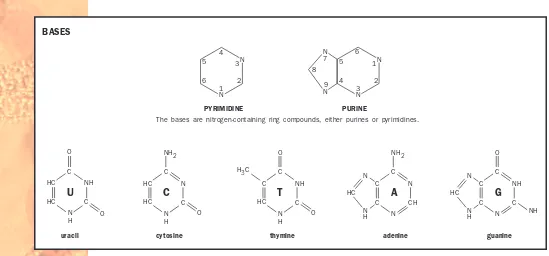
The following section provides information that is applicable to a num-ber of articles in this reference work. Included are a metric measurement and conversion table, geologic timescale, diagrams of an animal cell and a plant cell, illustration of the structure of DNA nucleotides, detail of DNA nucleotides pairing up across the double helix, and a comparison of the mol-ecular structure of DNA and RNA.
METRIC MEASUREMENT
To convert Into Multiply by
Acres Hectares 0.4047
Centimeters Inches 0.3937
Feet Meters 0.3048
Gallons Liters 3.7853
Grams Ounces 0.0353
Grams Pounds 0.0022
Hectares Acres 2.4710
Inches Centimeters 2.5400
Kilograms Pounds 2.2046
Kilometers Miles 0.6214
Liters Gallons] 0.2642
Meters Feet 3.2808
Miles Kilometers 1.6093
Ounces Grams 28.3495
STARTED
A TYPICAL ANIMAL CELL
Smooth endoplasmic reticulum
Rough endoplasmic reticulum Golgi apparatus
Ribosomes
Vacuole
Lysosome Plasma membrane Nuclear membrane Nucleolus Nucleus Chromosome Centrioles Mitochondrion Peroxisome Stalk
Basal body Rootlet
Cilium
A TYPICAL PLANT CELL
Endoplasmic reticulum
Golgi apparatus Chromosome Nucleolus Nucleus Nuclear membrane
Chloroplast
Ribosomes
Vacuole
O two DNA strands connected
by hydrogen bonds
Sugar-phosphate backbone of complementary DNA strand DNA NUCLEOTIDES PAIR UP ACROSS THE DOUBLE HELIX
3' 5'
H
O
H H
H
H HOCH
2 OH
H
OH
Deoxyribose
O
H H
OH
H
HOCH2 OH
H
OH
Ribose
O
C
C N
C
N C
O H
3C H
H H
Thymine
O
C
C N
C
N C
O
H
H
H H
Uracil
DNA RNA
A T
C G
G C
T A
A
C
G
U
Table of Contents
F
Genetic Control of Development . . . 131
Genetic Counselor . . . 135
History of Biology: Cell Theory and Cell Structure . . . 186
History of Biology: Inheritance . . . 189
History of Evolutionary Thought . . . 192
History of Medicine . . . 196
Lamarck, Jean-Baptiste . . . 23
Mimicry, Camouflage, and Warning Coloration . . . 93
Mitochondrion . . . 94
Mitosis . . . 98
Model Organisms: Cell Biology and Genetics . . . 101
Model Organisms: Physiology and Medicine . . . 102
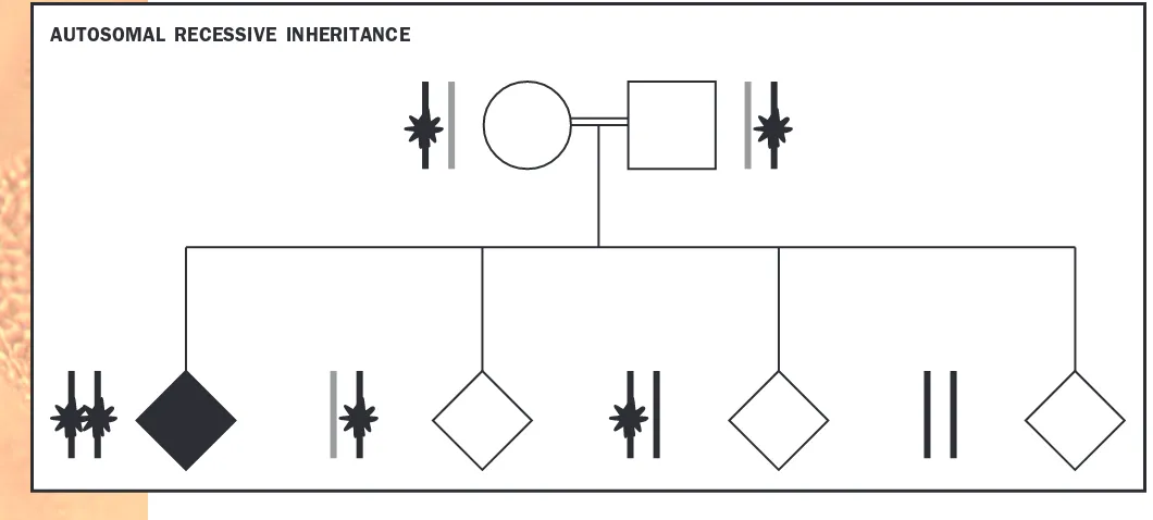
Pedigrees and Modes of Inheritance . . . 186
Peripheral Nervous System . . . 189
Pharmaceutical Sales Representative . . . 192
Pharmacologist . . . 192
Pheromone . . . 193
Photoperiodism . . . 195
Photosynthesis . . . 196
Physical Therapist and Occupational Therapist . . . 200
PHOTO ANDILLUSTRATION CREDITS . . . 243
Separation and Purification of Biomolecules . . . 93
Sex Chromosomes . . . 94
Sex Determination . . . 96
Sexual Reproduction . . . 98
Sexual Reproduction, Evolution of . . . . 101
Sexual Selection . . . 104
Sexually Transmitted Diseases . . . 106
Shoots . . . 110
Signaling and Signal Transduction . . . . 112
Skeletons . . . 118
Skin . . . 120
Sleep . . . 121
Slime Molds . . . 124
Smoking and Health . . . 126
Social Behavior . . . 127
Sociobiology . . . 131
Soil . . . 132
Speciation . . . 134
Species . . . 136
Spinal Cord . . . 137
Stress Response . . . 139
Structure Determination . . . 141
Symbiosis . . . 142
Synaptic Transmission . . . 145
T
T Cells . . . 148Taxonomy, History of . . . 151
Temperature Regulation . . . 154
Theoretical Ecology . . . 157
Thyroid Gland . . . 158
Tissue . . . 159
Torrey, John . . . 160
Touch . . . 161
Transcription . . . 162
Transfer RNA . . . 166
Transgenic Techniques . . . 167
Translocation . . . 168
Transplant Medicine . . . 172
Transposon . . . 174
Tropisms and Nastic Movements . . . 175
Tuatara . . . 176
Tundra . . . 177
Tunicate . . . 178
Turtle . . . 179
V
Vaccines . . . 180Vacuole . . . 182
van Helmont, Jan . . . 183
Vavilov, Nikolay . . . 183
Vesalius, Andreas . . . 184
Veterinarian . . . 185
Viral Diseases . . . 186
Virus . . . 187
Vision . . . 188
Vitamins and Coenzymes . . . 190
von Humboldt, Alexander . . . 192
W
Water . . . 192Water Cycle . . . 193
Water Movement in Plants . . . 193
Watson, James . . . 196
Wetlands . . . 197
Wildlife Biologist . . . 199
Wine-making, Botany of . . . 200
Wood and Wood Products . . . 201
Z
Zoology . . . 204Zoology Researcher . . . 204
PHOTO ANDILLUSTRATION CREDITS . . . 207
GLOSSARY . . . 215
TOPICOUTLINE . . . 235
CUMULATIVEINDEX . . . 245
I
Imaging in Medicine
As recently as in the 1970s, the diagnosis of some diseases often required exploratory surgery, opening a body cavity to “have a look around” for vis-ible disorders. The risks of infection, anesthesia, and imperfect healing weigh against exploratory surgery, but the diagnostic benefit may make the risk worth taking. In the last few decades, however, a variety of medical imag-ing techniques has made most exploratory surgery unnecessary and has greatly accelerated progress in medicine. Although the basic principles of some of these techniques have been known for much longer, they did not become clinically useful until computer technology had advanced enough to process data into clear images of the body, mostly since the 1970s.
Radiography
Radiography, use of X rays, is the oldest imaging technique. The term “X ray” can refer either to the type of radiation used or to the photographic image produced (the radiogram). X rays were discovered in 1885, and Marie Curie (1867–1934) trained military doctors in the use of X-ray machines in World War I. X rays are relatively simple and inexpensive to make, and they are commonly used in dentistry, mammography, chest examinations, and di-agnosis of fractures. They are best used for dense structures such as bone, but hollow organs can be visualized by filling them with a radiopaque sub-stance such as barium, given by swallow or enema to X ray the stomach or colon. Angiography is the X-ray visualization of blood vessels after injec-tion with a radiopaque dye.
Sonography
Sonography, or ultrasound imaging, is the second oldest imaging method, and the second most widely used. An outgrowth of the sonar technology developed in World War II, it uses a handheld probe to “bombard” the body with ultrasound waves and a computer to analyze the reflected signal into an image. Sonography avoids the harmful effects of X rays and is com-monly used to examine fetuses.
Computed Tomography (CT)
patient is moved through a machine that emits low-intensity X rays on one side and receives them with a detector on the other side. By imaging body slices as thin as a coin, CT scans show less overlap of organs than conven-tional X rays and thus produce sharper images. CT scans are useful for
iden-tifying tumors, aneurysms, cerebral hemorrhages, kidney stones, and other
disorders.
Magnetic Resonance Imaging (MRI)
With magnetic resonance imaging (MRI), a cylindrical device surrounds the body with a magnetic field three thousand to sixty thousand times as
Imaging in Medicine
A magnetic resonance imaging (MRI) scan of a human brain, cervical spine, and spinal marrow.
strong as Earth’s. Hydrogen atoms align themselves with this field. The
patient is then irradiated with radio waves. Hydrogen ionsabsorb this
en-ergy and align in a new direction. When the radio waves are turned off, they realign to the magnetic field and emit energy at rates that vary with the type of tissue. This emitted energy is received by a detector and ana-lyzed by a computer into an image of the body’s interior. MRI can see
through cranialand vertebral bone to visualize brain and spinal cord
tis-sue in finer detail than CT.
Positron Emission Tomography (PET)
Positron emission tomography (PET) is used to visualize the metabolic state
of a tissue. The patient receives an injection of radioactively labeled
glu-cose, which emits charged particles called positrons. When a positron and
electron meet, they annihilate each other and give off gamma rays that are picked up by a detector and analyzed by computer. The result is a color-coded image that shows which tissues were using the most glucose (that is, were most metabolically active) at the time. In cardiology, a PET scan can show the location and extent of dead heart tissue. In neuroscience, it can show which parts of the brain are active from moment to moment as a per-son engages in various sensory, motor, or intellectual tasks.
Functional MRI (fMRI)
A new variation of MRI, functional MRI (fMRI) detects the anaerobic
ac-tivity of active neuronsof the brain. It can pinpoint brain activity to within
1 or 2 millimeters, and is even more precise and useful than PET scans for studies of brain function. It also has the advantage of requiring no
injec-tions or radioactive isotopes, and it is much quicker than a PET scan. The
PET and fMRI techniques not only have been valuable for clinical diagno-sis but have added enormously to our knowledge of brain function, pin-pointing abnormalities correlated with depression, schizophrenia, and attention deficit disorder. They have also provided images of the mind at work, so to speak, identifying areas involved in consciousness, memory, thought, musical perception, reading, motor control, and speech.
Radiology is the medical specialty that embraces all of these imaging techniques. Nuclear medicine is a branch of medicine that uses radioiso-topes in the making of medical images, as in PET scans, and in the treat-ment of diseases such as cancer. Noninvasive techniques are those that require no break in the body surface whatsoever: conventional X rays; sonog-raphy; and CT, MRI, and fMRI scans. If a technique involves even such a slight invasion of the body as an injection or a barium swallow, it is con-sidered an invasive procedure (angiography and PET scans, for example). S E E A L S O Brain; Doctor, Specialist
Kenneth S. Saladin
Bibliography
Brant, William E., and Clyde A. Helms, eds. Fundamentals of Diagnostic Radiology,2nd ed. Philadelphia, PA: Lippincott, Williams and Wilkins, 1999.
National Institutes of Health, National Library of Medicine. Visible Human Project.
<http://www.nlm.nih.gov/research/visible/visible_human.html>.
Weisslader, Ralph, Mark J. Rieumont, and Jack Wittenberg. Primer of Diagnostic Imag-ing,2nd ed. St. Louis, MO: Mosby, 1997.
Imaging in Medicine
ion an electrically charged particle
cranial related to the cranium, or brain cavity
glucose simple sugar that provides energy to animal cells and is the building block of cellu-lose in plants
anaerobic without oxygen, or not requiring oxygen
neuron nerve cell
Immune Response
Among the many threats organisms face are invasion and infection by bac-teria, viruses, fungi, and other foreign or disease-causing agents. All organ-isms have nonspecific defenses (or innate defenses) that provide them with some of the protection they need. This type of defense exists throughout the animal kingdom, from sponges to mammals. Vertebrate animals, how-ever, have an additional line of defense called specific immunity. Specific immunity is also called acquired immunity, adaptive immunity, or, most sim-ply, an immune response.
Overview
One characteristic of specific immunity is recognition. Immune responses begin when the body recognizes the invader as foreign. This occurs because there are molecules on foreign cells that are different from molecules on
the body’s cells. Molecules that start immune responses are called antigens.
The body does not usually start an immune response against its own anti-gens because cells that recognize self-antianti-gens are deleted or inactivated. This concept is called self-tolerance and is a key characteristic that defines immune responses.
A second characteristic is specificity. Although all immune responses are similar, each time the body is invaded by a different antigen, the exact re-sponse is specific to that antigen. For example, infection with a virus that causes the common cold triggers a response by a different set of cells than infection with bacteria that causes strep throat.
A third characteristic is memory. After an antigen is cleared from the body, immunological memory allows an antigen to be recognized and re-moved more quickly if encountered again.
Antigen Presentation
Three groups of white blood cells are involved in starting an immune re-sponse. Although immune responses can occur anywhere in the body these
cells are found, they primarily occur in the lymphnodes and spleen. These
organs contain large numbers of antigen-presenting cells (APCs), T
lym-phocytes (or T cells), and B lymphocytes(or B cells).
APCs include macrophages, dendritic cells, and B cells. These cells en-counter the foreign invader and present the invader’s antigens to a group
of T cells called helper T cells (THcells). APCs do this by first engulfing
an invader and bringing it inside the cell. The APC then breaks the invader apart into its antigens and moves these antigens to its cell surface.
Receptors are cell surface proteinsthat can attach to antigens. Each TH
cell has a different receptor, allowing each cell to recognize a different
anti-gen. The APC “shows” the antigen to the TH cells until there is a match
between a THcell receptor and the antigen. The contact between the two
cells stimulates the THcell to divide rapidly. This process is called clonal
selection because only the THcells that recognize the foreign invader are
selected to reproduce. Stimulated TH cells also produce chemical
messen-gers called cytokines. Cytokines are made by all immune cells and control the immune response.
Immune Response
antigen foreign sub-stance that provokes an immune response
lymph pale fluid that circulates in the lym-phatic system, princi-pally composed of blood plasm and cell fluid
T cell white blood cell that controls the immune response
B lymphocyte white blood cell that makes antibodies
Antigen Clearance
The large numbers of THcells activate two other populations of white blood
cells: cytotoxic T cells (TC cells) and B cells. Like THcells, each TC cell
and B cell has receptors that match one antigen. This is why the immune system can recognize millions of antigens with specificity. The cells with the appropriate receptor encounter the antigen, preparing them for activa-tion. They receive the final signal necessary for clonal selection from TH cells and cytokines.
Cloned TCcells attach to invaders they recognize and release a variety
of chemicals that destroy the foreign cell. Because this must happen through cell-to-cell contact, it is called cell-mediated immunity (or cellular immu-nity). It is especially effective at destroying abnormal body cells, such as can-cerous cells or virus-infected cells.
Cloned B cells destroy foreign invaders differently. After activation by
THcells, B cells release proteins called antibodies. Antibodies travel through
the body’s fluids and attach to antigens, targeting them for destruction by
nonspecific defenses. This type of immune response is called antibody
-mediated immunity (or humoral immunity). It is especially effective at de-stroying bacteria, extracellular viruses, and other antigens found in body fluids.
Immunologic Memory
A primary immune response happens the first time that the body en-counters a specific antigen. It takes several days to begin and one or two
Immune Response
Antigen Presentation
antigen presenting cell
invader with antigens
TH cells with different receptors displayed
antigen
antibody immune system protein that binds to foreign mole-cules
weeks to reach maximum activity. A secondary immune response occurs if the body encounters the same antigen at a later time. It takes only hours to begin and may peak within a few days. The invader is usually removed before it has a chance to cause disease. This is because some of the cloned
TCcells and B cells produced during a primary immune response develop
into memory cells. These cells immediately become activated if the anti-gen appears again. The complex interactions among cells described above are not necessary.
In fact, this is what happens when an individual is immunized against
a disease. The vaccination (using weakened or killed pathogens) causes a
primary immune response (but not the disease) and the production of memory cells that will provide protection if exposed to the disease-causing agent.
Immune System Disorders
Studying immune responses also allows scientists to understand immune system diseases. For example, hypersensitivity disorders occur when the immune system overreacts to an antigen, causing damage to healthy
tis-sues. The result of this excessive antibody and TC cell activity can be
relatively harmless (as with allergies to pollen, poison ivy, or molds) or
deadly (as with autoimmune diseasesor allergies to bee venom and
an-tibiotics).
At the opposite end of the spectrum are immunodeficiency diseases, conditions in which the body does not respond effectively against foreign invaders. HIV (human immunodeficiency virus) infection causes AIDS
(ac-quired immunodeficiency syndrome) by attacking THcells. Occasionally an
individual is born with a deficient immune system, but these disorders are usually acquired (for example, from radiation treatment, chemotherapy, or infection with HIV). Whatever the cause, the individual has a more diffi-cult time fighting infections.
Because immune responses exhibit the characteristics of self-tolerance, specificity, and memory, a healthy body is well equipped to remove foreign invaders and prevent recurrent infections. Age, nutrition, exercise, and stress
all affect the ability of the body to fight disease. S E E A L S O AIDS; Antibody;
Autoimmune Disease; Nonspecific Defense
John M. Ripper
Bibliography
Beck, Gregory, and Gail S. Habicht. “Immunity and the Invertebrates.” Scientific American275, no. 5 (1996): 60–65.
Friedlander, Mark P., Jr., and Terry M. Phillips. The Immune System: Your Body’s Dis-ease-Fighting Army.Minneapolis, MN: Lerner Publications Company, 1998. Gustav, J. V. “Life, Death, and the Immune System.” Scientific American269, no. 3
(1993): 53–62.
National Institutes of Health. Understanding the Immune System. Washington, DC: National Institutes of Health, 1993.
Paul, William. E. “Infectious Diseases and the Immune System.” Scientific American
269, no. 3 (1993): 90–97.
Immune Response
A scanning electron micrograph of a cancer cell (red in image) being attacked by tumor-infiltrating lymphocytes.
pathogen disease-causing organism
Ingenhousz, Jan
Dutch physician and plant physiologist 1730–1799
Jan Ingenhousz was a pioneer in plant physiology and demonstrated that oxygen is produced during photosynthesis. Born in the Netherlands, In-genhousz practiced medicine in several European countries and served as a court physician to Empress Maria Theresa of Austria for twenty years. In-genhousz promoted vaccination against smallpox and helped develop a new vaccination procedure.
Ingenhousz used the gas-measuring techniques of his friend Joseph Priestley to study how plants alter the air. Priestley had shown that animals or burning candles “spoil” air, making it unfit for breathing. He had also reported that plants restore the air, but other experimenters could not repli-cate his results.
Ingenhousz attacked this problem systematically and meticulously. By placing different plant parts in sealed containers either exposed to or hid-den from sunlight, Ingenhousz showed that plants do restore the air by the production of oxygen (a gas that Priestley had recently discovered) and that the green leaves must be exposed to sunlight for this to occur. In this way, Ingenhousz began the scientific understanding of photosynthesis, a process elucidated further by Swiss agriculturist Nicolas de Saussure and others. In-genhousz contemplated using oxygen to treat patients but did not develop
the equipment to do so. S E E A L S O de Saussure, Nicolas;
Photosynthe-sis; Van Helmont, J. B.
Richard Robinson
Insect
Insects are a class of arthropods. Like other arthropods, they have
ex-oskeletons made from the carbohydrate chitin, segmented bodies, and
jointed appendages. Insects are distinguished by having three major body
segments (head, thorax, and abdomen), with three pairs of legs attached to the thorax. Ancestral head appendages have been modified to form anten-nae and mouth parts, while abdominal appendages are either absent or mod-ified to aid in reproduction. Most insects possess wings as adults, also attached to the thorax.
Sensory Systems
The insect head bears a single pair of compound eyes, composed of many individual units, called ommatidia, each of which senses a small portion of the visual field. Hunting insects such as the dragonfly may have thousands of ommatidia per eye, while others, such as ants, have many fewer. A sin-gle pair of antennae serves as chemical sensors to help find food or mates. In many species, including the tobacco hornworm moth, the female releases
airborne chemicals called pheromones that attract the male. The highly
branched antennae of the male moth can detect the molecules of the female pheromone, and can track the scent to find the female over very long
Insect
arthropods organisms with jointed appendages and exoskeletons, including insects, spi-ders, and crustaceans
exoskeleton external skeleton
chitin nitrogen-containing carbohydrate found in arthropod exoskeletons and fungus cell walls
appendage attached organ or structure
distances. Chemoreceptors are also located on the feet, allowing an insect to taste its food as it walks across a leaf or a table. The numerous hairs cov-ering the insect body are linked to mechanoreceptors, which aid its sense of touch. Some mechanoreceptors can sense changes in air pressure, useful for flying or evading a swooping predator. Receptors for carbon dioxide, water, and temperature also exist.
Ingestion, Digestion, and Excretion
Insect mouth parts vary tremendously in their shapes, reflecting adaptations to a wide variety of feeding habits. Mosquitoes, for instance, have a long hypodermic needlelike stylet, perfect for piercing skin to suck blood. But-terflies and moths, among others, have a very long, flexible strawlike mouth part, the proboscis, which they unfold to sip nectar from the base of flow-ers. Houseflies have a spongy tonguelike labrum for sopping up a variety of foods. Grasshoppers and beetles have small, sharp mouth parts adapted for chewing. The insect gut is divided into three regions, with most digestion occurring in the midgut. Suspended into the midgut are the Malpighian tubules, which filter nitrogenous waste from the blood and deposit it as
crys-Insect
tals within the gut, avoiding the water loss that urine formation would en-tail. In termites, the hindgut houses a complex group of protists and bacte-ria that digest wood.
Legs and Wings
Insect legs are used for walking and climbing. In some predatory species such as the praying mantis, the front pair of legs has been modified for cap-turing prey, with barbed surfaces that hold other insects tightly. Almost all insects have wings, although a few primitive forms do not. In the ants, only the reproductive members of the colony have wings, which they shed after their “nuptial flight,” in which they mate with members of the opposite sex.
Respiration and Circulation
Insects do not have lungs, but instead employ a highly branched network of internal tubes, called tracheae, to deliver oxygen to the tissues. Tracheae connect with the atmosphere through openings in the exoskeleton called spiracles. Insect circulatory systems transport nutrients and wastes in a fluid called hemolymph, which is pumped into and out of internal chambers sur-rounding the organs, an arrangement called an open circulatory system.
Reproduction and Development
Most insects reproduce sexually, although the aphids are a notable excep-tion. Aphids reproduce by parthenogenesis, in which the egg develops into
a new organism without fertilization. In honey bees and some other social
insects, only one female per colony reproduces, and males are haploid,
whereas females are diploid, a system called haplodiploidy. The queen
pro-duces new (diploid) females (workers, soldiers, and future queens) from fer-tilized eggs. Males are produced from eggs that are not ferfer-tilized, and thus males are haploid.
Insects vary in their degree of metamorphosis during development.
Butterflies, beetles, and flies, for example, undergo complete metamorpho-sis, in which the egg hatches into a feeding larva, which then pupates. Within the pupa, the larval tissues dissolve and rearrange into the adult form. In contrast, grasshoppers, cockroaches, and cicadas undergo incomplete meta-morphosis, emerging from the egg as a miniature adult, but minus the wings and genitals. To grow, all insects must molt, or shed their exoskeleton, which then reforms around the larger individual.
Metamorphosis often allows juvenile and adult individuals of the same species to avoid competition for food. Larval moths feed voraciously and can be significant agricultural pests, while adult moths either don’t feed or consume only nectar.
Diversity
Insects are the most diverse of all groups of organisms, with over 800,000 species named and many thousands, probably millions, yet to be discovered. Insect diversity may be linked to their close association with the angiosperms (flowering plants). The Coleoptera (beetles) are the most diverse of all in-sect orders, with at least 350,000 species, representing one fourth of all known animal species. (Asked what could be inferred about the work of the
Insect
fertilization union of sperm and egg
haploid having single, nonpaired chromosomes in the nucleus
diploid having pairs of chromosomes in the nucleus
Creator from a study of His works, British scientist J. B. S. Haldane is re-ported to have quipped, “an inordinate fondness for beetles.”) The evolu-tionary reasons for the mind-boggling diversity of this single order are not clear. Other major orders of insects include the Diptera (flies), Hymenoptera (bees and wasps), Hemiptera (true bugs), and Lepidoptera (moths and
but-terflies). Note that each name describes the wing (pterameans “wing”). For
instance, Diptera means “two wings,” referring to the presence of only one wing pair in this order. In the Coleoptera (“sheath wings”), the first pair of wings is modified into a hard covering for the rear pair, which is easily
ob-served in a lady beetle, for instance. S E E A L S O Angiosperms; Arachnid;
Arthropod; Biodiversity; Osmoregulation; Physiological Ecology; Plant Pathogens and Pests; Symbiosis
Richard Robinson
Bibliography
Berenbaum, May. Bugs in the System.New York: Addison-Wesley, 1995.
Daly, H. V., J. T. Doyen, and A. H. Purcell. Introduction to Insect Biology and Diver-sity.New York: Oxford University Press, 1998.
Evans, Arthur V., and Charles L. Bellamy. An Inordinate Fondness for Beetles. New York: Henry Holt & Company, 1996.
Fabre, Jean Henri. Fabre’s Book of Insects.Mineola, NY: Dover Publications, 1998.
Invasive Species
Animals, plants, and other organisms that are newly introduced into an area from another part of the world are sometimes referred to as “alien” or “exotic” species. These words are used to distinguish newly arrived species from the native species that have lived in the environment for very long periods of time. Although some people refer to all exotic species as in-vaders, some scientists believe it makes more sense to use the term “inva-sive species” only when referring to new species that are spreading rapidly and having a large negative impact on the environment, economic activi-ties, or human health.
Many of these invasive species have been introduced into new environ-ments by human activities. Sometimes they are introduced intentionally, such as European starlings, kudzu, and purple loosestrife, three species that spread very rapidly across the United States beyond their initial range of in-troduction and are believed to have reduced the abundance of native bird and plant species in many areas. However, most species introductions prob-ably occur inadvertently by humans, a byproduct of frequent movements around the globe. For example, small ocean organisms are commonly picked up in the ballast water of ocean ships. When the ships release their ballast water at a port in another part of the world, these organisms are introduced into a new environment. Logs and other wood and fiber products imported into the United States sometimes contain insects from their country of ori-gin, which accounts for the introduction of Chestnut Blight fungus in the United States.
In many cases, the new species do not spread very much nor do they have a large impact. However, many of these new species have created huge problems. Zebra mussels are reducing populations of native mussels in many
areas of the United States, and they are so numerous in places that they are clogging up water intake pipes of power plants and municipal water sup-plies. Leafy spurge, an introduced poisonous plant of grassland, has covered large regions of the northern Great Plains and threatens many of the live-stock operations in these areas.
One of the most famous ecological disasters associated with invasive species is the brown tree snake that was accidentally introduced on to the Pacific island of Guam. In just a few decades, through its hunting habits, the snake was responsible for the extinction of several of the island’s bird species that were found nowhere else on Earth. Problems produced by in-vasive species are believed to cost billions of dollars every year.
Scientists are working very hard to find out what factors facilitate these biological invasions in hopes of providing some help to those trying to con-trol their negative effects. It is clear that trying to prevent the introduction of new species into an area in the first place is the primary step to take. Some scientists are also trying to determine why some environments seem to be invaded more easily than others, or why some environments are
Invasive Species
invaded only at certain times. Some think that environments that have a high diversity of native species may be more resistant to invasions by new species, while others believe that disturbances and other factors that free up new resources are more important to opening an environment to invaders. There is still much work that needs to be done to increase scientists’
un-derstanding of the causes and effects of invasive species. S E E A L S O
Biodi-versity; Conservation; Extinction; Global Climate Change
Mark A. Davis
Bibliography
Elton, Charles S. The Ecology of Invasions by Animals and Plants.Chicago: University of Chicago Press, 2000.
Pimental, D., L. Lach, R. Zuniga, and D. Morrison. “Environmental and Economic Costs of Nonindigenous Species in the United States.” Bioscience50: 53–65.
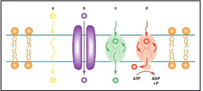
Ion Channels
Ions are charged particles such as Na+, H+, K+, Ca2+, and Cl-. Ions have a
significant effect on many cell processes and also influence the amount of
water in the cell. Cells use inorganicions for transmitting signals across the
cell membrane or along the surface of the cell. Other cellular functions as
diverse as secretionof hormones to fertilizationof egg cells require ion
transport across the cell membrane. However, ions have great difficulty pass-ing through the membrane by simple diffusion because cell membranes are
composed of hydrophobic phospholipids that oppose the passage of
hy-drophilic ions. Furthermore, the negatively charged phosphate head groups of the phospholipids tend to repel negatively charged anions and trap
pos-itively charged cations. Therefore, an ion as small as a hydrogen ion (H+)
requires a specific portal proteinto facilitate its transport through the
mem-brane. Such a protein molecule is called an ion channel.
Molecular Structure of Potassium and Sodium Channels
An ion channel is usually equipped with four basic parts: a central conduc-tion pathway (opening) for ions to pass through, an ion recogniconduc-tion site to allow passage of specific ions (selectivity filter), one or more gates that may open or close, and a sensor that senses the triggering signal and transmits it to the gate.
The Shaker-type voltage-gated potassium channel of nerve and mus-cle provides a good example of the four parts of the ion channel. The
name Shakerarises from the gene coding for this channel in the fruitfly
(Drosophila melanogaster), whose mutation causes the fly to shake. Humans have many potassium channels belonging to the Shaker family. This chan-nel is composed of four identical subunits arranged like a four-leaf clover, with the center serving as the ion conduction pathway. Each subunit has six segments that cross the membrane and are termed S1 through S6. The region between S5 and S6 segments from each subunit contributes to form the ion conduction pathway; hence, it is called the “pore” or “P-region.”
Ion Channels
inorganic not bonded to carbon
secretion material released from the cell
hormone molecule released by one cell to influence another
fertilization union of sperm and egg
hydrophobic “water hating,” such as oils
hydrophilic “water loving”
protein complex mole-cule made from amino acids; used in cells for structure, signaling, and controlling reactions
In the P-region, a few critical amino acidsfrom each subunit gather to form the selectivity filter that specifically recognizes only potassium ions. The S4 segment contains positively charged amino acids on every third po-sition and serves as a voltage sensor. When the potential on the internal surface of the membrane becomes more positive, the potential drives the S4 segment toward the outside. This movement triggers a channel gate to open. The voltage-gated sodium channel has a similar architecture, except that the four subunits are strung together in a long peptide chain like a train of parad-ing elephants linkparad-ing up trunk-to-tail. This channel is highly selective for sodium ions.
Biophysics
As the charged ions flow across the membrane, they generate an electric current. The amount of current flow is determined by three factors. First, when the gate of an ion channel opens, ions flow down the concentration
gradientfrom high to low across the membrane, which is typical of the pas-sive transport mechanism. Second, the flow of ions is controlled by the volt-age difference across the membrane. For instance, if the cell interior is
Ion Channels
Shaker Voltage-gated K+ Ion Channel
ex
cyt
chain
ball pore
amino acid a building block of protein
gradient difference in concentration between two places
already highly positive, less K+will flow in. Third, a channel may be highly selective for a specific ion (such as the voltage-gated sodium channel) or rather nonselective (such as the mechano-sensitive channel). Thus, the to-tal ion flow is influenced by the concentration gradient of the ions, the volt-age difference across the membrane, and the permeability of the ions.
The patch clamp technique developed in 1980 has enabled scientists to record current flow through a single ion channel. This technique uses ex-tremely fine glass electrodes attached to membranes to measure electrical activity in a very small part of the membrane. One of the most exciting re-sults from the development of the patch clamp technique is direct observa-tion of the opening and closing of a single channel, like observing the twinkle
of a little star in the night sky. The opening of a channel represents a
con-formationalchange of the channel molecule from a closed state to an open state. If the rate for such a conformational change is dependent on voltage, then the channel is said to be voltage-gated. A channel may stay in the open state for less than a millisecond to tens of seconds. The current flow through a single channel may range from less than a picoampere to hundreds of
pi-coamperes (a picoampere is 10-12ampere).
Drugs and Toxins Acting on Ion Channels
Nature produces a wide variety of highly potent toxins that target specific ion channels. The toxins are usually packaged in venom and delivered by stings or fangs. A large number of toxins have been isolated from scorpi-ons, sea anemones, cone snails, and snakes. They have been used for study-ing various ion channels. One of the most famous toxins is tetrodotoxin, which selectively blocks the sodium channel. It is contained in the poison-ous puffer fish, which ironically is the most expensive delicacy served in Japanese restaurants. Only chefs who have passed rigorous licensing exam-inations are allowed to prepare the fish. Tetrodotoxin is also commonly por-trayed in fictions and movies; it almost killed the fictitious Agent 007 James
Bond in From Russia with Love. Drugs have been developed to target ion
channels and to prevent the channels from conducting ions. They are widely used as local anesthetics, antiarrhythmic drugs to prevent irregular heart-beats, antihypertensive drugs to lower blood pressure, and anti-epileptic drugs to prevent seizures.
Genetic Defects of Ion Channels
Several genetic diseases exhibiting defects in the physiological functions of ion channels have now been shown to be caused by mutations in the genes coding for specific ion channels. For example, a cardiac potassium channel named HERG (human ether-a-go-go-related gene) acts to protect the heart against inappropriate rhythmicity. People lacking a functional HERG gene exhibit an abnormality on their electrocardiogram called “long Q-T syn-drome,” which predisposes them to sudden cardiac arrest when they are un-der stress. Cystic fibrosis results from mutations of a particular chloride
channel called the cystic fibrosis transmembrane conductance regulator. S E E
A L S O Membrane Proteins; Membrane Transport; Neuron; Synaptic Transmission
Chau H. Wu
Ion Channels
Bibliography
Hille, Bertil. Ionic Channels of Excitable Membranes,2nd ed. Sunderland, MA: Sinauer Associates Inc., 1992.
Neher, Erwin, and Bert Sakmann. “The Patch Clamp Technique.” Scientific Ameri-can266 (1992): 44–51.
Kidney
The kidneys of vertebrates have the vital function of removing metabolic wastes from the blood and otherwise maintaining its normal composition. The two kidneys of a normal human adult produce 1 to 2 liters (about 30 to 70 fluid ounces) of urine each day that contain wastes, excess water, and other unneeded molecules. Production of less than 0.4 liter (13.5 fluid ounces) of urine per day is insufficient to eliminate wastes and regulate the composition of blood. Such a condition is always fatal within a few weeks unless the underlying cause is corrected, a new kidney is transplanted, or
the blood is artificially cleared by dialysis.
The human kidney belongs to one of three kinds of kidneys that occur among different vertebrates at various developmental stages. The first type, called the pronephros, lies toward the front of some fishes and the embryos of many vertebrates. The mesonephros lies more posteriorly and occurs in most adult fishes and amphibians and in the embryo of humans and other mammals. The metanephros occurs still farther posteriorly and is the type of kidney in adult reptiles, birds, and mammals, including humans.
Each human kidney is about the size of a fist, shaped like a kidney bean, and located on one side of the lower abdomen toward the back. At any given
Kidney
K
Adrenal gland
Left renal artery
Left renal vein Left kidney
Aorta
Urinary bladder
Urethra Right ureter
Vena cava Renal pelvis
Medulla Cortex
time about one-fifth of the body’s blood is flowing through the kidneys. The blood enters each kidney from the body’s major artery, the aorta, by means of the renal artery. (The word “renal” refers to kidney.) Blood leav-ing the kidney enters the major vein, the vena cava, via the renal vein. Also connecting to the kidney is a third tube, the ureter, which conducts urine to the urinary bladder for temporary storage.
From this “plumbing diagram” one can get an overview of renal func-tion: blood enters the kidney, wastes and excess molecules are removed with the urine, and the blood is returned to the circulatory system. To appreci-ate how the kidneys function, however, one must take a microscopic view
of one of the million or so structures called nephronswithin each kidney.
Each nephron begins its work by producing a filtrate of blood. Filtration
occurs in a tuft of capillaries called the glomerulus. The lining of the
glomerulus is leaky enough to allow blood pressure to force water, ions, and
small molecules out while retaining cells and very large molecules in the blood. The filtrate, which is very much like the fluid portion of blood (plasma), enters Bowman’s capsule, which encloses the glomerulus like a hel-met. Bowman’s capsule conducts the filtrate into the first part of the nephron tubule, called the proximal convoluted tubule. In humans approximately 180 liters of filtrate (almost enough to fill a 50-gallon drum) make it this far each day. Fortunately, not all of it goes into urine. In the proximal tubule, many
of the inorganicions and almost all of the glucose and amino acids get
pumped out of the filtrate and go back into the blood. Most of the water in the filtrate is also drawn back into the blood.
The tubular fluid next passes through a hairpin turn called the loop of Henle, which helps the nephron return more water to the bloodstream rather than allowing it to be lost in the urine. How this works will be explained
later. Tubular fluid then enters the distalconvoluted tubule of the nephron.
Here further transport of particular ions may occur, depending on whether the concentration of that ion in the blood is too high or too low. For
ex-ample, if the pHof the blood is too low, hydrogen ions (H+) are transported
out of the blood and into the tubular fluid. If the pH is too high, H+ions
are transported from the fluid into the blood.
By the time the fluid has completed its journey through the distal con-voluted tubule, it is essentially dilute urine, called preurine. Preurine from several nephrons enters a tube called the collecting duct. As preurine passes through the collecting duct, more water can be removed and returned to the blood.
Water is drawn out of the collecting duct by osmosis due to an
in-creasing concentration of ions surrounding the collecting duct. The loops
of Henle produce this concentration gradient by a combination of
trans-port and diffusion of ions and urea. Urea is a molecule that temporarily
stores the nitrogen produced by the metabolismof proteins. After
help-ing to create the concentration gradient, urea is eventually eliminated with
the urine. S E E A L S O Blood; Drug Testing; Excretory Systems; Heart
and Circulation; Osmoregulation; Pituitary Gland
C. Leon Harris reabsorbs water in the kidneys, and so increases urine volume.
Saladin, Kenneth S. “The Urinary System.” In Anatomy and Physiology, 2nd ed. Dubuque, IA: McGraw-Hill Higher Education, 2001.
Supplemental Image Database, The Kidney. <http://www.kumc.edu/instruction/ medicine/pathology/ed/ch_16/mainframe.html>.
Kingdom
Kingdom is the highest category in the hierarchical classification of organ-isms created by Carolus Linnaeus around 1750. Linnaeus recognized two kingdoms, plants and animals, a scheme that worked reasonably well for large multicellular organisms but failed as microscopes revealed diverse uni-cellular organisms. In 1959 Robert Whittaker devised a five-kingdom sys-tem that maintained kingdoms Plantae and Animalia but added kingdoms Monera, Protista, and Fungi (see Table).
Kingdom
A COMPARISON OF THE FIVE KINGDOMS
Characteristic Monera Protista Plantae Fungi Animalia
Internal cell Absent Present Present Present Present
membranes (Prokaryotes) (Eukaryotes) (Eukaryotes) (Eukaryotes) (Eukaryotes)
Cell wall Present Present or Present Present Absent
Absent
Organization Unicellular Unicellular or Multicellular Mainly Multicellular
Multicellular multicellular
Mode of nutrition Autotrophs or Autotrophs or Autotrophs Heterotrophs Heterotrophs
Heterotrophs Heterotrophs
Representative Archaea, Protozoa, Mosses, ferns, Molds, yeasts, Animals with
groups eubacteria algae, slime seed plants mushrooms and without
molds backbones
Note: An autotroph is an organism that uses solar energy or energy from inorganic chemicals to make organic molecules. A heterotroph obtains organic molecules by consuming other organisms or their products.
Whittaker placed bacteria in their own kingdom, Monera, because of
fundamental organizational differences between prokaryotic bacterial
cells, which lack membrane-enclosed nuclei and organelles, and the
eu-karyotic cellsof other organisms that possess internal membranes. Plan-tae, Fungi, and Animalia consist of complex, multicellular eukaryotic organisms that differ from each other in details of cell structure and in how they secure and process energy. Protista is a collection of single-celled eukaryotic organisms and simple multicellular forms, some animal-like, some plantlike.
Molecular evidence, particularly from ribosomal ribonucleic acid (RNA), suggests that the five-kingdom scheme is also too simple. Some bi-ologists believe that Protista should be partitioned into three or more king-doms. Similarly, kingdom Monera contains two very biochemically distinct groups of prokaryotes: archaebacteria, and eubacteria. A proposed system acknowledges this ancient evolutionary split by creating a higher level of classification, domain, above kingdom. This system distinguishes three do-mains: Archaea, Eubacteria, and Eukarya (containing protists, plants, fungi,
prokaryotic without a nucleus
organelle membrane-bound cell compartment
and animals). S E E A L S O Animalia; Archaea; Eubacteria; Fungi; Linnaeus, Carolus; Plant; Protisa
Cynthia A. Paszkowski
Bibliography
Margulis, Lynn, and Karlene V. Schwartz. Five Kingdoms: An Illustrated Guide to the Phyla of Life on Earth.New York: W. H. Freeman and Company, 1998.
Krebs Cycle
When glucoseis converted to pyruvate during glycolysis, two adenosine
triphosphates (ATPs) are formed, but most of the energy in the original
glucose remains in pyruvate. In most aerobiccells, the pyruvate formed by
glycolysis is further degraded in a pathway called the Krebs cycle (also called the tricarboxylic acid cycle or citric acid cycle). In the Krebs cycle, the
car-bon of pyruvate is fully oxidized to carcar-bon dioxide in a series of
oxidation-reduction reactions. During these reactions, much of the energy in the original pyruvate is carried as high-energy electrons by the electron shut-tles NADH and FADH2. These electrons will ultimately be passed to the electron transport chain, where their energy will be used to synthesize ATP by oxidative phosphorylation. Much more ATP is made by the Krebs cy-cle and oxidative phosphorylation than by glycolysis alone.
In eukaryotic cells, pyruvate is transported to the mitochondrial
ma-trix, where the Krebs cycle takes place. Before entering the Krebs cycle, the
three-carbon pyruvate is oxidized to a two-carbon acetate molecule and car-bon dioxide, producing one molecule of NADH. The acetate joins to a mol-ecule of coenzyme A to form acetyl coenzyme A, which carries the acetyl group to the Krebs cycle. The acetate enters the cycle by combining with OAA (oxaloacetic acid) to form citric acid. At this point, two of the origi-nal three carbon atoms in pyruvate have been incorporated into citric acid and one has been oxidized to carbon dioxide, and one molecule of NADH has been produced.
As the reactions of the Krebs cycle continue, the two acetyl carbons are successively oxidized to carbon dioxide, forming two molecules of NADH and one of FADH2, which will provide electrons to the electron transport chain to form ATP. In addition, one guanosine triphosphate
(GTP) is formed directly by substrate-level phosphorylation, or transfer
of a phosphate directly from the reacting molecules. (The GTP eventu-ally transfers its phosphate to form ATP.) The final unoxidized product of the entire cycle is OAA, which can accept another acetyl group to start the cycle again.
The Krebs cycle occupies a central position in cellular metabolism. It
can break down the pyruvate produced in glycolysis, but these two path-ways do not form an isolated system in cells. Both are linked to other processes in many ways. Acetyl coenzyme A is produced by other means, notably by fatty-acid oxidation, and the Krebs cycle will oxidize this acetyl coenzyme A as readily as that produced from pyruvate.
Similarly, other substances are fed into the Krebs cycle at this and other points, either to be consumed as fuel or to be transformed for other
cellu-Krebs Cycle
glucose simple sugar that provides energy to animal cells and is the building block of cellu-lose in plants
pyruvate the ionized form of pyruvic acid, a key intermediate in cell metabolism
glycolysis initial stages of sugar breakdown in a cell oxidation is loss of elec-trons, and reduction is gain of electrons
lar needs. For example, amino acidscan be consumed by entering the Krebs cycle at several points. Conversely, several amino acids can be synthesized from intermediates of the Krebs cycle. Thus the Krebs cycle can serve ei-ther to degrade amino acids, releasing energy in the process, or to supply precursor molecules for amino acid synthesis. Which of these activities
pre-vails depends on the needs of the cell at any particular time. S E E A L S O
*Enzymes that catalyze these reactions: 1. Citrate synthase
2. Aconitase
3. Isocitrate dehydrogenase 4. ␣-Ketoglutarate dehydrogenase
5. Succinyl CoA synthetase 6. Succinate dehydrogenase 7. Fumarase
8. Malate dehdrogenase *CoA is often written as CoA-SH
CH2
The series of reactions that make up the Krebs cycle.
Glycolysis and Fermentation; Metabolism, Cellular; Mitochondrion; Oxidative Phosphorylation
David W. Tapley
Bibliography
Bodner, G. M. “The Tricarboxylic Acid (TCA), Citric Acid or Krebs Cycle.” Jour-nal of Chemical Education63 (1986): 673–677.
Hinkle, P. C., and R. E. McCarty. “How Cells Make ATP.” Scientific American238 (March 1978).
Racker, E. “The Membrane of the Mitochondrion.” Scientific American218 (Febru-ary 1968).
Laboratory Technician
Laboratory technicians do almost all of the hands-on work in scientific re-search, development, and analysis. One of the benefits of being a laboratory technician is being the first to see experimental outcomes, whether they are prize-winning projects or more routine medical exams.
The different types of jobs that laboratory technicians have and the skills and training required for those jobs can vary tremendously. For example, a laboratory technician working on a research project might operate an elec-tron microscope, isolate DNA (deoxyribonucleic acid), make behavioral ob-servations of animals, monitor pharmaceutical effects in test subjects, or monitor environmental quality. In a clinical laboratory, a laboratory tech-nician may examine blood samples for cell counts, examine tissue samples for parasites, or test fluids for chemical contaminants or drugs. In indus-trial production environments, laboratory technicians may conduct product quality tests and monitor product quality control. In all settings, laboratory technicians work with the most modern and sophisticated laboratory and computer equipment available. Potential employers include government and private research laboratories, universities, hospitals, and private industries.
These employers may have research, development, clinical, forensic, or
pro-duction-oriented objectives. With growth in technology, the job market for laboratory technicians is expected to expand.
Education and training for a laboratory technician is based in science and technology. Preparation in high school should include college prepara-tory courses that will support extensive college requirements for mathe-matics and science. Entry-level positions for laboratory technicians almost always require a two-year associate’s or a four-year bachelor’s degree in a scientific area (commonly biology, chemistry, physics, biotechnology, or natural resources). In some cases, a master’s of science degree or profes-sional certification program and exam must be completed. Almost all be-ginning laboratory technicians receive additional on-the-job training, and laboratory technicians should expect to continue updating their education
and training as technology advances. S E E A L S O Medical Assistant;
Mi-croscopist
Michael G. Scott
Bibliography
U.S. Department of Labor. Bureau of Labor Statistics. Occupational Outlook Handbook.
<http://stats.bls.gov/ocohome.htm>.
Laboratory Technician
L
parasite organism living in close association with another from which it derives most of its nutrition
Lakes and Ponds
Lakes and ponds are inland bodies of standing or slowly moving water. Al-though lakes and ponds cover only 2 percent of the world’s land surface, they contain most of the world’s fresh water. Individual lakes and ponds range in area from a few square meters to thousands of square kilometers. In general, ponds are smaller than lakes, though regional idiosyncrasies of naming abound—Henry David Thoreau’s famous Walden Pond in Massa-chusetts has a surface area of 64 acres. Lakes and ponds are an important source of fresh water for human consumption and are inhabited by a diverse suite of organisms.
Formation
Lakes and ponds are formed through a variety of events, including glacial, tectonic, and volcanic activity. Most lakes and ponds form as a result of glacial processes. As a glacier retreats, it may leave behind an uneven sur-face containing hollows that fill with water. Glacial activity at the end of the Pleistocene epoch (ten thousand to twenty thousand years ago) resulted in the formation of most of the lakes and ponds in the Northern Hemisphere, including the Great Lakes of North America. Some of the oldest lakes and ponds (more than three hundred thousand years old) were formed by tec-tonic activity related to movement of Earth’s crust. For example, Lake Baikal
in Siberia formed from the movement of tectonic platesand is the largest
freshwater lake by volume in the world. Volcanic activity can also lead to lake and pond formation. For example, the collapse of a volcanic cone of Mount Mazama in Oregon led to the formation of Crater Lake, the sev-enth deepest lake in the world.
Physical and Chemical Features
Light and temperature are two key physical features of lakes and ponds. Light from the sun is absorbed, scattered, and reflected as it passes through Earth’s atmosphere, the water’s surface, and the water. The quantity and quality of light reaching the surface of a lake or pond depends on a variety of factors, including time of day, season, latitude, and weather. The quality and quantity of light passing through lake or pond water is affected by prop-erties of the water, including the amount of particulates (such as algae) and
the concentration of dissolved compounds. (For example, dissolved organic
carbon controls how far ultraviolet wavelengths of light penetrate into the water.)
Light and wind combine to affect water temperature in lakes and ponds. Most lakes undergo a process called thermal stratification, which creates three distinct zones of water temperature. In summer, the water in the shal-lowest layer (called the epilimnion) is warm, whereas the water in the deep-est layer (called the hypolimnion) is cold. The middle layer, the metalimnion, is a region of rapid temperature change. In winter, the pattern of thermal stratification is reversed such that the epilimnion is colder than the hy-polimnion. In many lakes, thermal stratification breaks down each fall and spring when rapidly changing air temperatures and wind cause mixing. How-ever, not all lakes follow this general pattern. Some lakes mix only once a year and others mix continuously.
Lakes and Ponds
tectonic plate large segment of Earth’s crust that moves in rela-tion to other similar plates
The chemistry of lakes and ponds is controlled by a combination of physical, geological, and biological processes. The key chemical character-istics of lakes and ponds are dissolved oxygen concentration, nutrient
con-centration, and pH. In lakes and ponds, sources of oxygen include diffusion
at the water surface, mixing of oxygen-rich surface waters to deeper depths, and photosynthesis. Oxygen is lost from lakes and ponds during respiration by living organisms and because of chemical processes that bind oxygen. The two most important nutrients in lakes and ponds are nitrogen and phorus. The abundance of algae in most lakes and ponds is limited by phos-phorus availability, whereas nitrogen and iron are the limiting nutrients in the ocean. The acidity of water, measured as pH, reflects the concentration
of hydrogen ions. The pH value of most lakes and ponds falls between 4
and 9 (the pH value of distilled water is 7). Some aquatic organisms are ad-versely affected by low pH conditions caused by volcanic action, acid-releasing vegetation surrounding bog lakes, and acid rain.
Habitats and Diversity
Lakes and ponds are characterized by three main habitats: the pelagic zone, the littoral zone, and the benthic zone. The pelagic zone is the open water
Lakes and Ponds
A mountain lake in the Canadian Rockies. Although lakes and ponds cover only 2 percent of the world’s land surface, they contain most of the world’s fresh water.
pH measure of acidity or alkalinity; numbers below 7 are acid, above are basic
area of lakes and ponds. In large lakes, the pelagic zone makes up most of the lake’s volume. The littoral zone is the inshore area where light pene-trates to the bottom. This zone often contains large, rooted plants called macrophytes. The areas of the lake or pond bottom that are not part of the littoral zone are referred to as the benthic zone. This zone contains fine sediment that is free of plant life because light levels are too low to support plant growth.
Lakes and ponds typically contain a diversity of organisms that perform different ecological functions. Many of the organisms in lakes and ponds are quite small and can only be seen with a microscope. Plankton are micro-scopic aquatic organisms, including bacteria, algae, and zooplankton, that have little or no means of locomotion. In addition, there are many larger vertebrate animals that inhabit lakes and ponds, including fish and amphib-ians. Other organisms that use lakes and ponds for some activities include birds such as ducks, mammals such as beavers, and reptiles such as snakes.
Larger lakes can support as many as four or five different trophic
lev-els, or groups of organisms that get energy in the same way. For instance,
the major trophic levels in the pelagic zone, or open water areas, are
phy-toplankton, zooplankton, planktivorous (plankton-eating) fish, and pisciv-orous (fish-eating) fish. Microbes such as bacteria and protists are also important in lakes and ponds due to their role in decomposition and
nutri-ent recycling. The food webin the pelagic zone is connected to the inshore
food web because many mobile organisms from the pelagic zone (especially
fish) use the inshore areas for shelter and food. S E E A L S O Algae;
Ecosys-tem; Estuaries; Limnologist; Rivers and Streams; Wetlands
Janet M. Fischer and Katharine E. Yoder
Bibliography
Lampert, Winfried, and Ulrich Sommer. Limnoecology: The Ecology of Lakes and Streams.New York: Oxford University Press, 1997.
Moss, Brian. Ecology of Fresh Waters: Man and Medium.Oxford: Blackwell Scientific Publications, 1988.
Wetzel, Robert G. Limnology.Philadelphia, PA: W. B. Saunders Co., 1983.
Lamarck, Jean-Baptiste
French naturalist1744–1829
Jean-Baptiste Lamarck is best remembered for the incorrect hypothesis that evolutionary change occurs due to the inheritance of acquired characteris-tics. However, Lamarck’s contributions to biological thought are much more important than being the champion of a failed idea. He was the first really important thinker about evolution, and he established the central role of the environment in determining the adaptations of all types of organisms.
Born into a military family, Lamarck had a brief career as a soldier
before turning his attention to medicine and science. His Flore Française
(1778) on the plants of France brought him to the attention of French naturalist Comte de Buffon (Count Buffon), who became his sponsor in scientific circles. He was appointed professor at the National Museum of
Lamarck, Jean-Baptiste
trophic related to feed-ing
phytoplankton micro-scopic floating creatures that photosynthesize
Natural History, in charge of insects and “worms,” meaning all inverte-brates. Lamarck was the first to propose separating the arachnids (spiders), mollusks, and crustaceans from the insects, placing them in separate classes.
Lamarck’s appreciation of the enormous diversity of the invertebrates (a term he invented) strengthened his belief that species evolve over time. Lamarck proposed that environmental changes cause a change in an or-ganism’s needs, which leads to a change in behavior. For instance, scarce prey might lead to the need for a hawk to search the ground more carefully from a greater height. The increased use of its eyes would, according to Lamarck, improve the hawk’s eyesight. Furthermore, this acquired im-provement would be inherited by the hawk’s offspring over time. Alterna-tively, the disuse of an organ would cause it to shrink or weaken. Lamarck
published his hypothesis in his book Philosophie Zoologique (1809).
Lamarck also believed that all animals were becoming progressively
more complex and “perfect” over time, leading him to propose that
spon-taneous generation accounted for the appearance of the simplest of or-ganisms.
We now know that heritable change cannot be induced by use or dis-use, but can only arise through changes in an organism’s deoxyribonucleic acid (DNA); nor does spontaneous generation occur. Despite his incorrect mechanism for evolution, Lamarck focused evolutionary thought on the idea of adaptation to the environment, an idea that was to be central to English
naturalist Charles Darwin’s concept of natural selection fifty years later.
S E E A L S O Adaptation; Buffon, Count (Georges-Louis Leclerc); Dar-win, Charles; Evolution; Natural Selection
Richard Robinson
Bibliography
Magner, Lois E. History of the Life Sciences,2nd ed. New York: Marcel Dekker, 1994. Mayr, E. The Growth of Biological Thought.Cambridge, MA: Harvard University Press,
1982.
Landscape Ecology
Landscape ecology is the study of the causes and ecological consequences of spatial pattern in landscapes. While there is no specific spatial extent that defines a landscape, most landscape ecologists are interested in large areas ranging from a few square kilometers to entire continents. Within
land-scapes it is usually possible to define a series of different ecosystemtypes
occurring as patches within the greater landscape. For example, in an agri-cultural landscape the patches might be different fields, woodlots, hedgerows, buildings, and ponds. The goal of a landscape ecologist is to un-derstand and describe landscape structure; how this structure influences the movement of organisms, material, or energy across the landscape; and how and why landscape structure changes over time.
A landscape’s structure can be quantified by describing characteristics of patches, such as their number, size, shape, position, and composition. Landscape ecologists have defined measures to quantify each of these
at-Landscape Ecology
ecosystem an ecologi-cal community and its environment
spontaneous generation the theory that life began from nonliving matter