www.elsevier.com / locate / bres
Research report
Spontaneous and auditory-evoked activity of medial agranular cortex
as a function of arousal state in the freely moving rat: interaction with
locus coeruleus activity
a,b ,
*
a aToshikazu Shinba
, Laurent Briois , Susan J. Sara
a
´
Neuromodulation et Processus Cognitifs, Institut des Neurosciences, Universite P&M Curie, 9 quai Saint-Bernard 75005 Paris, France b
Department of Neurophysiology, Tokyo Institute of Psychiatry, 2-1-8 Kamikitazawa, Setagaya-ku, Tokyo 156-8585, Japan
Accepted 19 September 2000
Abstract
To characterize the electrophysiological properties of neurons in the medial agranular frontal cortex (Fr2) with respect to arousal level and locus coeruleus (LC) activity, we recorded spontaneous and auditory-evoked single unit activity in these areas simultaneously during different states of arousal in the rat. In the low-arousal state, as determined by EEG, 14 / 56 Fr2 neurons showed a tonic increase in spontaneous firing rate and 9 / 56 presented a clear inhibitory response to tone (onset latency 37 ms, duration 200 ms). The inhibitory response was not clear during the high-arousal state. Cross-correlation analysis of pairs of Fr2 and LC units, excluding activity during the actual tone, also showed a negative correlation from 120 ms before, to 170 ms after, Fr2 discharge in 5 / 63 pairs, only during low arousal. Significantly, 4 / 5 of the Fr2 neurons having this negative correlation with LC were included in that population which showed a tonic increase in spontaneous firing rate and inhibited to tone during low arousal. LC neurons, on the other hand, all showed excitation to tone stimulation (peak latency 30 ms), and response amplitude was not affected by changes in arousal level. The negative correlation in spontaneous activity, as well as their differential responses to tone, suggests an interaction between a select population of Fr2 neurons and the LC during the low-arousal state. Future studies with lesion or pharmacological manipulations would be necessary to confirm the presence of this interaction. 2000 Elsevier Science B.V. All rights reserved.
Theme: Neural basis of behaviour
Topic: Monoamines and behaviour
Keywords: Medial agranular frontal cortex; Locus coeruleus; Rat; EEG; Arousal; Auditory stimulation
1. Introduction properties of Fr2 neurons in awake animals, with the
exception of studies by Pirch et al. [14,18] showing both Anatomical studies have suggested that the medial excitatory and inhibitory conditioned responses in this agranular frontal cortex (Fr2) of the rat contains the region in awake rats. Based on these observations, it has homologue of the primate frontal eye field with extensive been proposed that the Fr2 is a frontal association cortex of cortico-cortical connections [16,17] and subcortical projec- the rat and is engaged in cognitive functions including tions to brain stem visuomotor centers [23]. Lesion studies attention.
show that unilateral damage of Fr2 in the rat is followed The locus coeruleus (LC) is a brain stem noradrenergic by neglect of the contralateral side [8] and appearance of nucleus that is also involved in attention [1,27] with ipsilateral response bias in a spatial reaction-time task [4]. diffuse projection to widely-distributed areas of the brain There are relatively few studies on electrophysiological [24]. Administration of drugs which inhibit LC activity or impair noradrenergic function such as clonidine and 6-hydroxydopamine produces a decrease in novelty-seeking
*Corresponding author. Present address: Dept. of Neurophysiology,
behavior and electrodermal response to orienting stimuli
Tokyo Institute of Psychiatry, 2-1-8 Kamikitazawa, Setagaya-ku, Tokyo
indicating the disturbance of attention [22,26].
Electro-156-8585, Japan. Tel.:181-3-3304-5701; fax:181-3-3329-8035.
E-mail address: [email protected] (T. Shinba). physiologically, LC neurons show an increase in firing
when arousal level is elevated [1], and are activated in a and each full rotation of the screw advanced the electrode burst in response to sensory stimuli when they are novel 400mm. During the recording sessions, the electrode was
[21] or task-relevant [2,20,25]. moved by increments of 50mm to find a stable unit firing
Luppi et al. [10] have revealed by anatomical tracing activity.
methods that the Fr2 is a cortical afferent to LC in the rat. Two stainless microelectrodes attached to the micro-Furthermore, the Fr2 is the only region, among multiple drive apparatus were implanted in the Fr2 (Bregma13.0, frontal areas investigated, that is antidromically activated L 1.25–1.5) and in the LC (Lambda 23.9, L 1.15), by LC stimulation [19]. Suppression of neuronal activity in respectively. The electrode tip in the Fr2 was set at the the Fr2 increases LC firing in anesthetized rats [19] and surface of the cortex. The LC electrode was lowered to a electrical stimulation of Fr2 elicits excitatory, inhibitory or depth of 5.0–6.0 mm from the cerebellar surface, and was mixed excitatory-inhibitory responses in LC [3,7,12]. held at the depth where the recorded cells were identified These studies suggest a functional relationship between as of LC with the following criteria: localization just below Fr2 and LC and this might be involved in modulation of the fourth ventricle, situation just medial to the mesence-attention. Simultaneous recording of unit activity in the phalic trigeminal cells that respond to jaw movement, low two regions allows further evaluation of this functional spontaneous discharge (below 2 Hz during surgery under relationship through analysis of correlation in the neuronal anesthesia), broad spikes (above 0.7 ms), biphasic excitat-activity. In anesthetized rats, correlative activity between ory–inhibitory response to paw pinch [5]. At the end of the Fr2 and LC has already been described using single each experiment, the recording site was confirmed his-unit recording [19]. Further analysis of these data by tologically by passing current through the electrode (Fig. cross-correlograms revealed a consistent phase difference 1A). The alpha 2 receptor agonist, clonidine (15 mg / kg, between the Fr2 and LC activity [9]. However, the i.p.) was injected following some LC recording sessions to presence of slow oscillations, perhaps induced by
anes-thesia, limits the functional interpretation of these data. The present study, therefore, was designed to investigate the interaction between spontaneous Fr2 and LC neuronal activity in the awake, freely-moving rat, as well as to compare the electrophysiological properties of Fr2 neurons to LC neurons in response to sensory stimulation during different states of arousal or attention.
2. Materials and methods
2.1. Animals and surgery
Four rats of Sprague–Dawley strain, obtained from IFFA CREDO, were used (316–430 g at the time of surgery). They were housed individually in Plexiglas cages in a temperature controlled vivarium with a 12 h light– dark cycle. They were handled daily, before and after surgery. All recordings and behavioral observations were carried out in the middle of the light cycle.
Under pentobarbital anesthesia (60 mg / kg), the skull was fixed with the ear bars in a stereotaxic apparatus with the upper jaw level being 7.3 mm below the inter-aural line.
2.2. Micro-drive for electrode manipulation
Fig. 1. (A) A histological picture showing the recording site of LC firing. The arrow indicates the lesion at the dorsal part of LC produced by
The micro-drive apparatus was made in our laboratory passing current to the electrode at the end of experiment. The LC is
using two metal screws (about 10 mm long) attached in situated medial to the mecencephalic nucleus of trigeminal nerve (Me5). The position of the motor nucleus of trigeminal nerve (M5) is also
parallel to an Amphenol strip (about 535 mm) to which a
presented. (B) Effect of clonidine injection (15 mg / kg, i.p.) on the
stainless steel microelectrode (2–5 MV) was glued for unit
spontaneous firing rate of a LC neuron. The arrow indicates the time of
recording. The shape of the screw tip was designed as to clonidine injection and each bar represents the number of LC firing stay firm but movable in the resin after being cemented on during 10 s period. Suppression of the firing activity was observed
pharmacologically control for the noradrenergic nature of synchronized waves (Fig. 2B), and indicates that the the cell from which the recording was made (Fig. 1B). arousal level was lower.
During the surgery, two stainless screw electrodes were
also placed on the frontal (Bregma 13.0, L 1.0) and 2.4. Data analysis occipital (Bregma 28.0, L 1.0) areas of the cortical
surface for EEG recording. The lead wires from the Using the Spike 2 software, off line analysis examined microelectrodes and the screw electrodes were connected wave forms and spike duration to verify the accuracy of to a socket. Then the micro-drives, screw electrodes, and the separation of single units. Spontaneous firing rate and the socket were firmly attached to the skull with resin tone-evoked response for both Fr2 and LC unit recordings
cement. were analyzed. Cross-correlation analysis evaluated the
relationship between the firing patterns of units in the two
2.3. Recording of unit activity and EEG regions.
For analyzing the tone-evoked response, peri-stimulus After several days for recovery, the recording session time histograms were generated and the response am-was initiated in a sound-attenuated chamber (25325340 plitude was calculated as the difference between the firing cm) where the rat could move freely. Each lead wire from rate during the response period and that during the baseline the electrode was connected to a pre-amplifier attached to which was the 100 ms period before the tone onset. When the socket for impedance reduction, and then through a the recording contained periods with synchronized and mercury commutator to an amplifier. The screw electrodes desynchronized EEG during tone stimulation, the effect of on the frontal and occipital cortices were used as the arousal level on the response amplitude was also analyzed reference for unit recording in the Fr2 and LC, respective- by making separate histograms for each state.
ly. EEG was recorded bipolarly between the frontal and Cross-correlograms were plotted as the distribution of occipital screw electrodes. A band-pass filter was set
between 300 and 3000 Hz for unit and between 1 and 300 Hz for EEG recording. Unit firing was sought by ad-vancing or retracting the microelectrode attached to the micro-drive apparatus. Neuronal activity was displayed on an oscilloscope and on an auditory monitor. Neuronal data were collected through the CED interface (1401plus, Cambridge Electronic Design) using Spike 2 software and stored on a computer. EEG activity was collected continu-ously on one channel. Two other Spike 2 channels collected the wave marks of neurons from the microelec-trodes in Fr2 and LC. Individual units were discriminated by establishing templates of the wave forms of the units recorded from each electrode before each recording ses-sion. In addition, the wave marks of all neuronal events which passed a predetermined amplitude were stored as unidentified neurons for later off line evaluation. Move-ment artifacts were noted and later removed from analysis. Other Spike 2 channels recorded the individual tones or other events.
For each daily session, spontaneous firing was recorded for about 20 min followed by the recording with intermit-tent auditory stimulation for about 20 min, using 8 KHz tones of 40 ms duration, presented with the inter-stimulus interval of 2.5 s through a speaker situated on the ceiling of the recording chamber. The intensity of the tone was adjusted so that orienting or startle movements of the rat
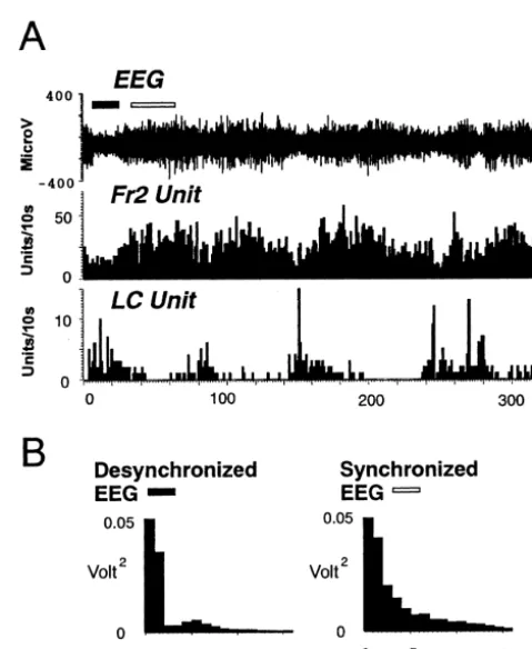
were not elicited in response to the tone. Fig. 2. (A) A pair of Fr2 and LC firings showing opposite relationships to
In order to examine the relation to arousal level, the data arousal level as indicated by EEG (top). When EEG was synchronized (open horizontal bar), this Fr2 neuron showed a tonic increase
accom-were collected both during synchronization and
de-panied by a decrease in firing of the LC neuron. When EEG was
synchronization of EEG. These two states of arousal were
desynchronized (filled horizontal bar), the relation was opposite with a
identified by the difference in EEG voltage and power decrease in firing rate of the Fr2 neuron and an increase in LC activity. spectrum with FFT analysis using Spike2 software. The Bin width 1 s. (B) Power spectrum of the EEG when it is desynchronized
LC unit activity, with the trigger being the Fr2 spikes. The range of analysis was60.5 s from the Fr2 trigger and the bin width was 10 ms. When the tone was presented, the cross-correlation analysis was conducted for the period excluding the 200 ms period after the tone onset.
Wilcoxon’s non-parametric test was employed to
statistically evaluate the difference between the data acquired during the periods with synchronized and de-synchronized EEG.
3. Results
3.1. Unit activity as a function of EEG
A total of 56 Fr2 neurons were recorded from four rats. All the neurons had a wide spike duration (0.8260.02 ms, mean6S.E.M.) and were most likely generated by pyrami-dal cells. They were located in the Fr2 area whose activation or inactivation changed the LC activity in previous studies [7,19]. The spontaneous firing rate was calculated for each neuron during the period with synchronized EEG and that with desynchronized EEG (Fig. 2A), and it was found that 25% (n514) of the Fr2 firings exhibited an increase in firing rate during the period with synchronized EEG. For these 14 neurons, the firing rate during synchronized EEG was 8.462.4 Hz and was higher than that during the desynchronized EEG (4.861.7 Hz; Z53.2958, P50.001). The other 42 neurons did not manifest a clear relationship with EEG synchronization or desynchronization.
Thirty seven LC neurons were recorded in the present
Fig. 3. Top: Auditory-evoked responses of two types of Fr2 neurons. (A)
study and all showed a lower firing rate during the period Fr2 neuron displaying an inhibitory response to tone (*) during synchron-with synchronized EEG (0.860.2 Hz) than during the ized EEG (left) and no response during desynchronized EEG (right) (B) Fr2 neuron displaying an excitatory response to tone during synchronized
period with desynchronized EEG (3.560.7 Hz, Fig. 2A;
EEG and a much attenuated response during cortical desynchronization.
Z53.4078, P50.001).
Bottom: Auditory evoked responses of LC neuron. The response is always excitatory and does not change as a function of the cortical EEG.
3.2. Responses to tone stimulation Response during synchronized EEG (left) and desynchronized EEG
(right). Tones were presented at 0 ms, and each bin in the peri-stimulus time histogram is 10 ms.
There were two types of tone-evoked response in the Fr2 neurons. The first type was inhibitory with a decrease in firing rate between the period from 37.266.2 to 238.8636.2 ms after the tone onset and was found in 16%
(n59) of the Fr2 units (Fig. 3, Fr2 A). The response desynchronized (9.062.1 Hz, n55, Z52.0226, P50.043). amplitude during the inhibitory period was 24.962.1 Hz Two units showed both excitatory and inhibitory responses when the EEG was synchronized, and20.761.1 Hz when (Fig. 5).
desynchronized. The response was more prominent during All the LC neurons showed an excitatory response with the period with synchronized EEG (n56, Z51.9917, P5 the onset, peak and offset latencies being 15.262.0,
0.046). 29.763.9 and 89.166.7 ms, respectively (Fig. 3, LC).
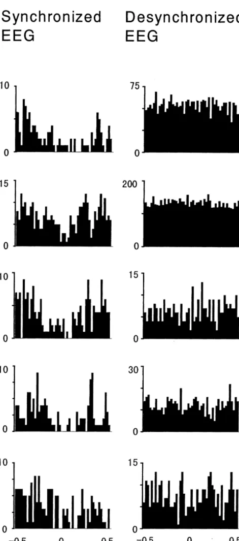
3.3. Relation between the Fr2 and LC activity 167.6651.6 ms after the Fr2 discharge, but only during the period with synchronized EEG (Fig. 4). This negative Cross-correlation analysis on 63 Fr2 / LC neuronal pairs correlation was not observed when EEG was desynchron-showed that in five pairs, the LC firing manifested a ized. Firing rates within the period of cross correlation negative correlation from 117.4648.6 ms before to analysis were further examined by measuring the ratio of the firing rate during the correlated period to the average firing rate during the period from20.5 to20.4 s and from 0.4 to 0.5 s after the Fr2 discharge. The ratio was 0.38060.058 during the EEG synchronization which was significantly smaller than the ratio during the EEG de-synchronization (0.96260.058, Z52.0226, P50.043). This ratio approaches 1 during desynchronized EEG which is merely a reflection of the fact that the correlogram is flat, as can be seen in Fig. 4. It should be noted that 4 / 5 Fr2 neurons having this negative correlation with LC discharge were included in the population which showed increase in spontaneous firing and inhibitory response to tone stimula-tion during low arousal i.e. synchronized EEG (Fig. 5).
3.4. Topographical distribution of Fr2 neurons
Fig. 5 presents the anatomical distribution of different types of Fr2 neurons described above. Among the data obtained with the four penetrations of electrodes in the present study, 8 / 9 inhibitory Fr2 neurons as well as the neurons manifesting a negative correlation with LC were found in the more medial region, although no marked regional localization was observed in the vertical direction. Neither Fr2 neurons showing an increase in spontaneous firing rate during low arousal, nor those with excitatory response presented noticeable topographical localization (Fig. 5).
4. Discussion
4.1. Spontaneous activity as a function of arousal state
In the present experiment, it was found that about one fourth of the Fr2 neurons presented an increase in firing rate during low-arousal state, defined by EEG. This variable relation to arousal level among Fr2 neurons is in contrast to LC activity, where all neurons show a low firing rate during low arousal state and an increase when the EEG becomes desynchronized. It is known that the effect of noradrenaline in the cortex released from termi-nals of the LC is principally inhibitory on the target cells [6,11], so there is a possibility that the low firing rate of Fr2 neurons during high arousal in the present study is related to LC activation during this state. However, the inhibitory effect of noradrenaline may not be sufficient to
Fig. 4. Five Fr2 neurons showing a negative correlation with LC firing. explain the relationship of Fr2 activity to arousal levels, Each row contains the data of one neuron with different EEG features
since only a portion of the population of Fr2 cells are
(synchronized and desynchronized). The negative correlation was
ob-suppressed during LC activation (desynchronized EEG) in
served only during the period with synchronized EEG. Each bar
areas being responsible for the appearance of reciprocal activity in these areas.
4.2. Evoked activity
The present study revealed that some of the neurons in the Fr2 cortex of the rat respond to auditory stimuli; both excitatory and inhibitory responses were observed as well as a majority of nonresponsive cells. This multiplicity of response types indicates that the Fr2 is not homogeneous regarding the processing of sensory information. As for the relation to arousal level, both inhibitory and excitatory responses were a function of the EEG, and were evoked significantly only during the low-arousal state. Further-more, six of the nine Fr2 neurons showing inhibitory evoked responses belong to the group of cells showing an increase in spontaneous firing rate during the low arousal state (Fig. 5). These results concerning the auditory-evoked responses of Fr2 neurons in relation to arousal level, together with the arousal-related change in sponta-neous firing rate, suggest that there is a group of neurons in the rat Fr2 whose activity depends on the low arousal level.
In contrast to this diversity of responses in Fr2 and the dependence upon arousal levels, the LC response to tone was homogeneous, excitatory in all cases, and all LC neurons responded to the tone. This response occurred under all levels of arousal.
4.3. Interaction between the Fr2 and LC
The correlograms revealed a decrease in LC firing from about 120 ms before to 170 ms after the Fr2 discharge, and indicated the presence of reciprocal firing pattern between
Fig. 5. Schematic diagram of recording sites in the Fr2. Thin vertical
the Fr2 and LC neurons. This relationship was conspicuous
lines represent the penetration tracks of recording electrodes in 4 rats.
only during low arousal state, reminiscent of the
tone-Each dot or number in the figure corresponds to the recording site of a
Fr2 unit firing showing different characteristics with respect to arousal evoked inhibition and excitation in Fr2 neurons described
level, auditory-stimulation, and correlation with LC activity; open circle: above. However, the negative relationship between these increase in spontaneous activity during low arousal, open square: negative
areas should not be the result of common auditory input
correlation with LC activity, 1: inhibition to auditory stimulation, 2:
because the period of evoked response was excluded from
excitation to auditory stimulation, 3: both inhibitory and excitatory
the analysis. The proportion of Fr2 neurons negatively
responses to auditory stimulation. Filled dots represent the neurons
showing no relation to arousal or auditory stimulation, and no correlation linked to LC unit activity (5 / 6357.9%) is strikingly
with LC activity. Some Fr2 neurons have both number and symbol(s) similar to the proportion of Fr2 cells found to be antid-since they had more than one type of characteristics described above. fmi:
romically driven by stimulation of the LC in anesthetized
forceps minor corpus callosum.
rats (52 / 70057.4%) [19]. Significantly, 4 / 5 of the Fr2 neurons having this negative correlation with LC were included in that population which showed a tonic increase There could be some specific cell types in the Fr2 cortex in spontaneous firing rate during low arousal and inhibited that are inhibited by noradrenaline, or it could be a to tone.
function of the cortical position being recorded from, given Thus, these observations in behaving rats are consistent ´
[4] V.J. Brown, E.M. Bowman, T.W. Robbins, Response-related deficits
anesthetized rats and the present results lies in the fact that
following unilateral lesions of the medial agranular cortex of the rat,
a relationship between the activities of neurons in the two
Behav. Neurosci. 105 (1991) 567–578.
structures is only found during a synchronized EEG. In the [5] J.M. Cedarbaum, G.K. Aghajanian, Activation of locus coeruleus former studies the relationship was only discerned when neurons by peripheral stimuli: modulation by a collateral inhibitory
both Fr2 and LC were firing in a synchronized, oscillating mechanism, Life Sci. 23 (1978) 1383–1392.
[6] S.L. Foote, R. Freedman, A.P. Oliver, Effects of putative
neuro-mode [9]. However, in order to confirm the presence of
transmitters on neuronal activity in monkey auditory cortex, Brain
interaction between these two areas, it would be necessary
Res. 86 (1975) 229–242.
to employ lesion or pharmacological manipulations in the [7] E. Jodo, C. Chiang, G. Aston-Jones, Potent excitatory influence of
future studies. prefrontal cortex activity on noradrenergic locus coeruleus neurons,
The functional significance of the present observations Neuroscience 83 (1998) 63–79.
[8] V. King, J.V. Corwin, Neglect following unilateral ablation of the
and their role in the putative attention functions attributed
caudal but not the rostral portion of medial agranular cortex of the
to both the Fr2 and the LC, as outlined in the introduction,
rat and the therapeutic effect of apomorphine, Behav. Brain Res. 37
remains to be elucidated. Other investigators have found (1990) 169–184.
that a small population of neurons in the same cortical [9] R. Lestienne, A. Herve-Minvielle, D. Robinson, L. Briois, S.J. Sara,
region, show conditioning-related increase in inhibitory Slow oscillations as a probe of the dynamics of the locus coeruleus-frontal cortex interaction in anesthetized rats, J. Physiol. (Paris) 91
responses during auditory conditioning in both awake and
(1997) 273–284.
urethane-anesthetized rats [14,15,18]. In the present
ex-[10] P.H. Luppi, G. Aston-Jones, H. Akaoka, G. Chouvet, M. Jouvet,
periments the inhibitory responses of Fr2 neurons to tone Afferent projections to the rat locus coeruleus demonstrated by stimulation were present only during low arousal state, retrograde and anterograde tracing with cholera-toxin B subunit and
when LC neurons showed excitatory responses in spite of Phaseolus Vulgaris leucoagglutinin, Neuroscience 65 (1995) 119– 160.
the low spontaneous activity. This suggests that the
[11] Y. Manunta, J.M. Edeline, Effects of noradrenaline on frequency
inhibitory response in Fr2 neurons projecting to the LC
tuning of rat auditory cortex neurons, Eur. J. Neurosci. 9 (1997)
region might serve to promote or permit oriented and 833–847.
conditioned LC phasic responses, by blocking the Fr2 [12] S. Nakamura, C. Tsai, K. Iwama, Recurrent facilitation of Locus
inhibitory influence. The critical question now is whether Coeruleus neurons of the rat, in: J. Hobson, M. Brazier (Eds.), The Reticular Formation Revisited, Raven Press, New York, 1980, pp.
the population of Fr2 neurons inhibiting to tone are the
303–314.
same cells which have terminals in the LC region and have
[13] J.M. Palacios, M.J. Kuhar, Beta-adrenergic receptor localization by
an inhibitory influence on LC activity. Future behavioral light microscopic autoradiography, Science 208 (1980) 1378–1380. studies, in which unit activity is recorded simultaneously [14] J.H. Pirch, M.J. Corbus, G.C. Rigdon, Single unit and slow potential
from both regions during discrimination learning, will responses from rat frontal cortex during associative conditioning, Exp. Neurol. 82 (1983) 118–132.
further elucidate the role of interaction between these areas
[15] J.H. Pirch, M.J. Corbus, G.C. Rigdon, Conditioning-related single
in control of attention.
unit activity in the frontal cortex of urethane anesthetized rats, Int. J. Neurosci. 25 (1985) 263–271.
[16] R.L. Reep, J.V. Corwin, A. Hashimoto, R.T. Watson, Efferent connections of the rastral portion of medial agranular cortex in rats, Acknowledgements
Brain Res. Bull. 19 (1987) 203–221.
[17] R.L. Reep, G.S. Goodwin, J.V. Corwin, Topographic organization in
This study was supported by CNRS UMR 7624, Fonds the corticocortical connections of medial agranular cortex in rats, J. pour la Recherche Medicale, and a CNRS ‘poste rouge’ to Comp. Neurol. 294 (1990) 262–280.
[18] G.C. Rigdon, J.H. Pirch, Nucleus basalis involvement in conditioned
T.S. The authors thank to Yves Moricard and Jacques
neuronal responses in the rat frontal cortex, J. Neurosci. 6 (1986)
Fuzellier for their technical support in electrode
prepara-2535–2542.
tion and histology and Roland Rouxel for support in
[19] S.J. Sara, A. Herve-Minvielle, Inhibitory influence of frontal cortex
electronics. on locus coeruleus neurons, Proc. Natl. Acad. Sci. USA 92 (1995)
6032–6036.
[20] S.J. Sara, M. Segal, Plasticity of sensory responses of locus coeruleus neurons in the behaving rat: implications for cognition,
References Prog. Brain Res. 88 (1991) 571–585.
[21] S.J. Sara, A. Vankov, A. Herve, Locus Coeruleus-evoked responses [1] G. Aston-Jones, F.E. Bloom, Activity of norepinephrine-containing in behaving rats: A clue to the roles of noradrenaline in memory,
locus coeruleus neurons in behaving rats anticipates fluctuations in Brain Res. Bull. 35 (1994) 457–465.
the sleep–waking cycle, J. Neurosci. 1 (1981) 876–886. [22] S.J. Sara, C. Dyon-Laurent, A. Herve, Novelty seeking behavior in [2] G. Aston-Jones, J. Rajkowski, P. Kubiak, T. Alexinsky, Locus the rat is dependent upon the integrity of the noradrenergic system,
coeruleus neurons in monkey are selectively activated by attended Cog. Brain Res. 2 (1995) 181–187.
cues in a vigilance task, J. Neurosci. 14 (1994) 4467–4480. [23] S.L. Stuesse, D.B. Newman, Projection from the medial agranular [3] L. Briois, S.J. Sara, Locus coeruleus responses to frontal cortex cortex to brain stem visuomotor centers in rats, Exp. Brain Res. 80
stimulation after local injection of GABA antagonists or lesion of (1990) 532–544.
the nucleus paragigantocellularis, Soc. Neurosci. Abstr. 23 (1997) [24] U. Ungerstedt, Stereotaxic mapping of the monoamine pathways in
[25] A. Vankov, A. Herve-Minvielle, S.J. Sara, Response to novelty and dopamine, Prog. Neuropsychopharmacol. Biol. Psychiat. 10 (1986) its rapid habituation in locus coeruleus neurons of the freely 723–728.
exploring rat, Eur. J. Neurosci. 7 (1995) 1180–1187. [27] K. Yamamoto K, N. Ozawa, Increased firing of locus coeruleus [26] K. Yamamoto, H. Kaneko, Sequential changes of skin conductance neurons associated with preparatory set in rats, Neurosci. Lett. 106