Cystic Meningioma in the Inter-Hemisferic Space Location
Introduction
There are few case reports available on cystic meningioma.1–7 Cystic meningioma is
an uncommon variant of menin¬gioma. This
disease is often difficult to distinguish from
other intra axial tumors, including necrotic gliomas. Jorge et al.4 reported that the cystic
meningioma simulates an arachnoid cyst on computed tomography (CT) scan and magnetic resonance imaging (MRI), while Qiu et al.7 also reported the presentation
of lateral ventricular cystic meningioma. However, cystic meningiomas located in the interhemispheric space are particularly rare
and may be difficult to distinguish from other
brain tumors. From all types of meningiomas of the central nervous system (CNS), cystic meningiomas are a distinct histological variant of meningiomas accounting for up to 1.6% of all CNS meningiomas.
This present case study describes a case of interhemispheric meningiomas exhibiting intratumoral or peritumoral cystic with MRI. Therefore, the purpose of this report was to emphasize that the cystic lesion might have
different histological findings, such as in
meningiomas.
Case
A case of 57 years-old female with weakness on the right extremities for one month followed by intermittent headache for the past three months is presented. Neurological examinations revealed hemiparesis of right
motor with increased physiology reflex and also pathologic reflex. General examination
showed no abnormality. CT-scan was then performed, identifying the presence of an isohypodense mass in the left frontal, with peritumoral edema. This tumor was compressing sulcy and giry. Ventricle and
sylvian fissures were also compressed. Cistern
was opened and the midline had shifted more than 5 mm to the right (Fig. 1). The magnetic Abstract Objective: The presentation of cystic meningioma in the inter-hemispheric
near the falx cerebri is uncommon. It is difficult to differentiate it from
intra-axial tumors, such as gliomas. Therefore, it is likely that it is misdiagnosed as other types of brain tumors.
Methods: In this study, we reported a cystic meningioma case in the inter-hemispheric location, showing an intramural nodule on magnetic resonance imaging scans.
Results: Patient underwent surgical treatment and pathological section
confirmation revealing meningioma. The patient was a middle-age woman
and had been misdiagnosed as suffering from glioma followed by slight hemipharesis on the right extremities.
Conclusions: Although this is a rare case, it will be good if we always consider cystic meningioma in inter-hemispheric space when diagnosing this type of cystic lesion if the magnetic resonance imaging (MRI) shows a cystic lesion mimicking glioma image presentation.
Keywords:Cystic meningioma, interhemispheric space, magnetic resonance imaging, diagnosis
Muhammad Zafrullah Arifin, Firman Priguna Tjahjono, Agung Budi Sutiono, Ahmad Faried
Department of Neurosurgery, Faculty of Medicine, Universitas Padjdajaran-Dr. Hasan Sadikin General Hospital
Case
Correspondence:
Agung Budi Sutiono, Department of Neurosurgery, Faculty of Medicine, Universitas Padjdajaran-Dr. Hasan Sadikin General Hospital
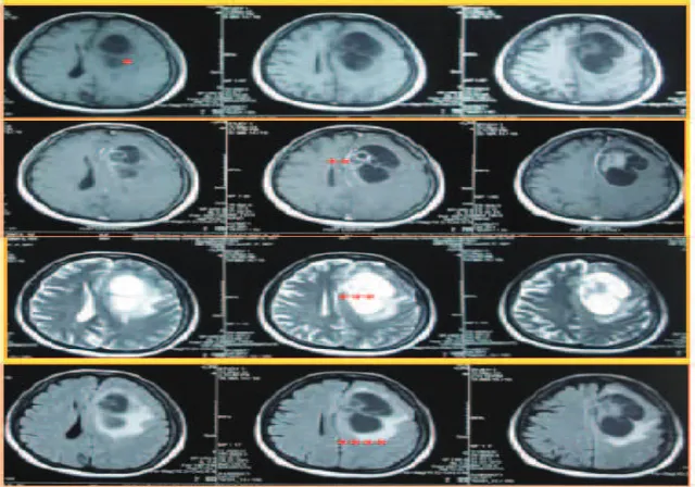
Isohypodense Mass in Left Frontal, with Peritumoral Edema. Sulcy, Giry, Ventricle, and Sylvian Fissure was Compressed. Cistern was Opened and Midline had Shifted more than 5 Mm to the Right
Fig. 1
Isohypointense Mass in Left Frontal on T1 (*). Inhomogenously Enhanced with
Positif Ring Enhanced on T1 with Contrast (**). Hyperintense Mass in Left Frontal on T2 (***) with Peritumoral Edema (****)
resonance imaging (MRI) confirmed this
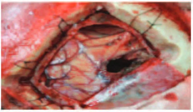
condition (Fig. 2). With these results, the tumor was then diagnosed as a space occupying lession (SOL) supratentorial in left frontal due to suspected high grade glioma. Craniotomy was performed under general anesthesia. Additionally, at the time of surgery, a mass located in interhemispheric space (Fig. 3) was completely resected (Fig. 4).
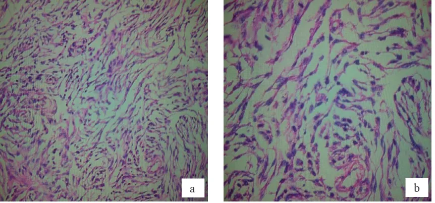
The pathologic specimen was submitted for microscopic analysis. The macroscopic features showed a cystic lesion containing a dense proteinaceous liquid (Fig. 5). The
confirmation of pathological section revealed
that it was meningioma (Fig. 6).
Discussion
Meningiomas are generally solid tumors, and their classical appearance on CT and MRI usually leads to a correct diagnosis.8 Mittal et al.5 mentioned that meningiomas generally
have the characteristic imaging appearance of a well circumscribed, solid, homogeneously enchancing extra-axial mass on both CT and MRI.
A problem with the diagnosis arises when meningiomas have cystic components that can be confused with other tumors, including glial or metastatic tumors with cystic or necrotic
Mass Located in Interhemispheric Space Fig. 3
Mass Completely Resected Fig. 4
changes.8 In solid meningiomas, the sensitivity of a CT-scan is virtually 100% and the specificity
is 90%; however, with cystic meningiomas, the diagnosis is made pre-operatively in less than 38% of cases.2 At MRI, cystic meningiomas can be difficult to differentiate from gliomas which
partially show enhancement after the injection of contrast media or metastases, due to the presence of cyst which does not enhance and also due to the focal edema. Contrast enhanced
MRI can distinguish cystic walls infiltrated
by tumor cells from those formed by gliotic tissue.4 Cyst formation in meningiomas is
thought to be a resultant of ischemic necrosis, cystic degeneration, intratumoral hemorrhage, trapping of CSF, peritumoral edema into cyst, active secretion by tumor cells, or due to glial reaction and transudation.1 The most
common location of cystic meningiomas is in the cerebral convexity, particularly in the
Macroscopic Features Showed a Cystic Lesion Containing a Dense Proteinaceous Liquid
Fig. 5
a
b
frontal and parietal regions. The cerebral falx is the second most frequent location.2 In
this case, location of cystic meningiomas in interhemispheric space make this case very rare.
Cystic meningiomas have been classified
into types 1 to 4. Type I is a central intratumoral cyst, type II is a peripheral intratumoral cyst, type III is a peritumoral cyst in the adjacent brain parenchyma, and type IV is a cyst between the tumor and adjacent brain parenchyma.3,6 Based on the fact that the cysts
were intratumoral, peritumoral with/without tumor lining cyst wall, and that the cyst is at the brain-tumor interface, the present case was of type 2.
In terms of therapy for cystic meningiomas, previous studies have shown that total
removal of tumor is the key for preventing irecurrence.9,10 In this present case, the tumor
was completely resected . The aim of this report was to highlight an extremely rare location of cystic meningioma for future differential diagnosis.
Imaging studies, including computed tomography and magnetic resonance imaging, had demonstrated a round, enhancing anterior interhemispheric mass, appearing to arise from the falx and caused mild mass effect. The patient underwent a craniotomy and removal of the anterior mass without complication, and her headache and hemiparesis resolved. A pathological examination of the specimen
confirmed the diagnosis of meningioma, not
glioma.11
References
1. Deb P, Sahni H, Bhatoe HS. Cystic angiomatous meningioma in the cerebellopontine angle mimicking hemangioblastoma. J Cancer Res Ther. 2010;6(4);560–3.
2. Guan TK, Pancharatnam D, Chandran H,
Hooi TK, Kumar G, Ganesan D. Infratentorial
benign cystic meningioma mimicking a hemangioblastoma radiologically and a lipocytic astrocytoma intraoperatively: a case report. J Med Case Rep. 2013; [cited 2014 Des 3]:[about 4 p.]. Available from: http://www. ncbi.nlm.nih.gov/pmc/articles/PMC3639851/. 3. Hu SL, Li F, Hu R, Cui G, Meng H, Feng H. Atypical
histopathologic type of cystic meningioma. Acta Neurochir. 2010;152(1):105–9.
4. Docampo J, Gonzalez N, Vazquez C, Morales
C, Gonzalez-Toledo E. Cystic meningioma
simulating arachnoid cyst; report of an unusual case. Case Rep Radiol. 2014; [cited 2014 Des 3]:[about 4 p.]. Available from: http://www. ncbi.nlm.nih.gov/pmc/articles/PMC4098774/. 5. Mittal A, Layton KF, Finn SS, Snipes GJ,
Opatowsky MJ. Cystic meningioma: unusual imaging appearance of a common intracranial tumor. Proc (Bayl Univ Med Cent). 2010;23(4):
429–31.
6. Nauta HJ, Tucker WS, Horsey WJ, Bilbao JM,
Gonsalves C. Xanthochromic cysts associated
with meningioma. J Neurol Neurosurg Psychiatry. 1979;42(6):529–35.
7. Qiu L, Lui S, Zou L, Yue Q, Gong QY. Lateral ventricular cystic meningioma: 2 rare case
reports. Exp Ther Med. 2014;7(5):1393–5.
8. Tabuchi S, Nakajima S. Usefullness of 320-Row area detector computed tomography for the diagnosis of Cystic Falx Meningioma. Case Rep Oncol. 2013;6(2):362–6.
9. Wan X, Jiang B, Ma Z, Wang J, Hou Y, Liu Y. Diagnosis and treatment of cystic meningioma.
Zhong Nan Da Xue Xue Bao Yi Xue Ban. 2010;
35(9):1009–12.
10. Ergun T, Torun E, Lakadamyali H. Cystic meningioma showing peripheral rim enhancement without mural nodule. Acta Neurol Belg. 2015;115(3):456–6.