GIANT LIPOMA OF THE LEFT PARAPHARYNGEAL SPACE : A CASE
REPORT
Kurniawan Lidya 1, Nangoi Stefanus 1, Christian Steven I.N.W.2
1
General Surgery Resident, Medical Faculty, Udayana University-Sanglah General
Hospital, Denpasar-Bali, Indonesia
2
Subdivision of Surgical Oncology, Surgery Departement, Medical Faculty, Udayana
University-Sanglah General Hospital, Denpasar-Bali, Indonesia
Abstract
Introduction
Lipomas are the most common neoplasms of mesenchymal origin, but less common in
the head and neck accounting for only 1 – 4,4 % of all benign tumors. Of the variety of
lipomatous benign tumors that occur, over 80 % are ordinary lipomas and only about 13 % of
these occur in the head and neck region, most commonly in the posterior neck. Rarely,
lipomas can occur in anterior neck, infratemporal fossa, oral cavity, pharynx, larynx and
parotid gland. Lipomas involving submandibular and parapharyngeal spaces have been
reported. Diagnosis is based on clinical and radiographic evidence and is confirmed with
histopathologic analysis.
Case Report
We reported a case of 55 years old male who presented with solitary swelling over the
inferior part of left maxilla and extend to the parapharyngeal space and to the anterior part of
neck at the level of thyroid cartilage which confirmed to be fibrolipoma on histopathological
examination after surgical excision.
Lipomas are common tumors of the head and neck region. Giant lipoma of the anterior
neck is rare. Total excision is the treatment of choice with satisfactory result.
Key words : Lipoma, head and neck, anterior neck, parapharyngeal space, total excision .
GIANT LIPOMA OF THE LEFT PARAPHARYNGEAL SPACE : A CASE
REPORT
Kurniawan Lidya 1, Nangoi Stefanus 1, Christian Steven I.N.W.2
1
General Surgery Resident, Medical Faculty, Udayana University-Sanglah General
Hospital, Denpasar-Bali, Indonesia
2
Subdivision of Surgical Oncology, Surgery Departement, Medical Faculty, Udayana
8
Introduction
Lipomas are the most common neoplasms of mesenchymal origin, they are benign and composed of mature fat cells grouped in lobules by connective tissue septa. These tumors are less common in the head and neck accounting for only 1 – 4,4 % of all benign tumors. Of the variety of lipomatous benign tumors that occur, over 80 % are ordinary lipomas and only about 13 % of these occur in the head and neck region, most commonly in the posterior neck.
Rarely, lipomas can occur in anterior neck, infratemporal fossa, oral cavity, pharynx, larynx and parotid gland. Lipomas involving submandibular and parapharyngeal spaces have been reported1. Fibrolipomas are a very rare subtype of the lipomas, composing 1,6 % of the facial lipomas2. Diagnosis is based on clinical and radiographic evidence and is confirmed with histopathologic analysis.
Lipomas tend to exhibit slow growth, are often asymptomatic, and on examination are generally well-circumscribed soft lesions. Radiographic imaging particularly computed tomography (CT) and magnetic resonance imaging (MRI) – can be suggestive of this diagnosis3.
Case Report
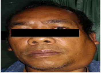
A 55 years old male presented with solitary painless swelling over his left face for last two years. On physical examination, the swelling was 10 cm x 4 cm in size, extending from the tragus of the ear to the upper border of thyroid cartilage and anteroposteriorly from the posterior border of mandible ramus to the anterior border of sternocleidomastoid muscle (Fig.1) . The swelling was soft and, mobile, and non-tender on palpation. On intraoral examination, we found oral extension of the swelling.
Figure 1. Pre operative profile photograph of the patient showing the location of swelling over his left face.
9
[image:9.612.80.243.71.179.2]
Figure 2a. Axial CT-Scan showing hypodense lesion filled the parapharyngeal space.
[image:9.612.78.241.236.349.2]
Figure 2b. Sagital CT- Scan showing hypodense lesion extending from the inferior part of maxilla to the anterior neck at the level of thyroid cartilage.
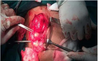
The tumor has been excised in toto, via extra oral sub mandibular approach under general anesthesia (Fig. 3a – 3c).
[image:9.612.89.279.457.576.2]
10
[image:10.612.71.259.251.366.2]
Figure 3b. The tumor was excised in toto.
Figure 3c. Post operative photograph of the patient showing the location of surgical approach.
Histopatological examination revealed lobules of mature adipose cells surrounded by thin fibrous connective tissue capsule. Mature adipose cells were round, vacuolated cytoplasm, with spindle nucleus eccentrically placed without atypia, and fibro collagen stroma among the cells. This histopatological report confirming the diagnosis of fibrolipoma.
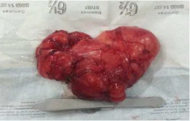
Figure 4. The tumor specimen (10cm x 10 cm x 4 cm in size).
[image:10.612.77.265.489.609.2]11
Lipomas are common benign tumors of the head and neck region. Giant lipoma of the anterior neck especially at the parapharyngeal space is rare. Among all the lipomas sub type, the fibrolipoma is a very rare subtype. Diagnosis is based on clinical and radiographic evidence and is confirmed with
histopathologic analysis. Total excision of the lipoma is the treatment of choice with satisfactory result.
References
1.
Gupta A
,Chopra V
, Lehl G
, et al:L
ipoma ofretromandibular space
.El Med J
2014
,2(2
):156
–158
.2.
Ozturk M, Ila K, Kara A, et al: Fibrolipoma of the nasal septum; report of the first case.
Ozturk et al. Journal of Otolaryngology - Head and Neck Surgery 2013, 42(11):1
–
4.
3.Mattiola MR
,Guerra de Sousa CI,
, Machado RB, et al: LaryngealL
ipoma – A Case Report.Intl.Arch.Otorhinolaryngol. São Paulo 2008. 1
2(1
):133–136
.4.
Phookan J
,Barman D,
Kumar S, et al: Retropharyngeal pleiomorphic lipoma presenting as a neck mass-a rare case.IOSR
Journal of Dental and Medical Sciences (IOSR-JDMS)2014. 13(10
):11–13
.5.
Som PM
,Scherl MP
, Rao VM, et al: Rare Presentation of Ordinary Lipomas of the Head and Neck : A Review. ANJR.7 1988. 7(1): 657-664.6.
Ashtiani MTK
,Yazdani N
, Saeedi M, et al: Large Lipoma of the Larynx : A Case Report. Acta Medica Iranica 2010, 48(5
): 353–356
.7.
Gong W, Wang E, Zhang B
, et al: A retropharyngeal lipoma causing obstructive sleep apnea in a child. J Clin Sleep Med 2006, 2(3
): 328–329
.8.
Le KR
,Bhatia KSS
: An Intramuscular Lipoma Developing Within an Anomalous Cleido-Occipitalis Cervicalis Muscle. Otolaryngology 2012, 2(116
): 1–3
.9.
Ono S
,Rana M, Takechi M, et al
:Myxolipoma in the tongue
–
A clinical case report and
review of the literature
.Head and Neck Oncology
2011
,3(50
): 1–5.