th th
October 11 - 12 , 2011
Graha Widya Bhakti
DRN Building
Proceedings
ISSN : 2089 - 6069
Serpong, October 11th ‐ 12th 2011
Gedung Graha Widya Bhakti Puspiptek
96
ISSN : 2089 ‐ 6069
Immunomodulator Activity Of An Isolate From
Artocarpus Champeden Spreng. On Cytotoxicity Function Of
Cd8
+Of Mice
Kartini
Department of Pharmaceutical Biology, Faculty of Pharmacy, University of Surabaya Corresponding author: Faculty of Pharmacy University of Surabaya,
Jl. Raya Kalirungkut Surabaya 60293, [email protected]
Abstract
Research of traditional medicine is important to maximize the utilization of natural
resources. Artocarpus champeden Spreng. is one of medicinal plants that must be
studied because a lot of diseases have been treated traditionally with this plant, such
as cancer. Phenolic compounds such as flavonoid, tannin, phenilpropane derivative
and simple phenol contained in medicinal plant have been proved for their
immunomodulator activity. This study was conducted to explore whether flavonoid of
Artocarpus champeden could also increase the activity of immunocompetent cell that
play an important role in immunity against cancer especially to Cytotoxic T
Lymphocyte (CD8
+). Assay of cytotoxicity of CD8
+by MLR (Mixed Lymphocyte
Response) showed that DE. 6.4 fraction (contain flavonoids) could not stimulate the
activation of CD8
+significantly (p<0.05) at all concentrations compared to the
control.
Keywords : Artocarpus champeden, flavonoid, CD8
+, cytotoxicity, immunomodulator
Introduction
Cancer is a disease caused by progressive expansion of single progenitor cell that may break away from the normal regulatory control mechanisms of cell division and homeostasis. Cancer is the second cause of death after cardiovascular disease, so nowadays some research have been conducted to develop bioactive ingredients from plants that have potential and selective anticancer properties (Baratawidjaja, 2004).
One of the Indonesian plants that deserve to be studied is Artocarpus champeden (Moraceae) or Cempedak. Although empirically this plant is often used for malaria, dysentery and skin diseases (Heyne, 1987), but it has been demonstrated that the prenylated flavones from Artocarpus sp. have shown to be a source of interesting biological activities including cytotoxic (Liou et al., 1993; Cidade et al., 2001), anticomplementary (Nascimento
et al., 1997), anti-platelets (Lin et al., 1993) and antimicrobial activities (Sato et al., 1996). Therefore, it needs to be further investigated whether the species Artocarpus champeden also effective as anticancer.
From the immunological aspect, there are three groups of cells involved in the response to cancer, namely: Cytotoxic T Lymphocyte/CTL/CD8+, NK cells and macrophages. In the immune system, the main function of CD8+ is to remove virus-infected cells, destroying malignant cells and histoincompatibel cells (Baratawidjaja, 2004).
This study was conducted to determine whether flavonoids from Artocarpus
champeden can increase the activity of
immunocompetent cells that play a role in immunity against cancer, especially of CD8+. It is necessary to obtain the scientific base of the use of Artocarpus champeden as immunotherapy for cancer.
Serpong, October 11th ‐ 12th 2011
Gedung Graha Widya Bhakti Puspiptek
97
ISSN : 2089 ‐ 6069
Material and Method
Equipment
Centrifus, open column chromatography, vacuum column chromatography, Pasteur pipette, culture tube, surgery equipment (scissor, knife, tweezers), autoclave, incubator, laminar air flow, filter membrane (0.22 m), microscope, micropipette, syringe, spectrophotometer, vortex, electric stirrer, vacuum evaporator and other glass equipment.
Material
Plant material was Artocarpus champeden cortex obtained from Bogor, West Java. Chemical included: ethanol (Merck), methanol (Merck), chloroform (Merck), n-hexane (Merck), dichloromethane (Merck), ethyl acetate (Merck), DMSO (Merck), silica gel 60 (0.063-0.200, for column chromatography) (Merck), silica gel 60 GF254 (for TLC) (Merck), TLC plate silica gel 60 F254 (Merck), RPMI 1640 (Sigma), mitomycin C (Sigma), thioglicolat, MTT Formazan (Sigma), PBS (Sigma), concanavalin A-Sepharose 4B (Sigma), histopaque-1077 (Sigma).
Test animal
Test animal was mice (2-3 month, 22-25 gram), ddy strain for isolation of TGPEC and
BALB/C strain for isolation of T Lymphocyte. Methods
1. Extraction, Fractionation and Isolation of Artocarpus champeden Cortex
1 kg of cempedak cortex was macerated successively with n-hexane, dichloromethane and methanol. Then, the dichloromethane extract (DE) was fractionated with vacuum column chromatography using n-hexane-ethyl acetate-methanol as mobile phase in gradient, with silica gel 60 GF254 as stationary phase. After that, each fraction was identified by TLC. Fractions having same chromatogram profiles were then collected. One fraction then was separated by column chromatography using silica gel 60 as stationary phase and CHCl3 -MeOH-ethyl acetate as mobile phase in various comparisons.
2. Identification of Isolate
Identification of the isolates were performed by TLC using silica gel stationary phase with mobile phase CHCl3-ethyl acetate (9:1) and stains reagent ammonia vapor. HPLC chromatogram of isolate also has been determined.
3. Preparation of Sample
1 mg isolate was dissolved in 100 l of DMSO, then added 900 l of 96% ethanol. Stock solution was then diluted using sterile RPMI to obtain a final concentration of K1, K2, K3 and K4 = 1, 10, 100 and 1000 g/ml.
4. Isolation of Mice Lymphocytes (Efector cells)
Isolation of lymphocytes of mice strain BALB/C as an effector cells was:
a. Mice were killed with ether, after that were inserted into a glass beaker containing 70% ethanol, soaked for 10 minutes. Once appointed, placed on a paper towel that has been moistened with 70% ethanol.
b. Peritoneal skin was cut, from tail to head, fed 70% ethanol on peritoneal layer to remove the stick hairs, then made an incision in the peritoneal layer, the entrails were removed and spleen was taken.
c. The spleen was placed in a petri dish, wrapped in sterile gauze, moistened with cold sterile PBS (5 ml). Spleen was rubbed against using tweezers.
d. Next, formed cell suspension pipetted, then made a layer above the ficoll hypaque solution in a test tube with the same volume, centrifuged for 15 minutes. Mononuclear layer was taken, then washed twice with PBS and set the density of 2 x 10⁶ cells/ml. The last, it was re-suspended into sterile RPMI 1640. 5. Isolation of Mice Peritoneal Exudate (TGPEC = Target Cells)
Isolation of peritoneal mice exudate (ddy strain) as target cells carried out as follows:
a. Ddy mice were injected intra peritoneal with 1.5 ml of 3%thioglicolat solution. b. 3-5 days after that, mice were killed with
Serpong, October 11th ‐ 12th 2011
Gedung Graha Widya Bhakti Puspiptek
98
ISSN : 2089 ‐ 6069
ml of sterile PBS and massaged his stomach.
c. Small incision was made in the abdomen of mice, peritoneal fluid taken with a sterile pipette and put into a sterile tube. d. Centrifuged (200 g, 10 min) and washed
twice with sterile PBS. Adjust cell density to 2-6 x 10⁷ cells/ml.
e. Added mitomycin C (0.5 mg/ml), 50
l/ml culture. After incubation (30 min, 370C), then centrifuged (200 g, 10 min) and washed twice with sterile PBS. Cell density was adjust into 2-6 x 10⁶ cells/ml, centrifuged again and re-suspended in sterile RPMI 1640.
6. Cytotoxicity Test of CD8+ Mice
a. 3 series of tubes was prepared, each series consists of 5 tubes. Into 5 first tubes (culture E/T) entered 400 l E cells and 400 l T cells, into the next 5 tubes (E culture) entered 400 l E cell and 400
l sterile RPMI, into the last 5 tubes (T culture) added 400 l T cells and 600 l of sterile RPMI.
b. Into a series of tubes E/T cells and E cells were added 100 l solution of concanavalin A (1 g/ml) and 100 l isolates (K1, K2, K3, K4). Into control were added 100 l of sterile RPMI in tube series E/T and E (Ko).
c. Cultures were incubated (72 h, 370C) and 4 h before the incubation period ends, into each culture tube was added a solution of 100 l MTT (5 mg/ml), then incubation was continued again.
d. After incubation was over, centrifuged (400 g), then washed with 2 ml PBS. Then, into each culture was added 1 ml isopropanol (0.01 N HCL) and vortexed for 2 minutes.
e. Optical Density (OD) of supernatant was observed by a spectrophotometer at 570 nm. Percentage of cytotoxicity was calculated with the formula:
Where OD E/T = Optical Density of culture effector and target cell mixture; OD E = Optical Density of effector cells dan OD T = Optical Density of target cells.
7. Analysis
Data analysis was performed with Anava (α=0.05).
Result and Discussion
From 1.2172 kg of Artocarpus
champeden cortex extracted with n-hexane,
dichloromethane and CHCl3 was resulted 14.21; 10.7; and 27.84 g of extract, respectively. Then dichloromethane extract was analyzed with TLC (figure 1).
Figure 1. TLC profile of dichloromethane extract of Artocarpus champeden cortex on silica gel 60 F254, with mobile phase CHCl3-ethyl acetate (9:1) and sprayer NH3
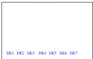
Next, 2.15 g of dichloromethane extract was fractionated using vacuum column chromatography and has been obtained 7 main fractions (DE1 – DE7). TLC chromatogram of 7 fractions can be seen at fig. 2.
Figure 2. TLC chromatogram of fractions yielded from vacuum column chromatography of dichloromethane extract of Artocarpus
champeden cortex on silica gel 60 F254, with mobile phase CHCl3-ethyl acetate (9:1) and sprayer NH3
DE6 fraction was 0.53 g. Its fractions (DE6.1 – DE6.7) with open column chromatography can be seen at fig 3.
% Cytotoxicity = 100 - (OD E/T-OD E) x 100 OD T
DE1 DE2 DE3 DE4 DE5 DE6 DE7
Serpong, October 11th ‐ 12th 2011
Gedung Graha Widya Bhakti Puspiptek
99
ISSN : 2089 ‐ 6069
Figure 3. TLC chromatogram of fractions ryielded from open column chromatography of DE.6 on silica gel 60 F254, with mobile phase CHCl3-ethyl acetate (9:1) and sprayer NH3
From fig. 3 can be seen that fractions DE6.3, DE6.4, DE6.5 have intensively yellow stains with ammonia vapor. This shows that these fractions contain flavonoids. Intense yellow stain on the fraction DE6.3, DE6.4 and DE6.5 each has a Rf 0.5, 0.4 and 0.23, respectively. Darmanto (2006) said that isolates with Rf 0.4 on similar TLC system identified as sicloheterofilin. From this study also concluded that sicloheterofilin of cempedak cortex inhibited the development stage of the malaria parasite (P. falciparum). Therefore, in this study immunomodulatory activity was conducted on DE6.4 fraction thought to contain sicloheterofilin.
DE6.4 fraction is composed of more than one stain fig. 3). This indicates that the fraction DE6.4 is still not a pure compound. To further ensure its purity, fractions DE6.4 was identified by HPLC using RP-18 stationary phase, mobile phase MeOH-H2O (1:10) with a
flow rate of 0.5 ml/min and detector UV 365 nm. The result can be seen at fig. 4.
Figure 4. HPLC chromatogram of DE 6.4 fraction
It has been shown that fraction DE6.4 contain several compounds, with 2 main compounds at
Rt 4.52 and 7.047 minute.
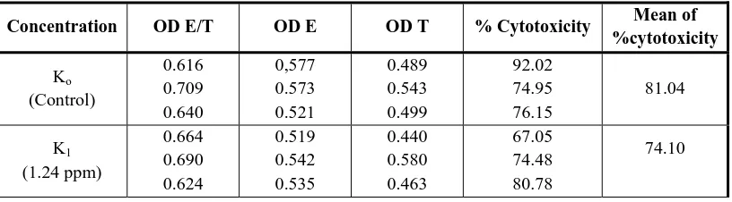
Activity of fraction DE6.4 on the cytotoxicity function by CD8+ with target cells TGPEC and the ratio between effector cell: target cells = 1:1 are presented in Table 1. In the table 1 we can see an increase in the percentage of cytotoxicity on the addition of fractions DE6.4 at K2 and K4 concentration compared with controls. To prove the existence of significant differences, statistical analysis was performed using ANAVA (α = 0.05) and showed that there were no significant differences between the control group with treatment at various concentrations. It can be concluded that DE6.4 fraction does not affect the function of cytotoxicity of T-cytotoxic lymphocytes (CD8+).
In addition to DE6.4 fraction, DE6.3 and DE6.5 fractions of dichloromethane extracts of cempedak also contain flavonoids (Fig. 3). Thus, development of the immunotherapy concept for cancer treatment can be directed to both factions.
Table 1. Toxicity test of DE6.4 fraction with ratio E/T=1/1 on cytotoxicity function of CD8+
Serpong, October 11th ‐ 12th 2011
Gedung Graha Widya Bhakti Puspiptek
100
The average of cytotoxicity caused by the addition of DE6.4 fraction on K2 concentration is higher than K3. According to Wagner & Jurcic (1991), "immunostimulating agents do not follow the normal dose-activity rule". Hence, it is possible that the higher concentrations increase immune response lower than the concentration of smaller ones.
Factors which may result in failure of the cytotoxicity of CD8+ (Whiteside, 1992) are expression of MHC class I from target cells is less strong, so it could not be recognized by cytotoxic T-lymphocytes (a); failure in the activation process (b); the decrease in IL-2 secretion by T-helper lymphocytes (c); the population of memory T lymphocytes is too small (d); failure to destroy target cells in cell culture in vitro (e) (Rose, 1992). Ratio between the number of effector cells with target cells also plays an important role in the cytotoxicity of CD8+. This is caused by the amount of antigen available for stimulation of the immune system depends on the size of the dose of infection (Bellanti, 1993).
Cytotoxicity testing of CD8+ using MTT method should be done with extreme caution, especially during cell washing. There is a possibility that cells drawn or less washing so there is still remaining MTT in the supernatant. This can lead to errors in the spectrophotometer readings. Other method for cytotoxicity of CD8+ is using target cells labeled with radioactive 51Cr and the reading of the results carried out by gamma scintillation counter. The advantage of this method is without washing so that the number of cells is maintained. Nevertheless, this method requires special handling because the radioactive material is dangerous.
Conclusion and Recomendation
The DE6.4 fraction of dichloromethane
Artocarpus champeden cortex containing
flavonoids did not affect the cytotoxicity function of T-lymphocyte cytotoxicity (CD8+) mice. From this study suggested for cytotoxicity testing function of CD8+ mice using other fractions that also contain flavonoids, namely DE6.3 and DE6.5 fractions.
Acknowledgement
The author greatly appreciate Project Leader of Improvement of Higher Education Research, Directorate General of Higher Education, for financial support in this research through a research program for young lecturer (contract number: 007/SP2H/PP/DP2M/2007, dated March 29th, 2007) as well as to Ir. Nunuk M. Januwati, MS, APU, agronomist from Balittro Bogor who provided plant material.
References
Bellanti, J.A., 1993, Immunologi III, Diterjemahkan oleh Samik Wahab, A., cetakan I, Yogyakarta: Gajah Mada University Press.
Baratawidjaja, K.G., 2004, Imunologi Dasar, Edisi ke-6, Jakarta: Balai Penerbit Fakultas Kedokteran Universitas Indonesia.
Cidade, H., Nascimento, M.S.J., Pinto, M.M.M., Kijjoa, A., Silva, A., Herz, W., 2001, Artelastocarpin and carpelastofuran, two new flavones, and cytotoxicities of prenyl flavonoids from Artocarpus
elasticus against three cancer cell lines,
Planta Medica, 67, 867-870.
Serpong, October 11th ‐ 12th 2011
Gedung Graha Widya Bhakti Puspiptek
101
ISSN : 2089 ‐ 6069
Heyne, K., 1987, Tumbuhan Berguna Indonesia
II, Jakarta: Badan Penelitian dan
Pengembangan Kehutanan, Departemen Kehutanan, Yayasan Sarana Wana Jaya, 669-670.
Lin, C.N., Shieh, W.L., Ko, F.N., Teng, C.M., 1993, Antiplatelet activity of some prenylflavonoids, Biochemical Pharmacology, 45, 509-512.
Liou, S.S., Shieh, W.L., Cheng, T.H., Won, S.J., Lin, C.N., 1993, Gamma-pyron compounds as potential anti-cancer drugs, Journal of Pharmacy and
Pharmacology, 45(9), 791-794.
Nascimento, M.S.J, Cidade, H., Pinto, M.M.M., Kijjoa, A., 1997, Anticomplementary activity of prenylated flavones from
Artocarpus elasticus, Pharmaceutical Pharmacological Letters, 7, 135-137.
Sato, M., Fujiwara, S., Tsuchiya, H., Fujii, T., Linuma, M.,Tosa, H., Ohkawa, Y.,
1996, Flavones with antibacterial activity against cariogenic bacteria,
Journal of Ethnopharmacology, 54,
171-176.
Wagner, H., 1986, Immunstimulantien und Phytotherapeutika, Zeitschrift für Phytotherapie, 7, 91-98.
Wagner, H., Jurcic, K., 1991, Assays for Immunomodulation and Effects on Mediators of Inflammation. In: Dey, P.M., Harborne, J.B., Methods in Plant
Biochemistry, Vol. 6, Assays for
Bioactivity, London: Academic Press, 195-217.
Whiteside, T.L., Rimedo, C.R., Heberman, R.B., 1992, Cytolytic Cell function, In: Rose, R.R., DeMacario, E.C., Fahey, J.L., Friedman, H., Penn, G.M., Manual of