Corresponding author:
Surgical outcome of scoliosis in Marfan
syndrome: a case series report
Komang Agung Irianto S*, Gestana R Wardana
Department of Orthopaedic and Traumatology, Airlangga University / Dr. Soetomo General Hospital Surabaya
DOI: http://dx.doi.org/10.19106/JMedSci005001201814
ABSTRACT
Scoliosis in marfan syndrome (MFS) manifests on 60% patients. Moreover, the scoliosis noticeable in earlier age is more progressive, refracted, and rigid compared to idiopathic adult scoliosis. The surgical correction provides notorious higher perioperative risk, whereas conservative treatment using brace is not effective to prevent progressivity of the scoliosis. In this a case report, we reported the surgical outcome of MFS scoliais patients with MFS who operated using posterior fusion instrumentation by mean of the
quality of life SF-36 questioner. This was a retrospective case series involving ive MFS
scoliosis patients who underwent posterior fusion instrumentation with initial Cobb angle of 87.417.57o and initial kyphotic angle of 32.8 ± 14.52o. Clinical, radiological and quality of life of the patients based on SF-36 questionnaire were evaluated within 6-36 months follow up. Post-operative showed the Cobb angle become 46.2 ± 16.3o and the kyphotic angle become 21.6 ± 9.94o. No intraoperative or post-operative complications were observed. After 6-36 months follow up, the Cobb angle became 45.2 ± 17.48o and the kyphotic angle became 21.6 ± 9.94o. In addition, all patients had physical and mental health scored similar to 2 years post-surgery scoliosis scoring according to SF-36 orthopedic scoring guidelines. I conclusion, the surgical outcome of posterior fusion instrumentation in MFS scoliosis showed good correction of Cobb angle and Kyphotic angle. The quality of life of the patients based on physical and mental health questionnaire is satisfactory.
ABSTRAK
Skoliasis diderita pada 60% penderita sindrom Marfan (SM). Selain itu, skoliasis yang diderita sejak usia muda lebih progresif, bias dan kaku dibandngkan dengan skoliais dewasa idiopatik. Penatalaksanaan melalui tindakan bedah memberikan risiko perioperative lebih tinggi, sedangkan penatalksanaan konservatif dengan penjepitan tidak efektif untuk mencegah progresivitasnya. Dalam laporan kasus ini disampaikan luaran tindakan bedah pasien SM dengan skoliais dan kualitas hidupnya berdasarkan pertanyaan dalam SF-36 setelah dilakukan tindakan dengan peralatan fusi posterior. Loran kasus serial retrospektif ini melibatkan lima penerita skoliosis dengan SM yang menjalani instrumentasi fusi posterior sudut Cobb awal 87,4 ± 17,57o dan sudut kifotic awal 32,8 ± 14,52o. Kondisi klinik, hasil pemeriksaan radiologi dan kualitas hidup berdasarkan kuisionair SF-36 dievaluasi selama pengamatan 6-36 bulan. Pasca operasi menunjukkan sudut Cobb menjadi 46,2 ± 16,30o dan sudut kifotik menjadi 21,6 ± 9,94o. Tidak dijumpai komplikasi intraoperasi dan pasca operasi selama pengamatan. Setelah dilakukan pengamatan selama 6-36 bulan, sudut Cobb menjadi 45,2 ± 17,48o dan sudut kifotik menjadi 21,6 ± 9,94o.Semua
pasien mempunyai skor kesehatan isik dan mental sama dengan skor skolastis setelah 2
bedah instrumentasi fusi posterior pada pasien scoliosis dengan SM menunjukkan koreksi yang baik terhadap suduk Cobb dan kifotik. Kualitas hidup pasien berdasarkan kuisionair
kesehatan isik dan mental memuaskan.
Keywords: scoliosis - Marfan syndrome – quality of life - questionnaire SF-36 – Cobb angle
INTRODUCTION
Marfan syndrome (MFS) is one of multi systemically disorder caused by generalized
collagen abnormality (FBN1; ibrillin-1) that is inherited in autosomal dominant.1,2 Other
than the excessive longitudinal growth on growth plate cartilage (hyperchondroplasia), thin and long extremity seen as spider-like
inger (arachnodactyly); the tangible sign
are the facial features (dolichocephalic, enophthalmos, down slanting palpebral issures, malar hypoplasia, retrognathia), and chest asymmetry (pectus excavatum/ carinatum).3,4 The condition is somehow
stigmatism, hamper the life insurance opportunity, as well as the psychosocial
burden2. The diagnosis criteria have been
revised for the purposes not to over diagnosed or under diagnosed it.2,3 Genetic evaluation is
not the only diagnosis tool, yet other ancillary technique is not always available and feasible for our community.
The undetected dilatation of aortic base such as aneurysm of ascending aorta might complicate the scoliosis surgery or the other way the abnormal thoracic cage would complicate the cardiac and pulmonary condition which could happened later in young adult age.1,2,3,4 The main management
for Marfan syndrome depends on which chief organ system involved.3,4,8,9 Scoliosis
prevalence in this syndrome is around 60 %.
Scoliosis in MFS usually occur and being
noticeable at younger age, more progressive, refractory, and rigid. It is also the main complaint of back pain in the later age.5,6,7
In common community, scoliosis is the disease, not just the symptom. Very seldom the underlying cause was investigated. In our former study in screening the junior high school student in Surabaya, 2.7% students indicate scoliosis and 3 neglected cases were found.10 Most of the cases are adolescent
idiopathic scoliosis (AIS); which surgery is performed when the cobb angel is more than 40°. But in MFS the early onset and progressive
scoliosis is the problem to be solved.3,4,5 The
use of brace in conservative treatment is not effective for preventing the progressivity of the scoliosis.5 Surgery by posterior fusion
instrumentation is one of the technique for scoliosis correction nevertheless the collagen
abnormality and anatomical deformity in MFS
might complicate the perioperative risk and the correction result.7,11,12 The purpose of this
study is to evaluate the surgical outcome not only the correction by clinical and radiology but also by using SF-36 questionnaires to value the physical and mental health of the MFS patient.
CASE REPORT
were managed by a single orthopedic surgeon,
same instrument, and same management
protocol. The MFS was diagnosed based on Revised Ghent criteria.2 The pre-operative
management was incorporating cardiac evaluation, ophthalmology evaluation, and MRI ofthe spine to evaluate the dural ectasia. None of the patients had aortic root aneurysm, ectopia lentis, or family history. But 4 out of 5 had thick myopia, the facial features, and >7/20 systemic features involvement.2
Database of initial patient condition were recorded from medical record, including the demographic data, clinical and radio imaging data. Pre and post Cobb angel and Kyphotic angel were compared to the follow-up measurement. The SF-36 questionnaire was performed by interviewing the patient on the follow up visit. Follow up was 6-36 months.
RESULTS
The 5 MFS patients were all female with the age range of 11–17 years (:13.6 years) at surgery; 13–18 years (:15.6 years) at the follow
up, and all were without cardiac or respiratory abnormalities. All of them were came when the Cobb angel were > 70°; 2 of them were
> 100° and 2 patients of double curves
(RT-LL). The mean Cobb angel before surgery patients were immobilized with brace for 3-6 months.
The length of the surgery averaged
(322±38.3) minutes (range:270-370 minutes).
The average blood loss was (495±44.7) cc
(range: 450–550 cc). There was absent of surgical complication during and after the surgery. On Follow-up after 6 – 36 months, there were almost no scoliosis nor kyphotic progression in all patients. The physical health status and mental health status from SF-36 questionnaire were good with the average of PHS 48.4 and MHS 49.
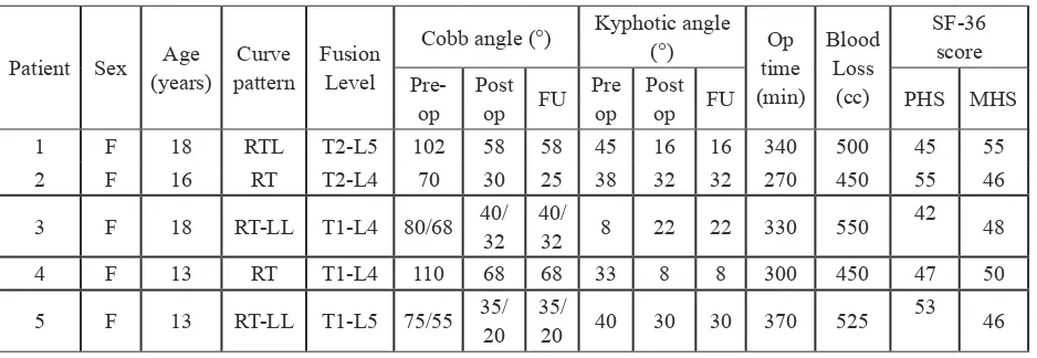
TABLE 1. Clinical, radiology, correction, and quality of life (SF-36) in Scoliosis Marfan syndrome patients
Patient Sex Age
Cobb angle (°) Kyphotic angle (°) Op
RTL: right thoraco-lumbal; RT: right thoracal; RT-LL: right thoracal-left lumbal; Pre-op: preoperative; Post op: post-operative; Op time: operative time; FU: follow up (6-36 months); PHS: Physical Health Status; MHS: Mental Health
FIGURE 1. Clinical appearance 18 years female with RT-LL curve treated with posterior fusion instrumentation, (a) initial pre-operative, (b) follow up post-operative after 1 year
(a) (b) (c)
FIGURE 2. Radiological imaging of 18 years female with RT-LL curve treated with posterior fusion instrumentation, (a) pre-operative, (b) post-operative, (c) follow up after 1 year
DISCUSSION
In this study the surgical outcome is not only about the physical outcome (cobb angel and kyphotic angel) but also about the quality of life. Since the main complaint from the patient’s site is the body contour, the self-esteem conident is also considered as one of the surgical outcome. The SF-36 do not have the cut-off point for good or bad result.13 The SF-36 forms have been used often in
examining orthopaedic patient populations.
The brief guide from Various Orthopaedic Procedures and Conditions comparing SF-36 pre and post-surgery reported similar result of scoliosis patient, PH 42.2 to 46.4 and MH 48.9 to 50.6.14
Surgical outcome of a reconstruction surgery is usually concern for degree of
correction. The degree of correction of the
cobb angel (40–44)°and kyphotic angel (6-29)
(44°) yet less than when using combine PSF
and Anterior (57°).11 Nonetheless blood loss and time of surgery were better in posterior spinal fusion only.6,7,11,12 When comparing
with surgery of AIS, the correction, blood loss, and time of surgery were similar and not signiicant.6,15
Surgical therapy is an effective choice of therapy for scoliosis in Marfan syndrome, bracing has been proven only success in 17% cases but eventually those cases need surgery as well, owing to the progressing curve.5,7,11,12
The use of growing rod is a good choice, anterior release is not necessary if surgery was not postponed; when the curve has progressed rapidly which is the notorious problem in MFS.6,7,11 Hook is not advisable due to the
underlying desmogenic disorder. Before surgery MRI should be assessed for possible dural ectasia, pedicle thinning, and dysplastic lamina.15
The surgery must cover all the major curve including pelvis fusion involvement when necessary to avoid the re-surgery.11,12,15 We
fused 15-17 level and after up to 3 years follow up the progression was none to minimal (5°) in
one case. The invention of pedicle screws gives important progression in scoliosis correction. Pedicle screws, using the strongest part of vertebral body as an anchor, provide the spine surgeon with an enhanced three-dimensional deformity correction. Pedicle screws that placed in the vertebral body have 30 % greater moment arm for applying corrective forces than posterior hooks. Posterior segmental instrumentation with a powerful pedicle screw anchor offers satisfactory correction without signiicant loss of curve correction even in severe deformity cases.12 Posterior fusion with
instrumentation has been widely used for the surgical treatment of scoliosis in the Marfan syndrome, particularly in the curves ranging beyond 40o–50o that tend to progress more
after skeletal maturation.7,12
This study need to be continued with larger samples and multicenter to give orthopedic surgeons precise and merit planning in surgical management of scoliosis in MFS. Scoliosis in MFS would not be too complicated if planned in the current knowledge of underlying the multi systemically disorder as MFS.
CONCLUSION
The surgical outcome of posterior fusion
instrumentationin MFS scoliosis shows good
Cobb angle and Kyphotic angle correction. The blood loss, time of surgery, and surgical complication is all satisfying and comparable to other study with larger samples. The quality of life of the patients based on physical and mental health questionnaire (SF-36) is similar to other various orthopedic procedure and condition.
ACKNOWLEDGEMENTS
We would like to thank all patients who involved in this study.
REFERENCE
1. Herring, JA. Tachdjian’s Pediatric
Orthopaedics 5th Edition. Philadelphia:
Saunders Elsevier; 2012
2. Loeys BL, Dietz HC, Braverman AC, Callewart BL, de Backer J, Devereux RB, et al. The revised ghent nosology for the marfan syndrome. J Med Genet 2010; 47(7):476-85. http://dx.doi.org/10.1136/jmg.2009.072785 3. Sponseller PD, Erkula G, Skolasky RL,
Venuti KD, Dietz HC. Improving clinical recognition of marfan syndrome. J Bone Joint Surg Am 2010; 92(9):1868-75.
http://dx.doi.org/10.2106/JBJS.I.00892 4. Dean JCS. Marfan syndrome: clinical
http://dx.doi.org/10.1038/sj.ejhg.5201851 5. Sponseller PD, Bhimani M, Solacoff D,
Dormans JP. Results of brace treatment of scolosis in marfan syndrome. Spine 2000; 25(18):2350-4.
http://dx.doi.org/10.1097/00007632-200009150-00013
6. Liang W, Yu B, Wang Y, Li Z, Qiu G, Shen J, et al. Comparison of posterior correction results between marfan syndrome scoliosis and adolescent idiopathic scoliosis-a retrospective case-series study. J Orthop Surg Res 2015; 10:73.
http://dx.doi.org/10.1186/s13018-015-0210-z 7. Qiao J, Xu L, Liu Z, Zhu F, Qian B, Sun Xu,
et al. Surgical treatment of scoliosis in marfan syndrome: outcome and complications. Eur Spine J 2016; 25(10):3288-93.
http://dx.doi.org/10.1007/s00586-016-4579-0 8. Tinkle BT, Saal HM. Health supervision for
children with marfan syndrome. Pediatrics 2013; 132(4):1059-72.
http://dx.doi.org/10.1542/peds.2013-2063 9. Pyerit RE. Evaluation of the Adolescent
or Adult with Some Features of Marfan Syndromes. Genet Med 2012; 14(1):171-7. http://dx.doi.org/10.1038/gim.2011.48
10. Komang Agung IS, Dwi Purnomo SB, Susilowati A. Prevalence rate of adolescense idiopathic scoliosis; result of school base screening in Surabaya, Indonesia. Malaysian Orthopaedic Journal 2017; 11(3):17-21. http://dx.doi.org/10.5704/MOJ.1711.011
11. Zenner J, Hitzl W, Meier O, Auffarth A, Koller H. Surgical outcomes of scoliosis surgery in marfan syndrome. J Spinal Disord Tech 2014; 27(1):48-58.
h t t p : / / d x . d o i . o r g / 1 0 . 1 0 9 7 / BSD.0b013e31824de6f1
12. Li ZC, Liu ZD, Dai LY. Surgical treatment of scoliosis associated with marfan syndrome by using postorior-only instrumentation. J Pediatr Orthop B 2011; 20(2):63-6.
h t t p : / / d x . d o i . o r g / 1 0 . 1 0 9 7 / BPB.0b013e328341bcc9
13. McHorney CA, Ware JE, Lu JFR, Sherbourne CD. The MOS 36-item short-form health across diverse patient groups. Med Care 1994; 32(4):40-66.
http://dx.doi.org/10.1097/00005650-199401000-00004
14. Nicholas CL, Ron DH, Bhattacharyya T. Scoring the SF-36 in orthopaedics. a brief guide. J Bone Joint Surg Am 2015; 97(19): 1628-34.
http://dx.doi.org/10.2106/JBJS.O.00030 15. Gjolaj JP, Sponseller PD, Shah SA, Newton
PO, Flynn JM, Neubauer PR, et al. Spinal
deformity correction in marfan syndrome
versus adolescent idiopathic scoliosis: learning from the differences. Spine 2012; 37(18):1558-65.
http://dx.doi.org/10.1097/BRS.0b013e