Diagnostic Value of Narrow Band Imaging in Diagnosing
Nasopharyngeal Carcinoma
Debbi Yuniserani,1 Bethy S. Hernowo,2 Agung Dinasti Permana3
1Faculty of Medicine Universitas Padjadjaran, 2Department of Anatomical Pathology Faculty of Medicine Universitas Padjadjaran/Dr. Hasan Sadikin General Hospital Bandung, 3Department of
Otorhinolaryngology-Head and Neck Surgery Faculty of Medicine Universitas Padjadjaran/Dr. Hasan Sadikin General Hospital Bandung
Abstract
Background: Carcinoma is the most common tumor in nasopharynx. Endoscopy is used to determine the presence of lesions suspected of malignancy. Narrow Band Imaging is an endoscopic technique that uses
narrow–band spectrum as a filter to determine any mucosal vascular changes in carcinoma. Narrow Band Imaging can early detect superficial mucosal lesions that are difficult to detect with conventional endoscopy,
so that diagnosing is more accurate and occurrence of unnecessary biopsies can be reduced. This study aimed to determine diagnostic value of Narrow Band Imaging in diagnosing nasopharyngeal carcinoma.
Methods: This diagnostic test study was conducted at Department of Otorhinolaryngology-Head and Neck Surgery Dr. Hasan Sadikin General Hospital, Bandung from September to October 2014. Twenty four patients
with clinical signs and symptoms of nasopharyngeal carcinoma that fulfilled the study criteria were assigned
using consecutive sampling to examine with Narrow Band Imaging and histopathology examination. The
data were then analyzed with 2x2 table to determine sensitivity, specificity, positive predictive value,
negative predictive value, and accuracy.
Results: In this study, the results of sensitivity, specificity, positive predictive value, negative predictive
value, and accuracy in diagnosing nasopharyngeal carcinoma using Narrow Band Imaging were 93.75%, 62.5%, 83.3%, 83.3%, and 83.3%, respectively.
Conclusions: Sensitivity of Narrow Band Imaging is 93.75%. [AMJ.2017;4(1):133–7]
Keywords: Diagnostic test, narrow band imaging, nasopharyngeal carcinoma
Correspondence: Debbi Yuniserani, Faculty of Medicine, Universitas Padjadjaran, Jalan Raya Bandung-Sumedang Km.21, Jatinangor, Sumedang, Indonesia, Phone: +62 83816734032 Email: [email protected]
Introduction
Carcinoma is type of tumor that is mostly discovered in nasopharynx. In Indonesia, prevalence of nasopharyngeal carcinoma is about 6 cases per 100,000 with 12,000 new cases per year. Nasopharyngeal carcinoma is one of five highest frequencies of malignancy in Indonesia.1 It is often discovered in adult than children and the increasing starts at the age 30 with peak age between 40 and 60 years old.2,3 Nasopharyngeal carcinoma is also more common in men than women with ratio 2–3:1.3
Difficulty in diagnosing nasopharyngeal carcinoma from clinical symptom often happens because the location of nasopharynx and the symptom is not specific.4 Definitive diagnosis that is used for nasopharyngeal carcinoma is biopsy-endoscope combination.5 Endoscopy is usually used to detect any
possible malignancy of lesion.5 However, conventional endoscopy has limitation in detecting superficial lesion that makes unnecessary biopsy often happen.6,7 Narrow Band Imaging is an endoscope technique that uses narrow–band spectrum as a filter to increase the visualization of vascular pattern. Narrow Band Imaging can detect superficial lesion, so that unnecessary biopsy can be reduced.7,8 Although study about Narrow Band Imaging is increased like in digestive system, it is not in head-neck region including nasopharynx.9 This study aimed to determine the diagnostic value of Narrow Band Imaging in diagnosing nasopharyngeal carcinoma.
Methods
technique was a consecutive sampling method with sample size was 24 samples. This study was conducted from September 1 to October 31, 2014 at Department of Otorhinolaryngology-Head and Neck Surgery, Dr. Hasan Sadikin General Hospital, Bandung and was approved by Health Research Ethics Committee.
Population of this study was patients with clinical signs and symptoms of nasopharyngeal carcinoma at Department of Otorhinolaryngology-Head and Neck Surgery, Dr. Hasan Sadikin General Hospital, Bandung. Inclusion criteria were new cases of suspected nasopharyngeal carcinoma,
while exclusion criteria were patients with 1) recurrent nasopharyngeal carcinoma, 2) history of radioteraphy, 3) history of hematologic diseases, and 4) history of biopsy in nasopharynx.
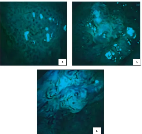
After informed consent, the subjects were given nasal decongestion containing lidocaine 10%. Then, they were examined with Narrow Band Imaging through nose and mucosa of nasopharynx that were obsessed and assessed based on predetermined criteria. Positive criteria of Narrow Band Imaging are: 1) absence of the normal pattern of nasopharyngeal mucosa, 2) irregular vascular pattern with Figure 1 Nasopharyngeal Carcinoma (+) a. Brownish Spot, b&c. Irregular Vascular Pattern
with Brown Spot
A B
brown, and/or 3) irregular follicle pattern with pale peripheral and central brownish. Negative criteria of Narrow Band Imaging are: 1) regular follicle pattern, 2) pale central and dark peripheral follicle pattern.10 Biopsy procedures would be conducted and samples were examined histopathologically. Data from both examinations that had been collected would be tabulated and converted into 2x2 tables to determine sensitivity, specificity, positive predictive value, negative predictive value, and accuracy.
Results
Data characteristics including age group and
sex can be seen in Table 1 and Table 2. The data were divided into two groups; carcinoma and non-carcinoma based on histopathological diagnosis. In carcinoma group, there were 1) non-keratinizing and undifferentiated nasopharyngeal carcinoma. Meanwhile, in non-carcinoma group, there were lymphoma and inflammation of the nasopharynx.
Generally, the highest frequencies were from age group 41–50 years and 51–60 years, which consist of 7 subjects each, and the lowest frequencies were from the age group 61–70 years old and 81–90 years, which consist of 1 subject each. In carcinoma cases, the most common was revealed from age group 41–50 years, which consists of 7 subjects.
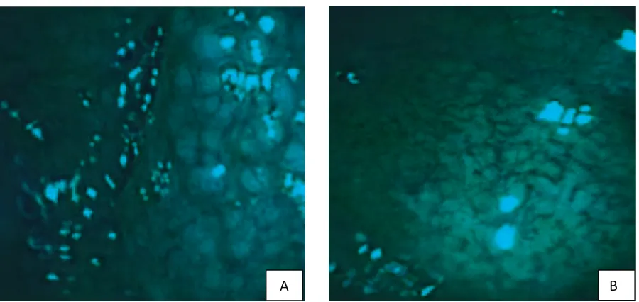
From all cases, the sex characteristics were Figure 2 Nasopharyngeal Carcinoma (–) a. Regular Follicle Pattern, b. Regular Vascular
Pattern
Table 1 Age Group Characteristics
Age Group Histopathology Total
Carcinoma Non-Carcinoma
<31 2 1 3
31–40 2 1 3
41–50 7 0 7
51–60 4 3 7
61–70 1 0 1
71–80 0 2 2
81–90 0 1 1
Total 16 8 24
higher in men (16 samples) than women (8 samples).
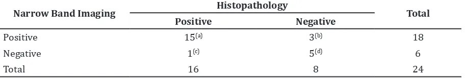
In this study, the sensitivity was 93.75%, specificity was 62.5%, positive predictive value was 83.3%, negative predictive value was 83.3%, and accuracy was 83.3%.
Discussion
From 16 nasopharyngeal carcinoma patients, most of subjects were from age group 41–50 years old consisting of male patients (10 samples) which was higher than female patients (6 samples). These data are similar to literatures that mention the age peak–of nasopharyngeal carcinoma at the age 40–60 years and more common happen in men than women.2,3 Nasopharyngeal carcinoma is more common in men, which is caused by prolonged exposure to risk factor like smoking which can increase the disease.11,12
In this study, the sensitivity was 93.75%, specificity was 62.5%, positive predictive value was 83.3%, negative predictive value was 83.3%, and accuracy was 83.3%. The sensitivity result showed that ability of Narrow Band Imaging to detect patients with nasopharyngeal carcinoma is about 93.75%. This result is similar to Wang’s13 study that the result is about 97.1% and Wen’s14 study that the result is about 93.9%. Specificity result showed that ability of Narrow Band Imaging to eliminate patients with non-nasopharyngeal carcinoma is about 62.5%. Compared to previous studies (Wang13 is 93.33% and Wen14 is 94.1%), the specificity from this study is lower. Thus, it might be due to inability
of Narrow Band Imaging to differ types of malignancy (like lymphoma and carcinoma) or total of sample is less compared to previous studies.
Positive predictive value result showed that the probability of subjects with positive Narrow Band Imaging will actually have nasopharyngeal carcinoma by 83.3%. Negative predictive value result showed that the probability of subjects with negative Narrow Band Imaging will actually not have nasopharyngeal carcinoma by 83.3%. Both values are also similar to previous studies.13,14 Accuracy result showed that the accuracy of Narrow Band Imaging in the diagnosing of nasopharyngeal carcinoma is 83.3% .
Based on this study, it can be concluded that the sensitivity of Narrow Band Imaging is 93.75%.
Limitation of this study was the samples used in this study were too small that affect Narrow Band Imaging value. In addition, subjects in this study were mostly at late stage.
It is recommended for further study to discuss about Narrow Band Imaging in wider population.
References
1. Adham M, Kurniawan AN, Muhtadi AI, Roezin A, Hermani B, Gondhowiardjo S, et al. Nasopharyngeal carcinoma in Indonesia: epidemiology, incidence, signs, and symptoms at presentation. Chin J Cancer. 2012;31(4):185–96.
2. Thompson LR. Update on nasopharyngeal carcinoma. Head Neck Pathol.
Woman 6 2 8
Total 16 8 24
Table 3 Diagnostic Values
Narrow Band Imaging Histopathology Total
Positive Negative
Positive 15(a) 3(b) 18
Negative 1(c) 5(d) 6
2007;1(1):81–6.
3. Barnes L, Eveson JW, Reicchart P, Sidransky D, editors. World Health Organization Classification of Tumours: pathology and genetics of head and neck tumours: Lyon: IARC Press;2005.
4. Cengiz K, Kumral TL, Yildirim G. Diagnosis of pediatric nasopharynx carcinoma after recurrent adenoidectomy. Case Rep Otolaryngol. 2013;2013:653963.
5. Chan AT, Gregoire V, Lefebvre JL, Licitra L, Hui EP, Leung SF, et al. Nasopharyngeal cancer: EHNS–ESMO–ESTRO clinical practice guidelines for diagnosis, treatment and follow–up. Ann Oncol. 2012;23 Suppl 7:vii 83–5.
6. Tabuchi K, Nakayama M, Nishimura B, Hayashi K, Hara A. Early detection of nasopharyngeal carcinoma. Int J Otolaryngol. 2011;2011:638058.
7. Piazza C, Del Bon F, Nicolai P. Narrow band imaging in endoscopic evaluation of head and neck mucosal cancer. J ENT Masterclass.2012; 5(1):59–64.
8. Lukes P, Zabrodsky M, Plzak J, Chovanec M, Betka J, Foltynova E, et al. Narrow band imaging–endoscopic method for detection of head and neck cancer. In: Amornyotin A, editor. Endoscopy. Intech; 2013. Available from: https://www.intechopen.com/
books/endoscopy/narrow-band-imaging- nbi-endoscopic-method-for-detection-of-head-and-neck-cancer
9. Piazza C, Dessouky O, Peretti G, Cocco D, De Benedetto L, Nicolai P. Narrow band imaging: a new tool for evaluation of head and neck squamous cell carcinomas. Acta Otorhinolaryngol Ital. 2008;28(2):49–54. 10. Thong JF, Ranjini KS, Loke D, Mok PKH.
Use of narrow–band imaging in detection of nasopharyngeal carcinoma. J Laryngol Otol. 2013;127(2):163–9.
11. Kumar S. Epidemiological and etiological factors associated with nasopharyngeal carcinoma. ICMR Bull. 2003;33(9):1–9. 12. Nesic V, Sipetic S, Vlajinac H, Stosic–Divjak
S, Jesic S. Risk factors for the occurrence of undifferentiated carcinoma of nasopharyngeal type: a case–control study. Srp Arh Celok Lek. 2010;138(1–2):6–10. 13. Wang WH, Lin YC, Lee KF, Weng HH.
Nasopharyngeal carcinoma detected by narrow–band imaging endoscopy. Oral Oncol. 2011;47(8): 736–41.