SYNTHESIS OF COPPER OXIDE NANO PARTICLES BY USING Phormidium
cyanobacterium
Abdul Rahman1,*, Amri Ismail2, Desi Jumbianti3, Stella Magdalena3.4,and Hanggara Sudrajat4 1
Department of Mechanical Engineering, Malikussaleh University, Jl. Tgk. Chick Ditiro No. 26 Lancang Garam, Po.Box.141, Lhokseumawe 24351, Nanggroe Aceh Darussalam, Indonesia
2
Department of Chemical Engineering, Malikussaleh University, Jl. Tgk. Chick Ditiro No. 26 Lancang Garam, Po.Box.141, Lhokseumawe 24351, Nanggroe Aceh Darussalam, Indonesia
3
Biomolecular Engineering Research Institute, 6-2-3, Furuedai, Suita 565-0874, Japan
4Faculty of Technobiology, Atma Jaya Catholic University of Indonesia, Jl. Jenderal Sudirman 51, Jakarta 12930, Indonesia
Received May 26, 2009; Accepted October 1, 2009
ABSTRACT
In this paper, we report a suitable method for extracellular synthesis of copper oxide nano particles by using Phormidium cyanobacterium. We hypothesize that synthesis of copper oxide nano particles is believed to occur by extracellular hydrolysis of the cationic copper by certain metal chelating anionic proteins/reductase secreted by bacteria under simple experimental conditions like aerobic environment, neutral pH and room temperature. Proteins not only reduce Cu (II) into copper oxide nano particles (CONPs) but also plays significant role in stabilization of formed nanoparticles at room temperature. Further TEM, SEM, XRD and FTIR analysis have confirmed the synthesis of nano particles through microbial route. Extracellular induction of metal chelating proteins/reductase was analyzed by SDS-PAGE.
Keywords: Synthesis, copper oxide nano particles, Phormidium cyanobacterium
INTRODUCTION
Materials at nano scale exhibit unique electronic and optical properties, which are different to that at their bulk level [1]. Synthesis of nano materials comprises mainly wet chemical/physical routes without much involvement of biological methods. In recent decades, researchers have been able to demonstrate controlled marriage between biology and chemistry; hence a new interdisciplinary area of “Bio-inspired” Physical/Chemical method is evolving in present nanomaterial synthesis protocols. Metal oxide nano particles are important because of their superior electronic [2], electrochemical [3], paint/ink materials [4], Catalytic [5] and magnetic [6] properties.
Among transition metal oxide, attention have been paid to copper oxide because of their potential commercial applications viz. biocidal activity [7], magnetic phase transitions [8], gas sensors [9], catalysis [10], and superconductivity [11] and CONP have been studied with great detail in this regard. Thus, development of protocols for synthesis of CONP is an issue of considerable primary interest.
Various chemical and physical methods so far reported for CONPs synthesis involves sol gel methods [12], chemical vapor deposition [13], precipitation-pyrolysis method [14], sonochemistry [15],
electrochemistry [16], one-step solid-state reactions [17], cathodic vacuum arc [18] and solvothermal reactions [19]. All employ toxic chemicals and energy intensive routes which makes these choices eco-hazardous and preclude their applications in biology, medicine and clinical applications. Thus, developing environment friendly protocols is the need of hour in nanomaterials synthesis.
It has been well established that when microbes were kept in harsh toxic metal environment, they evolve mechanism of bioremediation to survive in stringent conditions by transforming toxic metal ions into their corresponding non-toxic forms like metal sulfide/oxide [20]. As evident from recent reports, the attention is now being shifted to synthesis of metal nano particles using microbes [21], though details of mechanism (s) involved in nano scale transformation is yet to be established. While there are quite a few reports on bacterial mediated metal oxide nano particle synthesis [20-21], the literature on synthesis of magnetite nano particles is scanty [22].
In the present work, we report that when bacterial
biomass was incubated at 28 °C with aqueous CuSO4
salt solutions, CONPs are formed extracellularly, under aerobic conditions. The formation of CONPs could be mediated by the induction of secreted proteins by
Phormidium cyanobacterium that might have converted
* Corresponding author. Tel/Fax : +62-645-41373/645-44450 Email address : [email protected]
Indo. J. Chem., 2009, 9 (3), 355 - 360 356
Abdul Rahman et al.
copper ions to copper oxide. The nano particles thus formed were found to be stable at 28 °C (room temperature) for over 30 days in contrast to chemically and physically synthesized nanoparticles. Bacterial mediated synthesis of CONPs opens many promising avenues including enhancing the biocompatibility and large-scale production of nano particles for chemical and biomedical applications. To the best of our knowledge the present study is first report on stable, extracellular synthesis of CONPs using bacteria without the incorporation of any extra stabilizing/capping agent.
EXPERIMENTAL SECTION
Synthesis of CONPs
Phormidium cyanobacterium was cultured as described elsewhere [23]. For the synthesis of CONPs,
Phormidium cyanobacterium was grown in 500 mL Erlenmeyer flask containing 100 ml of citrate minimal medium (CMM) [24] and incubated for 15 h at 150 rpm at 28 °C. After incubation, two grams (wet weight) bacterial biomass was collected by centrifugation at 5000 rpm at 5 °C for 10 min, washed thrice under sterile
conditions and resuspended in 100 mL of 10-3 M
aqueous CuSO4 solution in 500 mL Erlenmeyer flask,
incubated at 150 rpm for 42 h at 28 °C. The biologically synthesized CONPs were collected in a supernatant under sterile condition by centrifugation at 5000 rpm for 10 min and characterized with different techniques like SEM, TEM, XRD, and FTIR. Supernatant was dialyzed
to remove un-reacted CuSO4. The progress of reaction
i.e. extra cellular synthesis of nanoparticles was monitored by observing the peak at 600 nm, at different time intervals, which is the characteristic of CONP [12-19].
Characterization of CONPs
Biologically synthesized CONPs were characterized by XRD, UV-vis-NIR spectrophotometry, TEM, SEM, and FTIR. Sample for TEM analysis was prepared by drop coating on to a carbon-coated copper grid. Selected area electron diffraction (SAED) analysis was also carried out for above prepared TEM samples. Measurements were performed on a JEOL model 1200EX instrument operated at an accelerating voltage of 120 kV. SEM measurement of CONPs synthesized by
Phormidium cyanobacterium biomass was carried out on a Leica Stereoscan-440 scanning electron microscope equipped with a Phoenix EDAX attachment. UV-vis-NIR spectroscopy measurement was carried out on a JASCO dual beam spectrophotometer (model V-570) operated at a resolution of 2 nm. XRD measurements of solution cast films of as-prepared biogenic CONPs on glass
substrates were carried out on a Phillips PW 1830 instrument operated at a voltage of 40 kV and a current
of 30 mA with Cu Kα radiation. FTIR measurements
were carried out on a Perkin Elmer Spectrum-One spectrometer operated at a resolution of 4 cm-1.
Protein separation and protein profile
To analyze the bacterial protein(s) responsible for
hydrolysis of CuSO4 salt precursor to copper oxide, 2 g
of bacterial biomass was suspended in aqueous 10-3 M
CuSO4 solution for a period of 42 h at 28 °C at 150
rpm. Secreted proteins were precipitated by TCA [24]. The protein profile on SDS-PAGE (10%) of the supernatant containing CONPs was compared with suitable controls, as explained below [24].
Control Experiments
In similar experiments for control bacterial biomass was suspended in deionised water in absence
of CuSO4 and supernatant obtained was analyzed by
XRD and FTIR for presence of CONP and SDS-PAGE for protein profile analysis. Result obtained did not show CONPs formation. In another set of control
experiment, CuSO4 was dissolved in absence of
bacterial biomass, and result obtained did not show CONPs formation.
RESULT AND DISCUSSION
UV-vis spectroscopy
Fig. 1a shows progress of biosynthesis of CONPs as a function of time. The UV-vis spectra indicate that
cells when added in the CuSO4 aqueous solution
initiated the synthesize CONPs after 24 h as is evident from the peaks obtained at different time intervals at 600 nm. Till 24 h there is no peak, and afterwards distinct peaks are observed around ~600 nm. owing to
the formation of Cu2O particles [25].
To ascertain the hydrolytic role of bacterial proteins/reductase in formation of CONPs, a parallel
control experiment was performed with CuSO4 Solution
without biomass under same shaking condition and UV-visible spectra recorded after 24 h do not show any CONPs. After 36 h of shaking of biomass and aqueous salt solution under aerobic condition, absorption peak shows blue shift and interestingly two UV-vis bands are observed at ~542 and ~310 nm which indicate a mixed
Cu2O and CuO phase in solution mixture. This clearly
indicates that after 36 h reaction, a substantial amount
of Cu2O changes to CuO phase due to on going
Abdul Rahman et al.
Figure 1. a) UV-visible spectra of the copper oxide nanoparticles measured as a function of time during the reaction of 10-3 M CuSO4 and Phormidium cyanobacterium. The spectra were measured after 24, 36 and 42 h. respectively (curve 1 to 3 as indicated by 24 to 42 h. towards the direction of ascending arrow). b) X-ray diffraction pattern of copper oxide nanoparticles synthesized by the reaction between 10-3 M aqueous CuSO4 and Phormidium cyanobacterium under the aerobic condition after 36 h reaction.
Figure 2. a) X-ray diffraction pattern of copper oxide nanoparticles synthesized by the reaction between 10-3 M aqueous CuSO4 and Phormidium cyanobacterium under the aerobic condition after 42 h. b, c) TEM micrograph of CONPs synthesized by Phormidium cyanobacterium at room temperature under aerobic conditions after 42 h of reaction. d)
SAED pattern recorded from representative CONPs.
may arise due to change in particle size after 36 h reaction. Further shaking for about 42 h, absorption
band due to Cu2O phase at ~542 nm disappears while
there is a little shifts in plasmone band occurring at ~310 nm to ~307 nm with increased intensity. These
results clearly suggest complete Cu2O→CuO phase
transition that is in conformity with available literature [26]. This probably occurs due to shaking of bacterial biomass under aerobic conditions and rather unstable
nature of Cu2O further makes it susceptible to convert to
CuO nano particle.
During the course of reaction color of solution
varies from light blue→green→light brown and finally
faint black at completion of reaction. UV-vis spectra showed that formed CONPs were stable in solution at room temperature for more than three weeks, suggesting stabilization of the particles by protein capping secreted by bacteria extracellularly. Interestingly, during the entire course of reaction, we did not receive characteristic peak for Cu (0) nano particles.
XRD Analysis
Fig. 1. b) shows X-Ray Diffraction recorded from copper oxide powder after 36 h of bacterial reaction gives strong bags reflection that could be indexed
based on mixed Cu2O and CuO nano particles.
Reflection peaks (●) at 2θ = 35.4°, 38.7°, 58.3°, 65.7°
and 68.0°are indexed as [002], [111], [202], [022] and
[220] planes of CuO phase with cubic symmetry.
Reflection peaks (○○) at 2θ = 48.7°, 53.4°, 58.3° and
72.4° are indexed as [202], [020], [202] and [311]
planes of Cu2O phase. Higher intensity at 2θ = 35.4°
and 38.7°respectively indicates that mixed phase has
major proportion of CuO with highly oriented crystalline CuO phase. Thus, above results suggest that bacterial mediated synthesis comprises mixed phase of both
Cu2O and CuO after 36 h of bacterial reaction and are
in good agreement with literature available [27].
Fig 2. a) shows X-Ray diffraction recorded after
42 h of reaction shows a major peak at 2θ = 38.7°
which can be indexed as [111] plane with d value
2.32Ǻ which is characteristic peak of CuO phase
indicating that after 42 h of reaction pure CuO phase
exist without any trace of Cu2O being present. High
intensity strongly suggests that CuO particles are highly oriented.
TEM Analysis
Transmission electron microscopy (TEM) was carried out to analyze the shape, size and protein coating copper oxide nano particles. Fig. 2 b, c shows representative TEM images obtained after 42 h of
bacterial reaction with aqueous CuSO4 salt solution.
Indo. J. Chem., 2009, 9 (3), 355 - 360
Abdul Rahman et al.
358
Figure 3. a, b) SEM images recorded from two different regions of drop coated films of copper oxide obtained after 42 h. reaction of CuSO4 with Phormidium cyanobacterium. c)
CONPs present over Phormidium cyanobacterium biomass (arrows). d) EDAX spectrum recorded from a drop-coated film of an aqueous solution incubated with Phormidium cyanobacterium and treated with CuSO4 ions for 42 h.
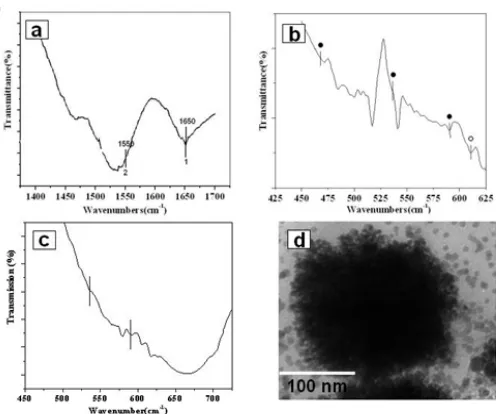
Figure 4. a) FTIR spectra of copper oxide nano particle in amide bond (I and II) frequency region b) FTIR spectra recorded after 36 h. showing CuO and Cu2O vibrational mode (see text for details and peak assignments). c) FTIR spectra recorded after 42 h. reaction showing CuO vibrational mode.
d) TEM micrograph showing clusters of CONPs formed after the reaction of CuSO4 with induced protein of molecular weight 52 kDa.
SEM Analysis
Fig. 3. a, b Shows Scanning Electron Microscopy
(SEM) Images of CONPs after Phormidium
cyanobacterium cells exposed to precursor ions after 42 h of reaction. Centrifuged sample coated on Si wafers and SEM images thus taken clearly shows formed nano particles are embedded and uniformly distributed in protein/surfactant matrix indicating that nano particles formed by the reaction of copper sulfate ions with bacterial biomass are bound by the proteins secreted by bacteria extracellularly.
Fig. 3 c shows that small clusters of CONPs
present over Phormidium cyanobacterium cells may be
due to weakly bound CONPs, which have not been dislodged during first round of centrifugation and preparation of sample for SEM investigation which recalls further second round centrifugation of the sample.
Fig. 3 d shows EDAX (Energy Dispersive Analysis of X-Rays) spectrum recorded by scanning in the spot profile from densely populated CONPs region of embedded nano particle protein matrix and also from the
Phormidium cyanobacterium cell surface. Strong signals from copper oxide protein matrix are observed while weaker signals from S, C, P, O, Mg and Na are also recorded. The S, C, P, O, Mg and Na signals are expected to due to X-Ray emission from
proteins/enzyme/surfactant in the supernatant and disruption of cell wall of bacterial biomass.
FTIR Analysis
To confirm the presence of protein moiety and capping of CONPs, Fourier transform infrared (FTIR) spectroscopy analysis was performed. CONPs obtained after 36 h of bacterial reaction shows (Fig. 4
a) two bands at 1650 and 1550 cm-1 respectively
indicating presence of amide I and amide II bands of protein due to capping the CONPs [21]. This confirms the presence of protein in the sample as proposed capping and stabilizing agent. Further, detailed protein profiles have been studied later in this section.
Moreover, CuO and Cu2O vibrational modes are also
observed (Fig. 4 b, ● Peaks CuO an ○ Peaks for
Cu2O).
For CuO nano particles three main vibrational
modes are observed at 468, 535 and 590 cm-1. High
frequency mode at 590 cm-1 is reported due to Cu-O
vibration stretching along [⎯101] direction. Further,
active vibrational mode due to Cu2O nano crystals is
observed at 610 cm-1 [16,28]. FTIR spectra recorded
after 42 h. reaction clearly shows corresponding band
at 610 cm-1 due to Cu2O (fig 4 b, ○ Peak) are absent
while ٧Cu-O vibrational mode (Fig 4 c, ● Peaks) are
Abdul Rahman et al.
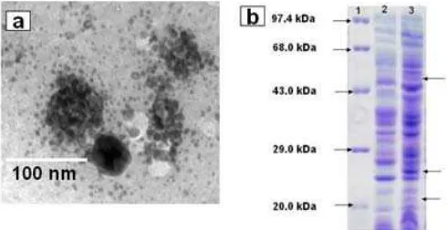
Figure 5. a) TEM micrograph showing clusters of CONPs formed after the reaction of CuSO4 with 22 kDa. induced protein fraction. b) SDS-PAGE protein profile of bacterium in the absence (Lane 2) and presence (Lane 3) of precursor CuSO4 solution. Arrows indicate induction of three proteins due to heavy metal (Cu2+) stress. Lane 1 shows standard protein molecular weight marker.
features appearing in IR spectra may be due to interaction of precursor ions with the protein/surfactant/enzymes secreted by bacteria in course of reaction.
Protein Profile
To identify the bacterial protein(s) secreted extracellularly that were responsible for hydrolysis of Cu ions to CONPs, the extracellular extracts obtained from
Phormidium cyanobacterium in presence and absence of salt precursor were purified and protein profiles were analyzed by 10% SDS-PAGE. Exposure of cationic precursor to bacteria resulted in induction of three
proteins with molecular weight ≈ 22 kDa, ≈ 25 kDa and ≈
52 kDa (Lane 3, fig. 5 b). These were absent in bacterial protein extract profile obtained without precursor salt. In another experiment, the purified fraction of protein when analyzed by native PAGE showed three similar bands as corresponding to those obtained in SDS-PAGE. This clearly indicated that these proteins were not the subunits of a single protein but three separate proteins induced under heavy metal stress. (data not shown for brevity).
In a separate experiment, to check the role of these proteins in hydrolysis of copper cationic complex, the dialyzed fraction containing mixture of proteins (in presence of copper precursor) were eluted from native gel and their hydrolytic activity was checked against
CuSO4 salt solution. Only two fractions of ≈ 22 kDa and
≈ 52 kDa tested positive for hydrolysis of precursor ions
(fig. 4 d and 5 a) while third fraction of 25 kDa does not show any hydrolytic activity. It might be possible that 25 kDa protein fraction expressed in heavy metal stress condition has a role as capping agent but not in hydrolysis of copper cation. Thus, the role of 25 kDa
protein fraction is yet to be established. Though there are some reports indicating the involvement of extra cellular fungal proteins in nano particle synthesis like silica and titania particles [29].
From SDS-PAGE profile, it was clear that proteins were over expressed in heavy metal environment as it was well established that few proteins were newly expressed, over expressed or modified in presence of heavy metals stress [30]. Currently studies are under way to establish amino acid sequence homology of these proteins to other hydrolyzing proteins responsible for biosilicification/nano particle formation to develop a methodology for recombinant protein expression for biosynthesis of commercially important nano particles.
CONCLUSION
In conclusion, facile biosynthesis of copper oxide nano particles has been shown by hydrolysis of copper
precursor using bacteria Phormidium cyanobacterium.
This is benign and environment friendly approach to synthesize CONPs without involving any toxic chemical and stabilizing agent. Proteins induced under metal stress play a dual role of hydrolysis of precursor salt to CONPs and stabilizing agent, as particle solution is stable at room temperature for more than a week. This bacterial mediated biosynthesis provides promising approach for scaling up of commercially and technologically important metal nano particles and metal oxide synthesis.
REFERENCES
1. Klabunde, K.J., 2002, Nanoscale Materials in
chemistry, Wiley, New York, USA. p. 1.
2. Kondo, R., Okimura, H., and Sakai, Y., 1971, Jpn.
J. Appl. Phys.,10, 1547.
3. Su, L.M., Grote, N., and Schmitt, F., 1984,
Electron. Lett., 20, 716.
4. Merikhi, J., Jungk, H.O., and Feldmann, C., 2000,
J. Mater. Chem., 10, 1311-1314.
5. Skarman, B., Wallenberg, L.R., Larsson, P.O.,
Andersson, A., and Helmersson, H., 1999, J.
Catal.,181, 6-15.
6. Ammar, S., Helfen, A., Jouini, N., Âvet, F.F., Rosenman, I., Villain, F.E., Molinie, Â., and Danot,
M., 2001, J. Mater. Chem., 11, 186-192.
7. Gabbay, E., 2006, J. Ind. Tex., 35, 4, 323-335.
8. Sukhorukov, Y.P., Loshkareva, N.N.,
Samokhvalov, A.A., Naumov, S.V., Moskvin, A.S.,
and Ovchinnikov, S., 1998, J. Magn. Magn. Mater;
183, 356.
9. Eranna, G., Joshi, B.C., and Runthala, D.P., 2004,
Indo. J. Chem., 2009, 9 (3), 355 - 360
Abdul Rahman et al.
360
10. Carnes, C.L. and Klabunde, K.J., 2003, J. Mol.
Catal. A., 194, 227.
11. Dai, P.C., Mook, H.A., Aeppli, G., Hayden, S.M., and
Dogan, F., 2000, Nature, 406, 965.
12. Punnoose, A, Magnone, A., and Seehra, M.S., 2001,
Phys. Rev. B., 64, 14120.
13. Serin, N., Serin, S., Horzum, A., and Elik, Y.C.,
2005, Semicond. Sci. Technol., 20, 398-401.
14. Fan, H., Yang, L., Hua, W., Wu, X.F., Wu, Z., Xie,
S., and Zou, B., 2004, Nanotechnol., 15, 37-42.
15. Kumar, R.V., Elgamiel, R., Diamant, Y., Gedanken,
A., and Norwig, J., 2001, Langmuir, 17, 1406.
16. Borgohain, K., Singh, J.B., Rama, M.V.R., Shripathi,
T., and Mahamuni, S., 2000, Phys. Rev. B., 61,
11093.
17. Xu, J.F., Ji, W., Shen, Z.X., Tang, S.H., Ye, X.R.,
Jia, D.Z., and Xin, H.X., 2000, J. Solid State Chem.,
147, 516.
18. Gan, Z.H., Yu, G.X., Tay, B.K., Tan, C.M., Zhao,
Z.W., and Fu, Y.Q., 2004, J. Phys. D: Appl. Phys.,
37, 81–85.
19. Ghosh, M. and Rao, C.N.R., 2004, Chem. Phys.
Lett., 393, 493-497.
20. Neis, D.H., 1999, Appl. Microbiol. Biotechnol., 51,
730-750.
21. Sastry, M., Ahmad, A., Khan, M.I., and Kumar, R.,
2003, Curr. Sci., 85, 2, 25.
22. Bharde, A., Wani, A., Shouche, Y., Pattayil, A.J.,
Bhagavatula, L.V., and Sastry, M., J. Am. Chem.
Soc.,127, 9326-9327.R
23. Ronald, M. A., 2003, Handbook of Microbiological
Media, 2nd edition. CRC Press, New York, USA.
24. Purwanto, E., 2008, Microbial Response to heavy
metal toxicity at the biochemical level, Dissertation, Department of Biological Science and Technology, Tokai University, Japan.
25. Yin, M, Wu, C.K., Lou, Y., Burda, C., Coberstein,
T., Zhu, Y., and O’Brien, S., 2005, J. Am. Chem.
Soc., 127, 9506-9511.
26. Zhu, J., Li, D., Chen, H., Yang, X., Lu, L., and
Wang, X., 2004, Mat. Lett., 58, 3324-3327.
27. The XRD patterns were indexed with reference to the crystal structure of gold from the JCPDS-ICDD, PCPDF WIN version 1.30 [chart reference no.
450937 (CuO) and 330480(Cu2O)].
28. Kumar, T.P., and Geckeler, K.E., 2006, Small, 2, 5,
616-620.
29. Bansal, V., Rautaray, D.l Bharde, A., Ahire, K.,
Sanyal, A., Ahmad, A., and Sastry, M., 2005, J.
Mater. Chem., 15, 2583–2589.
30. Sharma, S., Chandra, H., Baweja, R., and Gade,
W.N., 2003, Trends Clin. Biochem. Lab. Medicine,