SYNTHESIS OF FLUORESCENCE CARBON NANOPARTICLES FROM ASCORBIC ACID
Sri Sugiarti
*and Noviyan Darmawan
Department of Chemistry, Bogor Agricultural University, Jl. Agatis, Kampus IPB Darmaga, Bogor 16680, Indonesia
Received January 15, 2015; Accepted April 16, 2015
ABSTRACT
Carbon nanoparticles (C-dot) are materials that belong to a class of 0-dimensional nanoparticles with a photoluminescence property. C-dot can be synthesized from a variety of organic acids through bottom-up synthetic methods or from other source of inorganic carbons through top-down synthetic methods. Herein, we report a simple and fast synthesis method for the formation of C-dot from ascorbic acid using microwave heating technique. The success of this synthesis was shown by visual analysis, UV-Vis spectroscopy, fluorescence spectroscopy, and transmission electron microscopy (TEM). Visual analysis showed an increased in fluorescence intensity with the addition of urea as a passivation agent. The maximum fluorescence emission of the C-dot was a blue green color at wavelength 500 nm, after the C-dot was excited at a wavelength of 400 nm. The TEM analysis showed that the synthesized C-dot had an average size of 18 nm. We also conducted a preliminary study on the synthesized C-dot as a heavy ion metal sensor and found selective identification of Cr metal.
Keywords:carbon nanoparticles; ascorbic acid; urea; microwave; metal ion sensor
ABSTRAK
Nanopartikel karbon (C-dot) merupakan material yang termasuk ke dalam kelas nanopartikel 0 dimensi yang bersifat fotoluminesen. C-dot dapat disintesis dari berbagai sumber asam-asam organik melalui metode sintesis bottom-up, atau dari sumber karbon anorganik lainnya melalui metode top-down. Kami melaporkan metode sintesis yang cepat dan sederhana untuk sintesis C-dot dari asam askorbat menggunakan teknik pemanasan microwave. Keberhasilan sintesis C-dot ditunjukkan dari analisis visual, spektroskopi UV-Vis, spektroskopi fluoresen, dan TEM. Analisis visual menunjukkan peningkatan intensitas fluoresen dengan penambahan agen pasifasi urea. Emisi maksimum fluoresen C-dot yang diperoleh berada pada panjang gelombang sinar tampak yaitu 500 nm dan berwarna hijau, setelah dieksitasi pada panjang gelombang 400 nm. Analisis TEM menunjukkan bahwa C-dot yang diperoleh memiliki ukuran rata-rata 18 nm. Kami juga melaporkan aplikasi pendahuluan dari C-dot yang dihasilkan sebagai sensor ion logam, yang menunjukkan selektivitas terhadap ion logam Cr.
Kata Kunci:nanopartikel karbon; asam askorbat; urea; microwave; sensor ion logam
INTRODUCTION
Carbon nanodots (C-dots) are materials that belong to a class of 0-dimensional carbon nanoparticles [1]. These materials are analogous to quantum dots and have structural similarity with graphite oxide particles [2]. C-dots have similar characteristics with heavy metal based quantum dots but have a number of advantages such as good solubility in water, cheap starting materials, do not contain heavy metals, efficient synthetic process, having strong photoluminescence properties, low toxicity, and good photostability [3]. These materials are widely used as photocatalysts, sensors, lasers, LED, and tools for converting or storing energy [2,4]. C-dots can be synthesized from any starting materials that contain carbon atoms, and different sources of starting material will give different fluorescence. C-dots have been synthesized from citric
as bulky carbon allotropes do not emit fluorescence. Many researchers have suggested that C-dot is a nanoparticle consisting of graphite and graphite oxide [11].
The fluorescence mechanism of C-dots is poorly understood, however, the consensus is that they involve an energy trapping species such as occurs in quantum dots, therefore the addition of a surface passivation agent, which should be a nitrogen containing organic molecule, can enhance emission intensity of the particle [11]. Here, we used various concentrations of urea as the passivation agent. Finally, a preliminary study was conducted on the synthesized C-dots to test their ability as a sensor for identification of metal ions.
EXPERIMENTAL SECTION
Materials
All chemicals were purchased and used without further purification. Chemicals used in this study were ascorbic acid (Merck), urea (Merck), and deionized water.
Instrumentation
Instrumentation used in the synthesis of C-dots was common laboratory glassware, analytical balance, a Sharp R-222Y 700 W microwave oven, laboratory oven, and PLC series centrifuge. Identity and purity of the products were examined by UV-Vis spectrophotometer 1700 Shimadzu, a 366 nm UV light source, and an Ocean Optic USB4000 spectrophotometer. The morphology of the products was examined by transmission electron microscope (TEM) JEM-1400 JEOL instrument.
Procedure
Synthesis of C-dot
A solution was made by diluting 1 g of ascorbic acid and 1 g of urea in 10 mL of deionized water, which was then carbonized by heating in a microwave oven for 4 min [2]. During heating, the color of solution changed from colorless to brown, and eventually a brownish black solid phase precipitated from the solution. Then, the solution containing C-dot precipitate was heated in an oven for 1 h at 60 °C. The C-dot precipitate was purified
0, 10, 25, 50, and 75%. All C-dots from the varying urea concentrations were characterized and analyzed using UV-Vis and fluorescence spectrophotometers, and TEM.
Application of C-dots as metal ion sensor
As much as 0.025 g of C-dots precipitate was added to a 25 mL volumetric flask, and then diluted with deionized water up to the marking volume. 1 mL each of this solution was added to 6 small vials. Then, 1 mL each of 1000 ppm of Fe2+, Mn2+, Cr6+, Pb2+, and Hg2+solutions were added to separate vials containing the C-dot. Intensity of luminescence of each vial was then observed under the 366 nm UV light source.
RESULT AND DISCUSSION
Synthesis of C-dots
The bottom-up method of C-dot synthesis has been reported to include electrochemical, microwave/ultrasonic, combustion oxidation, hydrothermal, or acidic oxidation techniques. The downside of most of these techniques is that they required harsh conditions including high pressure, temperature, and acidity [11]. The microwave technique is an alternative method of C-dot synthesis under mild conditions. However, in this method, only a limited variety of precursors can be used to synthesize C-dots, with citric acid being the most commonly studied precursor. The physical and chemical similarities between citric acid and ascorbic acid led us to choose ascorbic acid as a precursor for the synthesis of C-dot. We successfully adapted the microwave oven method used by Qu et al. for citric acid, to synthesize C-dot from ascorbic acid.
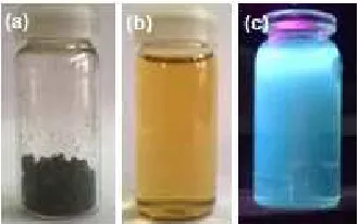
Fig 1. C-dot synthesized from ascorbic acid (a); C-dot aqueous solution (b); blue luminescent emission of C-dot (c)
Fig 3. Increasing intensity of luminescent emission from C-dots with the lowest concentration of urea, 0%, to the one with highest concentration of urea, 75%. (a) 0%, (b) 10%, (c) 25%, (d) 50%, (e) 75% urea
Fig 2.FTIR spectrum of C-dot synthesized from ascorbic
acid Fig 4.synthesized C-dotUV-Vis spectrums of (a) ascorbic acid, and (b)
the synthesis of C-dot resulted in higher emission intensity. The highest emission intensity was shown by C-dot synthesized by adding 75% of urea to the precursor solution (Fig. 3). However, microwave heating the urea solution without ascorbic did not resulted in the formation of C-dot thus it can be concluded that the main precursor in this synthesis is ascorbic acid.
Spectroscopic Characterization of C-dots
UV-Vis spectroscopy analysis was done on a 100 ppm C-dot solution. The absorption spectrum of the synthesized C-dot was very different from the absorption spectrum of ascorbic acid solution (Fig. 4). The greatest difference was the increased absorption in the 300–400 nm region of C-dot spectrum which may indicate of the presence of conjugation system on the graphite oxide structure [2,4]. This result is in agreement with a previous report on C-dot synthesized from orange peel [8]. We found that the absorption peak for the C-dots and its precursor, ascorbic acid, are similar (265 nm) which indicates that there are still organic functional groups on the surface of the C-dots in agreement with
the FTIR spectrum mentioned earlier. Fig. 5 shows that the higher the urea concentration in the reaction mixture, the lower its absorption intensity. This is because with the higher the concentration of urea there is a lower concentration of C-dots, for example a sample with 75% urea, only contains 25% C-dots in the mixture and this will reduce the intensity of C-dots absorption by 75%. However, the C-dots with 75% urea showed higher luminescence intensity as noted earlier.
Fig 5. Effect of urea concentration on the normalized absorbance of C-dots, urea concentrations are given in color code
Fig 6. Fluoresence emission spectrum of C-dots synthesized with the addition of 75% urea
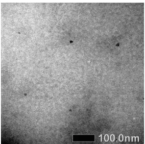
Fig 7.Representative TEM image of C-dots
Fig 8. Selective identification of Cr6+ ion. The order of metal ions solutions used in the study is (a) C-dot, (b) Mn, (c) Cr, (d) Fe, (e) Pb, (f) Hg. Solutions on top were not irradiated with UV light.
Characterization of C-dots Using TEM
The analysis was only done for C-dots with the brightest luminescence, the one with 75% urea. Fig. 7 shows that although some of the synthesized C-dots were round shaped, most were irregular in shape. The average size of the synthesized C-dots was 18 nm. This is in contrast with the homogeneous 1 to 5 nm round shaped C-dots synthesized from citric acid by Qu et al. [2]. This difference may be because the centrifuge technique we used in this synthesis is not very efficient to separate the particles. The use of other separating technique such as electrophoresis may yield smaller and more homogeneous C-dots. TEM analysis showed that the synthesized C-dot is amorphous, like graphite oxide. This is proved by the absence of diffraction pattern, and is similar to the the results in previous reports [2].
Preliminary Studies on the Application of C-dots as Metal Ions Sensor
Fig 9. fluorescent emission spectrum of C-dots in the presence of Cr6+ion
Fig 10. C-dot solution in the presence of Cr3+ ion (1), Cr6+ ion (2), and without metal ion (3); without (a) and with (b) UV light irradiation
Another test was done on Cr3+ ion to observe the effect of using a different source of Cr ion. Fig. 10 showed that there is no significant response between Cr6+ and Cr3+ ions. This result showed that the resulted C-dot selectively identified Cr metal, however cannot differentiate the type of Cr ions.
CONCLUSION
Blue green fluorescent emitting C-dots were synthesized quickly and easily by microwave heating ascorbic acid. Emission intensity increased with the
addition of urea as a passivation agent in the reaction process. Preliminary study showed the potential for the synthesized C-dots to serve as heavy metal sensors.
ACKNOWLEDGEMENT
The authors would like to thanks Ms. Awalia Khairunnisa for her help in the preliminary study, and Mrs. Lilis Sulistiawaty for her help in the analysis of C-dot using TEM.
REFERENCES
1. Li, H., Kang, Z., Liu, Y., and Lee, S.T., 2012, J. Mater. Chem., 22(46), 24230–24253.
2. Qu, S., Wang, X., Lu, Q., Liu, X., and Wang, L., 2012, Angew. Chem. Int. Ed., 124(49), 12381–12384
3. Yang, Z., Li, Z., Xu, M., Ma, Y., Zhang, J., Su, Y., Gao, F., Wei, H., and Zhang, L., 2013,Nano-Micro Lett., 5(4), 247–259.
4. Zhu, C., Zhai, J., and Dong, S., 2012, Chem. Commun., 48(75), 9367–9369.
5. Liu, H., Ye, T., and Mao, C., 2007,Angew. Chem. Int. Ed., 46(34), 6473–6475.
6. Wang, J., Wang, C.F., and Chen, S., 2012,Angew. Chem. Int. Ed., 51(37), 9297–9301.
7. Pandey, S., Thakur, M., Mewada, A., Anjarlekar, D., Mishra, N., and Sharon, M., 2013, J. Mater. Chem. B, 1(38), 4972–4982.
8. Prasannan, A., and Imae, T., 2013, Ind. Eng. Chem. Res., 52(44), 15673–15678.
9. Jiang, J., He, Y., Li, S., and Cui, H., 2012, Chem. Commun., 48(77), 9634–9636.
10. Hsu, P.C., and Chang, H.T., 2012, Chem. Commun., 48(33), 3984–3986.
11. Baker, S.N., and Baker, G.A., 2010,Angew. Chem. Int. Ed., 49(38), 6726–6744.