BENZYLISOQUINOLINE ALKALOIDS FROM BARK OF
Cryptocarya rugulosa
Nurdin Saidi
1,*, Hiroshi Morita
2, Marc Litaudon
3, Mat Ropi Mukhtar
4, Khalijah Awang
4, and A. Hamid
A. Hadi
41
Department of Chemistry, Faculty of Mathematics and Natural Sciences
,
Syiah Kuala University, Banda Aceh, 23111, Indonesia 2Faculty of Pharmaceutical Sciences, Hoshi University, Ebara 2-4-41 Shinagawa, Tokyo 142-8501, Japan 3
Institut de Chimie des Substances Naturelles, Centre Nationale des Recherches Scientifique, 91198, Gif-sur Yvette, Cedex, France
4
Department of Chemistry, Faculty of Science, University of Malaya, 50603, Kuala Lumpur, Malaysia
Received May 30, 2010; Accepted November 29, 2010
ABSTRACT
Seven known Benzylisoquinoline alkaloids, Papraline 1, (+)-norcinnamolaurine 2, (+)-codamine 3, (+)-6-methoxy-1-(3’-methoxybenzyl)-N-methyl-7-isoquinolinol 4, (+)-reticuline N-oxide 5, (+)-reticuline 6 and (-)-N-methylisococlaurine7were isolated from bark of Cryptocarya rugulosa. A combination of 1D-NMR (1H, 13C, DEPT, NOE), 2D-NMR (COSY, NOESY, HMQC, HMBC), MS (HRESI+, FAB+and EI) spectroscopy used to elucidate the structure.
Keywords:Cryptocarya rugulosa, lauraceae, benzylisoquinoline alkaloid
INTRODUCTION
Cryptocarya rugulosa (Lauraceae) is a medium sized to large tree up to 40 m tall. The species can be found in the lowlands and hill forest, mainly in Southeast Asia and Latin America [1]. This species has never been studied for its chemical constituents, but the chemical analysis of genus Cryptocarya revealed many types of compounds such as flavonoids, chalcone, lactone, α -pyrone and mainly alkaloids.
Isolation from the bark of C. crassinervia yielded two phenantrenes alkaloids, 2-hydroxyatherosperminine and N-demethyl-2-methoxyatherosperminine [2]. Benzylisoquinoline alkaloids, (+)-orientaline and laudanidine, were isolated from C. amygdalina [3]. Investigation on the bark ofC. ferreawere isolated three aphorpine alkaloids, (-)-o-methylisopiline, (+)-norlirioferine and (+)-lirioferine [4].
There are two types of alkaloid, a benzylisoquinoline i.e. romneine; and proaporphines i.e. cryptochine, cryptochinone, amuroline, amuronine and hydrostepharine were isolated from leaves and bark of C. chinensis [5]. Three quaternary pavine alkaloids, caryachine N-metho salt, neocaryachine N-metho salt and crychine N-metho salt were isolated from a callus culture ofC. chinensis [6]. Investigation on the leaves of C. chinensis resulted in the isolation of ten alkaloids. They are (-)-isocaryachine-N-oxide, Isoboldine-β -N-oxide, 1-hydroxy-cryprochine, (+)-isocaryachine, (+)-caryachine, (-)-caryachine, (-)-isocaryachine, isoboldine, (-)-munitagenine and bisnorargemonine [7].
Seven pavine alkaloids, neocrayachine, (-)-caryachine, (-)-eschscholtzine, and (+)-eschscholtzidine were isolated from bark ofC. chinensis[8].
Three benzylisoquinoline, (+)-(1R, 1aR)-1a-hydroxymagnocurarine, (+)-oblongine and (-)-8-o-methyloblongine and one aporphine, xanthoplanine were isolated from C. konishii [9]. Fifteen alkaloids have been isolated from leaves, bark and root of C. longifolia, reticuline N-methylcoclaurine, coclaurine, longifolidine, norisocorydine, laurotetanine, N-methyllaurotetanine, isoboldine, laurolistine, norargemonine, bisnorargemonine, scoulerine, longifolonine, cryptoleurospermine and thalifoline. They were divided into 5 types, namely benzylisoquinoline, aporphine, isoquinoline, pavine and protoberberine [10]. From dried bark of C. foveolata were isolated reticuline, a benzylsioquinoline [11].
EXPERIMENTAL SECTION
Materials
screening. The industrial and analytical reagent grade solvent was used for extraction and column chromatography.
Bark of C. rugulosa was collected from Segari, Perak, Malaysia in December 1996 and was identified at the Herbarium Chemistry Department, Faculty of Science, University of Malaya. The specimen was deposited at the Chemistry Herbarium, Faculty of Science, University of Malaya under the accession number KL-4672.
Instrumentation
Melting points were taken on a hot stage Gallen Kamp melting point apparatus and were uncorrected. The optical rotations were recorded on Jasco P1010 with tungsten lamp. The mass spectra were measured on a JMS 700 spectrometer using NBA as the matrix for FAB analysis. The Automass Thermofinnigan was used for HR ESI+ and ESI- analysis. The EIMS spectra were obtained on Shimadzu GC-MS QP2000A spectrometer 70 eV. The UV spectra were measured on a UV visible recording spectrophotometer, Model Shimadzu UV-160A with methanol as a solvent. The IR spectra were recorded on the Perkin Elmer 1600 Series FTIR using CHCl3 as solvent. The 1-D and 2D-NMR spectra were recorded in chloroform-D, methanol-D and acetone-D on a JOEL JNM-FX400. Chemical shift were reported in ppm and the coupling constants were given in Hz.
Procedure
Extraction
The dried and milled bark of C. rugulosa (1.9 kg) was macerated with n-hexane overnight (3 L x 3) at room temperature. The n-hexane extract was then evaporated under reduced pressure. The residue was dried and moistened with 10% NH3 and left overnight. The residue was then successively extracted with dichloromethane (5 L x 5) and then check with a Mayer’s reagent test after each extraction to make sure the extraction was completed. The extract was then evaporated under reduced pressure. The residue was macerated with of methanol (3 L x 3) and then check with a Mayer’s reagent test after each extraction to make sure the extraction was completed.
Dichloromethane extract were concentrated under reduced pressure to a volume of about 500 mL and tested for alkaloids content using TLC and spraying with Dragendorff’s reagent. The dichloromethane extract were extracted with a solution of 5% hydrochloric acid (10 L) until Mayer’s test negative. The combined extract were then basified with 10% ammonia solution to about pH 11 and then re-extracted with dichloromethane. The extract of dichloromethane (alkaloids) fraction were dried
with sodium sulphate anhydrous (500 g) and evaporated under reduced pressure.
Isolation and Purification
Extract of alkaloids (5 g) was placed on silica gel column and eluted with dichloromethane gradually enriched with methanol. The ratio of the solvent between CH2Cl2 and CH3OH were (100:0; 99:1; 98:2; 97:3; 96:4; 95:5; 94:6; 93:7; 92:8; 90:10; 85:15; 80:20 and 50:50). Fractions were collected every 100 mL and each fraction was tested with aluminium TLC plate for their alkaloids. The alkaloid spots were first detected by UV light (254 and 366 nm) and confirmed by spraying with Dragendorff’s reagent. Fraction having spots with the same Rf values and stains were combined and treated as a group. The combined groups were purified with CC and preparative TLC.
Isolation and purification (5 g) of alkaloid yielded 90 fractions and based on pattern of TLC they were divided into six fractions. Fraction 2 (67 mg), CH2Cl2 -CH3OH; 98:2, was then purified with CC to gave papraline 1 (15 mg). Fraction 4 (600 mg) eluted with CH2Cl2-CH3OH (95:5) was further separated using CC and preparative TLC, CH2Cl2-CH3OH; 93:7, to gave (+)-norcinnamolaurine2 (56.5 mg), (+)-codamine3 (57 mg), (+)-6-methoxy-1-(3’-methoxybenzyl)-N-methyl-7-isoquinolinol4 (21 mg) and (+)-reticulineN-oxide5(63 mg). (+)-reticuline 6 (298 mg) and (-)-N-methylisococlaurine 7 (40 mg) were obtained from fraction 5 (585 mg) by means of preparative TLC with CH2Cl2-CH3OH (92:8).
Papraline 1. Colourless amorphous solid: MS (EI, 70 eV), m/z 173 (100), 128, 115, 88, 62; IR (KBr)νmaxcm
-1
2917, 2621, 1593; UVmax(MeOH), nm, 325, 317, 312; 1
H NMR (CDCl3) δ 8.96 (1H, s, H-1); 8.34 (1H, d, J = 5.60 Hz, H-3), 7.47 (1H,d, J =5.60 Hz, H-4), 7.05 (1H, s, H-5), 7.17 (1H, s, H-8), 6.08 (2H, s, OCH2O);
13 C NMR (CDCl3)δ 150.34 1), 142.21 3), 120.30 (C-4), 126.01 (C-4a), 102.64 (C-5), 151.04 (C-6), 148.60 (C-7), 103.36 (C-8), 134.47 (C-8a), 101.78 (OCH2O). (+)-Norcinnamolaurine 2. Crystalline needles; m.p. 195-197 °C; [α]D
25
+70° (c = 0.03, MeOH); IR
(KBr) νmaxcm -1
3500, 2900; MS (EI, 70 eV), m/z 283, 176, 107; (HRESI)+ MS 284.1 (calcd for C17H17NO3, 284.1280); UVmax(MeOH), nm 302;
1
Table 1.1H NMR (in CDCl3, 400 MHz) and
(+)-Codamine 3. Brownish amorphous solid; [α]D 25 ppm, see Table 1.
(+)-6-methoxy-1-(3’-methoxybenzyl)-N -methyl-7-isoquinolinol 4. Brownish amorphous solid; [α]D
116.00 (C-4’), 112.08 (C-5’), 117.20 (C-6’), 56.08 (OMe-6), 56.19 (OMe-3’), 42.78 (N-Me).
(+)-ReticulineN-Oxide 5. White amorphous solid; [α]D 25
+55° (c=0.02, MeOH); IR (KBr) νmax cm -1
3424, 2093, 1516; MS (EI, 70 eV), m/z 192 (100), 177, 176, 151, 148, 137; MS (FAB+)346.1617 [M+H]+, (calcd for C19H23NO5, 346.1654); UVmx (MeOH), nm 296;
1 H and 13
C NMR (CD3OD)δ, ppm, see Table 2.
(+)-Reticuline 6. Brownish amorphous solid; [α]D 25
+ 62° (c = 0.23, MeOH); IR (KBr) νmaxcm
-1
3392, 2918, 1592; MS (EI, 70 eV), m/z 329, 192 (100), 177, 137, 122; (HRESI)+MS 330.1691 (calcd for C19H23NO4, 330.1695); UVmax (MeOH), nm 293;
1
H NMR (CDCl3) δ 3.66-3.69 (1H,t,H-1), 2.99-3.04 (2H,m, H-α), 2.74-2.79 (2H,m, H-3), 2.55-2.60 (2H,m, H-4), 6.51 (1H,s, H-5), 6.32 (1H,s, H-8), 6.73 (1H, d,J = 1.72, H-2’), 6.69 (1H, d, J = 8.32, H-5’), 6.54 (1H,dd,J1= 8.28,J2= 1.96, H-6’), 3.81 (3H, s, OMe-6), 3.81 (3H,s, OMe-4’), 2.44 (3H,s,N-Me);13C NMR (CDCl3) δ 64.45 (C-1), 40.85 (C-α), 46.48 (C-3), 24.65 (C-4), 128.75 (C-4a), 110.54 (C-5), 145.31 (C-6), 143.31 (C-7), 113.70 (C-8), 124.71 (C-8a), 132.72 (C-1’), 115.61 2’), 145.05 3’), 145.26 4’), 110.43 (C-5’), 120.90 (C-6’), 55.84 (OMe-6), 55.79 (OMe-4’), 42.12 (N-Me).
(-)-N-methylisococlaurine 7. Brownish amorphous solid; [α]D25 -15° (c = 0.02, MeOH); IR (KBr) νmaxcm-1 3364, 2917, 1597; MS (EI, 70 eV), m/z 192 (100), 177, 176, 148, 107, 92; (HRESI)+ MS 300.2 (calcd for C18H21NO3, 300.2000); UVmax (MeOH), nm 301;
1 H NMR (CDCl3)δ3.67-3.79 (1H,t,H-1), 2.80-2.86 (2H,m, H-α), 2.98-3.03 (1H, dd, Ha-3), 2.74-2.77 (1H,m, Hb-3), 3.17-3.24 (1H,m, Ha-4), 2.53-2.60 (1H,m, Hb-4), 6.51 (1H,s, H-5), 6.29 (1H,s, H-8), 6.86-6.88 (1H,d,J= 8.28, H-2’), 6.54-6.56 (1H, d, J = 8.28, H-3’), 6.54-6.56 (1H, d, J = 8.28, H-5’), 6.86-6.88 (1H,d,J= 8.28, H-6’), 3.74 (3H,s, OMe-6’), 2.37 (3H, s, N-Me); 13C NMR (CDCl3)δ 64.65 (C-1), 40.45 (C-α), 46.00 3), 24.21 4), 124.47 (C-4a), 110.71 (C-5), 145.49 (C-6), 143.42 (C-7), 113.97 (C-8), 130.36 (C-8a), 129.30 (C-1’), 130.32 (C-2’), 115.46 3’), 154.78 4’), 115.46 5’), 130.32 (C-6’), 55.74 (OMe-6), 41.75 (N-Me).
RESULT AND DISCUSSION
Papraline 1
The molecular formula of compound 1 was deduced as C10H7NO2 by the molecular ion peak at m/z 173 (100) in the EIMS. The fragmentation at m/z 115 indicated the loss of C2H2O2 in the structure. Other fragmentation peaks were observed at m/z 88 and 62 and the possibility of fragmentation pattern of1is shown in Scheme 1. The UV spectrum showed absorption bands at 325, 317 and 312 nm, which were characteristic of simple isoquinoline chromophore containing methylenedioxyl
Scheme 1.The possibility fragmentation pattern of1
Fig 1.Structure of benzylisoquinoline alkaloids fromC. Rugulosa
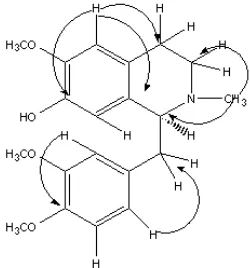
Fig 2. 1H-13C coupling pattern observed in HMBC spectrum of3
group [12]. The IR spectrum indicated the presence of a conjugated C=N group at 2621 cm-1, C-H aromatic at 2917 cm-1and an aromatic moiety at 1593-1463 cm-1.
characteristic of simple isoquinoline with fully aromatic, were observed at δ 8.34 and 7.47 with the coupling constants of 5.60 Hz. The methylenedioxy protons revealed at δ 6.08 as a singlet signal. The downfield chemical shifts for H-1 and H-3 were due to the deshielding effect of the iminic nitrogen [12].
The 13C NMR spectrum showed the presence of ten carbons, which belonged to one methylene, five methines and four quaternary carbons. The methylenedioxy peak was displayed at δ 101.78 and it was supported by HMQC data which showed the correlations between methylene proton at δ 6.08 and methylene carbon at δ 101.78. Finally, unambiguous assignment of all proton and carbon signals using DEPT, HMQC, and COSY and by comparison with literature data [12-13] showed that the alkaloid 1 was papraline. Alkaloid1 has previously been isolated from aerial parts ofFumaria indica, Fumariaceae [12].
(+)-Norcinnamolaurine 2
Alkaloid 2 was isolated as a white crystalline needles with [α]D25 +70° (c = 0.02, MeOH) and melting point 195-197 °C. The UV spectrum exhibited maxima at 302 nm. The absolute configuration at C-1 is in accordance with that norcinnamolaurine and determined as S [14-15]. The IR spectrum gave a broad band between at 3500 cm-1due to the presence of OH group.
The mass spectrum (HRESI)+ showed the [M+H]+ peak at m/z 284.1. The EI mass spectrum displayed a weak molecular ion signal at m/z 283 corresponding to a molecular formula of C17H17NO3. A strong signal at m/z 176 (100) appeared as a base peak was due to the loss of [C10H10NO2]
+
. Another peak displayed at m/z 107, was due to the loss of [C7H7O]
+ [16].
The 1H-NMR spectrum showed a singlet peak representing two protons at δ 5.84 indicated the presence of a methylenedioxyl group. Two aromatic protons revealed as a singlet at δ 6.64 and 6.49 which can be assigned to H-5 and H-6, respectively. Four peaks of an A2B2system of aromatic protons centered at
δ6.94 (H-3’and 5’,d, J = 8.32 Hz), 6.63 (H-2’and H-6’, d, J = 8.32 Hz), were assigned to four protons of 4-hydroxybenzyl portion of the molecule. The aliphatic protons gave a multiplet signals between δ 2.63 to 3.99 ppm.
The 13C-NMR and the DEPT spectra established the presence of 17 carbon signals which belonged to four methylenes, seven methines and six quaternary carbons. The analysis of COSY showed correlation between H-1/H-α, H-3/H-4, H-2’/H-3’ and H-5’/H-6’. Finally, based on the assignment of DEPT, HMQC, COSY and HMBC and by comparison with literature data [16-18], the structure of 2 was indeed norcinnamolaurine. This alkaloid for the first time has
been isolated from genus of Cinnamomum, Lauraceae [18].
(+)-Codamine 3
The EIMS spectrum of alkaloid 3 revealed a molecular ion peak at m/z 343 suggested a molecular formula of C20H25NO4. A base peak at m/z 192 [M-137]
+
due to the loss of [C8H9O2] +
, indicated that the ring A of benzylisoquinoline has two substituent, i.e. OCH3 and OH groups [18,28]. Other significant peaks were found at m/z 177, 176, 151 and 148. The UV spectrum showed two absorption peaks at 298 and 244 nm, which indicated the existence of the conjugated aromatic system. The IR spectrum showed absorption band at 3390 cm-1, which indicated the presence of a hydroxyl group in the molecule.
Its 1H-NMR spectrum (Table 1) displayed three singlet peaks at δ 3.76, 3.77 and 3.78 which were attributed to the methoxyl groups positioned at C-6, C-3’ and C-4’, respectively and five aromatic proton resonated at δ 6.75 (d, J = 1.68 Hz, H-2’), δ 6.65 (d, J = 8.32 Hz, H-5’), δ 6.49 (dd, J1 = 8.08 Hz, J2 = 2.20 Hz, H-6’),δ6.48 (s, H-5) andδ6.24 (s, H-8). The chemical shift values of the aliphatic proton appeared as a multiplet between δ 3.63-2.52 and N-methyl proton revealed atδ2.41.
There are 20 carbon signals appeared in the 13
C-NMR (Table 1) and experiment DEPT which showed four methyls, three methylenes, six methines and seven quaternary carbons in this molecule. The COSY spectrum revealed the correlations between H-5’/H-6’, H-3/H-4 and H-1/H-α. Inspection of its DEPT, HMQC, HMBC, NOE-differential and comparison of the spectral data obtained from literature values [14] allowed us to assemble the alkaloid 3. The absolute configuration at C-1 was in accordance with that of alkaloid3and determined asS[14].
(+)-6-methoxy-1-(3’-methoxybenzyl)-N -methyl-7-isoquinolinol 4
The UV spectrum of alkaloid4showed absorption maxima at 302 nm, which indicated the existence of aromatic system. The IR spectrum exhibited absorption bands at 3482 cm-1 (broad OH), 2915 cm-1 (CH) and 1598 cm-1 (benzene ring). The EIMS spectrum exhibited a molecular ion peak at m/z 313, corresponding to C19H23NO3. The peak at m/z 192 [M-137]+, appeared as a base peak, were due to the loss of [C8H9O2]
+ ion.
J2= 1.72 Hz, H-6’),δ6.55 (s, 1H, H-5) andδ6.42 (s, 1H, H-8). Two overlapped methoxyl proton signals resonated atδ 3.73 which can be assigned to OMe-6 and OMe-3’, oneN-methyl proton appeared at δ2.73 (s, 3H,N-CH3) and the aliphatic protons resonated as multiplets betweenδ2.46 to 3.62.
The 13C NMR spectrum revealed the presence of 19 carbons, including three methyl, three methylenes, seven methines and six quaternary carbons. The COSY spectrum verified the proposed structure, showing correlation between H-5’/H-6, H-2’/H-3’,H-2/H-3 and H-1/H-α. The1
H and 13C NMR data for 4 were identical as reported data in the literature [19-20].
(+)-ReticulineN-Oxide 5
Alkaloid 5, with [α]D 25
+55° (c=0.02, MeOH) was obtained as a white amorphous solid. The UV spectrum showed absorption maxima at 296 nm, which indicated the existence of highly conjugated system. The IR spectrum exhibited broad absorption at 3424 cm-1(OH), 2093 cm-1(CH) and 1516 cm-1(benzene ring).
The mass spectrum (FAB+) showed a medium molecular ion peak at m/z 346.1617 [M+H]+, (calcd for C19H23NO5, 346.1654). The EIMS showed the absent of any molecular ion peaks, but the mass range peaks was a peak formed by the loss of a hydroxyl from a molecular ion at m/z 328. The fragmentation ion at m/z 192 (base peak) showed that one methoxyl and one hydroxyl present in the structure [21]. The other significant signals were observed at m/z 177, 176, 148 and 137.
The prominent peak of methyl singlet proton in the 1
H NMR spectrum (Table 2) resonated at δ 3.30 (N-methyl) correlating with the downfield carbon signal at
δ 54.48 in HMQC spectrum due to the fact that the methyl group is attached to N-oxide. Five aromatic protons resonated atδ 6.28 (s, 1H, H-8), δ 6.71 (s, 1H, H-5), 6.58 (dd, J1 = 8.32 Hz,J2= 1.96 Hz, H-6’), δ 6.82 (d, J = 8.04 Hz, H-5’) and δ6.63 (d, J = 1.96 Hz, H-2’). Furthermore, chemical shift of H-1 at δ 4.53-4.59 (dd, J1 = 8.56 Hz, J2 = 2.68 Hz) and H-3 δ 3.42-3.49 (m) correlating with carbons signals at 79.26 (C-1) and 62.52 (C-3) were characteristic features ofN-oxide [22].
The 13C NMR spectrum (Table 2) showed the presence of 19 carbon signals, indicating one N-methyl, two O-methyl, three methylene, six methine and seven quaternary carbons. The data of DEPT, HMQC, COSY, HMBC and comparative literature data [22-27], support the structure of alkaloid 5. Reticuline-N-oxide has been obtained previously from leaves, stems and roots of Pochygone ovata(family Menispermaceae) [22].
(+)-Reticuline 6
Compound 6 was a major alkaloid from the bark of C. rugulosa. This compound was isolated as a brownish amorphous solid. The UV spectrum showed absorption band at 293 nm. The IR spectrum showed absorption at 3392 cm-1 indicated the presence of hydroxyl group in the structure. This alkaloid exhibited an [M+H]+ in the (HRESI)+ mass spectrum at m/z 330.1691. The EI mass spectrum showed a molecular ion peak at m/z 329 corresponding to a molecular formula of C19H23NO4. A base peak at m/z 192 [M-137]+, was due to the loss of [C8H9O2]
+ , a characteristic of benzylisoquinoline [28].
The1H NMR spectrum of6revealed two methoxyl groups overlapped to each other at δ 3.81 corresponding to 6-OMe and 4’-OMe. In addition, there are five aromatic protons appeared at δ 6.73 (d, J = 1.72 Hz, 1H, H-2’), δ 6.69 (d, J = 8.32 Hz, 1H, H-5’),δ6.54 (dd,J1= 8.28 Hz,J2 = 1.96 Hz, 1H, H-6’),
δ 6.51 (s, 1H, H-5), δ 6.32 (s, 1H, H-8) and one N-methyl proton resonated as a singlet atδ2.44.
The 13C NMR spectrum showed there were 19 carbon resonances, which is in agreement with the molecular formula of reticuline. The DEPT spectrum showed the appearance of three methyls, three methylenes and six methines and seven quaternary carbons in the molecule skeleton. The assignment of carbon and hydrogen in the structure was further confirmed by HMBC and HMQC experiment. The HMBC spectrum confirmed the peak correlations between H-1/C-α, C-1’, C-8a and C-4a, H-3/C-4a and C-1, 4/C-3 and C-8a, 5/C-4, C4a, C-6 and C-7, H-8/C-6, C-7, C-8a and C-1, H-2’/C-4’, C-1’ and C-α, H-5’/C-3’, C-4’ and C-1’, H-6’/ C-α, N-Me/C-1 and C-3. The reported values data of [17,19,29-30] support the structure of alkaloid 6. However, this compound is not new due to the alkaloid6had been isolated from many kinds of plants [17,22,29,31].
(-)-N-methylisococlaurine 7
(-)-N-methylisococlaurine 7 was isolated as a brownish amorphous solid with [α]D
25
The 1H-NMR spectrum showed one methoxyl signal at δ 3.74 appeared as a singlet which most probably attached to C-6. Protons H-5 and H-8 peaks appeared as a singlet atδ6.51 andδ6.29, respectively. Four aromatic protons resonated at δ 6.54-6.56 (H-3’ and H-5’,d,J = 8.28 Hz) andδ6.86-6.88 (H-2’ and H-6’, d, J = 8.28 Hz). Another signal presence as a singlet at
δ 2.39 was belonged to N-methyl group. The aliphatic protons appeared as multiplets at the region of
δ 2.53–3.70. The COSY spectrum indicated the correlation between H-3’/H-5’ and H-2’/ H-6’.
The 13C NMR spectrum displayed 18 carbon signals in the molecule. The DEPT spectrum showed there are one methoxyl, one N-methyl, three methylenes, seven methines and six quaternary carbons. In structure1 NOE-differential experiment was used to confirm the position of OMe-7. Irradiation of OMe-7 showed an enhancement of proton H-8. Therefore, the OH group was attached to C-6. The HMBC spectrum revealed correlation of H-5 to C-7 and C-8a further support the position of OMe group. Assignment of all proton and carbon signals, DEPT, HMQC, COSY, NOE-Diff. and HMBC and by comparison with literature values [15,22,31-33], confirmed to the structure. The absolute configuration at C-1 is in accordance with the known compound and it was determined as R [15]. As it can be found in previous research work, we have noticed that alkaloid7, has been isolated previously from leaves of Nelumbo nucifera, Nymphaceae [33].
CONCLUSION
Seven known Benzylisoquinoline alkaloids, Papraline 1, (+)-norcinnamolaurine, 2, (+)-codamine 3, (+)-6-methoxy-1-(3’-methoxybenzyl)-N-methyl-7-isoquino linol4, (+)-reticulineN-oxide5, (+)-reticuline6and (-)-N-methylisococlaurine 7 were isolated from bark of Cryptocarya rugulosa.
ACKNOWLEDGEMENT
This work was supported by Technological and Professional Skill Development Sector Project, TPSDP, No. 465/J11/SPMU-TPSDP/2000 (Indonesian Government) and Vot F, No. F0142/2003B, PPF/FP092/2005C and SAGA 66-02-03-0036 (Malaysian Government).
REFERENCES
1. Ng, F.S.P., 1989, Tree Flora of Malaya; A Manual for Forester, Longman Malaysia, Kuala Lumpur, 132–138.
2. Khalijah, A., Hadi, A.H.A., Saidi, N., Mukhtar, M.R., Morita, H., and Litaudon, M., 2008,Fitoterapia, 79, 308–310.
3. Borthakur, N., Mahanta, P.K., and Rastogi, R.C., 1981,Phytochem., 20, 501–504.
4. Saidi, N., Hadi, A.H.A., Khalijah, A., and Mukhtar, M.R., 2009,Indo. J. Chem., 9, 3, 461–465.
5. Lee, S.S., Chen, C.H., and Liu, Y.C., 1993,J. Nat. Prod., 56, 2, 227–232.
6. Chang, W.T., Lee, S.S., Chueh, F.S., and Liu, K.C.S., 1998,Phytochem., 48, 1, 119–124.
7. Lin, F.W., Wu, P.L., and Wu, T.S., 2001, Chem, Pharm.Bull., 49, 10, 1292–1294.
8. Lee, S.S., Liu, Y.C., and Chen, C.H., 1990,J. Nat. Prod., 53, 5, 1267–1271.
9. Lee, S.S., Lin, Y.J., Chen, C.K., Liu, K.S., and Chen, C.H., 1993, J. Nat. Prod., 56, 11, 1971– 1976.
10. Bick, I.R.C., Sevenet, T., Sinchai, W., Skelton, B.W., and White, A.H., 1981, Aust. J. Chem., 34, 195–200.
11. Lamberton, J.A., and Vashist, V.N., 1972, Aust. J. Chem.,25, 2737–2738.
12. Atta-Ur-Rahman, Ahmad, S., Bhatti M.K., and Choudhary, M.I., 1995, Phytochem., 40, 2, 593– 596.
13. Richard, A.M., Janssen, M., Robert, J., Lousberg, J.Ch., Wijkens, P., Kruk, C., and Theuns, H.G., 1989,Phytochem., 28, 10, 2833–2839.
14. Debourges, D., Roblot, F., Hocquemiller, R., and Cave, A., 1987,J. Nat. Prod., 50, 5, 852–859. 15. Kashiwada, Y., Aoshima, A., Ikeshiro, Y., Chen,
Y.P., Furukawa, H., Itoigawa, M., Fujioka, T., Mihashi, K., Cosentino, L.M., Morris-Natschke S.L., and Lee, K.H., 2005, Bioinorg. Med. Chem., 13, 443–448.
16. Gellert, E., and Summons, R.E., 1970, Aust. J. Chem.,23, 2095–2099.
17. Chowdhury, B.K., Sethi M.L., and Lloyd, H.A., 1976,Phytochem., 15, 1803–1804.
18. Gellert, E., and Summons, R.E., 1969,Tetrahedron Lett., 58, 5055–5058.
19. Richard, A.M., Janssen, M., Wijkens, P., Kruk, C., Biessels, H.W.A., Menichini F., and Theuns, H.G., 1990,Phytochem., 29, 10, 3331–3339.
20. Marsaioli, A.J., Ruveda, E.A., and Reis, F. de A.M., 1978,Phytochem., 17, 1655–1658.
21. Atta-Ur-Rahman, Wahab, A.T., Ahmad, S., Nawaz, S.A., and Choudhary, M.I., 2004, Chem. Pharm. Bull.52, 7, 802–806.
22. Dasgupta, S., and Ray, A.B., 1979, J. Nat. Prod., 42, 4, 399–406.
24. Kametami, T., and Ihara, M., 1980, J. Chem. Soc., Perkin Trans. 1, 629–631.
25. Ohiri, F.C., Verpoorte, R., and Svendsen, A.B., 1983,Planta Med.,47, 87.
26. El-Shabrawiy, A.O., Schiff, Jr, P.L., Slatkin, D.J., Dasgupta, S., Ray, A.B., and Tripathi, V.J., 1984, Heterocycles, 22, 993.
27. Leet, J.E., Freyer, A.J., Minard, R.D., and Shamma, M., 1985,J.Chem.Soc.,Perkin Trans. 1, 1565. 28. Al-Amri, A.M., Smith, R.M., EL-Haj, B.M., and
Juma’a, M.H., 2004, Forensic Sci. Int., 140, 175– 183.
29. Castro, O.C., Lopes, J.L. and Vergera, A.G. 1985, Phytochem., 24, 1, 203–204.
30. Jendrzejewski, S., 1990, Phytochem., 29, 1, 135– 139.
31. Brochmann-Hanssen, E., and Nielsen, B., 1965, Tetrahedron Lett.,18, 1271–1274.
32. Bhakuni, D.S., Satish, S., and Dhar, M.M., 1972, Tetrahedron, 28, 4579–4582.