A STUDY ON
KALANJAHA PADAI
Dissertation Submitted To
THE TAMIL NADU DR.M.G.R Medical University
Chennai – 32
For the Partial fulfillment for The Award of Degree of
DOCTOR OF MEDICINE (SIDDHA)
(Branch – III, SIRAPPU MARUTHUVAM)
DEPARTMENT OF SIRAPPU MARUTHUVAM
Government Siddha Medical College
Palayamkottai – 627 002
March - 2008
INTRODUCTION
The skin is a protective covering of the body. It, with all its specialised derivatives makes up what is called the integument [Latin- covering], which covers the entire surface of the human body.
The human skin shows wide regional variations in structure like scalp, face, earlobes, back, palms and soles etc., In thickness of the skin the number of sebaceous glands, collagen fibre and vasculature.
Diseases of the skin are a common occurrence. About 10-20% of patients seeking medical advice suffer from skin diseases. While infections are more common in the tropics, chemical and psychogenic dermatoses are common in western countries. Diseases of the skin account for a great deal of misery, suffering, incapacity and economic loss. Besides this, they are a great handicap in society because they are visible.
According to Siddha system, our human body is made up of pancha-boothas. It leads to 96 thathuvas. The entire universe is also made up of pancha boothas. So any change in the cosmos will reflect in the human body. This was quoted in Sattamuni Gnanam by Sattamuni as follows,
mz;lj;jpy; cs;sNj gpz;lk; gpz;lj;jpy; cs;sNj mz;lk; mz;lKk; gpz;lKk; xd;Nw mwpe;J jhd; ghh;f;Fk; NghNj"
Pancha boothas give birth to seven thathus of body. This was quoted by Thirumoolar in Thirumandhiram as,
“,ujk; cjpuk; ,iwr;rp Njhy; Nkij kUtpa tj;jp thOk; nghL kr;ir gutpa Rf;fpyk; ghohk; cghjp cUtk yhYly; xd;nwdyhNk”
- jpUke;jpuk;
Among the pancha bootha, earth and fire together gives birth to the thathu ‘Thol’ (skin).
According to great Yugimuni, skin disorders are classified into 18 varieties. Yugi had not explained “Kalanjaga padai” as a separate disease. Instead he had made just mention in the vatha diseases as kalanjaga vatham which resembles “virpodaga kuttam”. The clinical features of Kalanjaga padai correlated with Psoriasis as described in Modern dermatology.
Siddhars had explained the line of treatment into two major modes, one is internal medicine and other is external medicine. As per the Siddha medicine, both internal and external medicines are 32 in numbers.
“Nth; ghU jio ghU kpQ;rpdf; fhy; nky;y nky;y gw;g nre;J}uk; ghU”
- Fzghlk; jhJ tFg;G From the above stanza, it is ideal to choose herbal preparations. The author had selected Neeradimuthu Rasayanam as internal medicine and
Moreover, it is important to reduce mental stress, which is the main aggravating factor of Psoriasis. Life style modifications, Yoga therapy, transitional meditations, diet modifications etc., are given special emphasis to reduce the mental stress.
AIM AND OBJECTIVES
1. To collect the various Siddha literatures and modern text books as literal evidences regarding disease “Kalanjaga padai”.
2. To expose the talent of Siddhars in diagnostic principle.
3. To know the extent of correlation of aetiology, classification, symptomatology and diagnostic methods.
4. To have an idea of an incidence of Kalanjaga padai with reference to age, sex, socio-economic status, habits, and family history related to any psychosomatic problems and paruva kaalams (Seasons).
5. To have a complete study of the disease, under the topics of mukkutram, poripulangal, udal kattugal, enn vagai thervugal etc., in order to evaluate the pathology of Kalanjaga padai.
6. To have a detailed clinical investigation and to utilize the possible diagnostic tools in the confirmation of the diagnosis and prognosis of the disease.
7. To have a clinical trial Kalanjaga padai with NEERADIMUTHU RASAYANAM as internal medicine and AADUTHEENDAPALAI VEMBU THYLAM as external application.
8. To study the biochemical analysis and pharmacological analysis of the selected drugs.
9. To highlight the factors like land where they live, climate changes, diet and mental stress of human beings. To make an awareness among the patients in order to avoid further recurrence of the disease.
REVIEW OF LITERATURE
SIDDHA ASPECTS
In the Siddha medicine, the pancha bootha (five elements) are elaborately described. Our body which is formed by the combination of five boothas. “Thol"(skin) is a part of Prithvi. Skin is the largest organ in our body. It has got a lot of functions, each of which is important for the normal physiological functioning of whole human body, are regulation of body temperature, excretion, protection from pathogens etc.,
According to the Siddha text "Siddha maruthuvanga surukkam" skin is divided in to six types. They are
y Skin containing water. y Skin having blood.
y Skin which produces sirangu. y Skin which produces kuttam. y Skin producing tumour.
y Skin which produces severe pain during an injury.
Man is very much influenced by his environment. Since skin serves as a link between man and Universe, it is the first organ to be influenced by the change also, since it is related intimately to the mind, it responds even to the slightest change in mind. Any disease affecting the skin causes a socio economic problem, mental torture and social stigma to the patient.
Definition (iyal)
Kalanjaha padai is a non contagious, non infectious, inflammatory disease of the skin characterized by well defined dry erythematous plaques with large adhered silvery or ivory mica like scales.
Aetiology (Noi varum vazhi)
Among the available Siddha literatures only Yugi Vaithiya Chinthamani and Thirumoolar Vaithiyam are the sources of informations on aetiology and clinical features of the eighteen types of skin diseases. There is no specific obvious information about the specific factors causing Kalanjaha padai.
Thirumoolar quotes as,
“gay; nkhopaPh; jpNufj;jpy; fpUkp jhNd gue;J jphp Fl;lk; Nghy; Gs;sp fhZk; kayJTk; fpUkpAe;jh ele;J Gf;fpy; NkdpaJ rurnud ntbj;J Gz;zhFk;”
- FU ehb
“Fl;lKld; jpNufnky;yhk; gwf;Fk; NghJ
FopFopaha; fpUkpapdhy; nfhs;Sk; Gs;sp”
- FU ehb “fpUkpahy; te;j Njhlk; ngUfTz;L
Nfl;fyjpd; gphptjid fpukkhf nghUkptUk; thAnty;yhq; fpUkpahNy
nrUkp tUk; gTj;jpuq;fs; fpUkpahNy NjfkJ nrhwpf;Fl;lk; fpUkpahNy JUkp tUQ; RNuhzpjq; fpUkpahNy
R+l;rKld; fphpirg;ghy; njhopy; nra;tPNu”
- FU ehb The text book of “Sirappu Maruthuvam” describes the following aetiological factors of Kalanjaha padai,
y guk;giu Nehahf %thpy; xUtUf;Fk; - of unknown aetiology and may be genetic
y yRd jhgpjk; - Tonsillitis
y Gg;Grg; gzpfs; - Respiratory diseases y xt;thik - Allergic disorder
y kd cisr;ry; - Psychological disturbances y mjph;r;rp - Depression , anxiety
y fhykhWghLfs; - due to changes in humidity NkYk;>
y jhk;gpur; nre;J}uk; ( red oxide of copper) y ,sk;gps;isj; jLg;G kUe;J ( polio vaccine) y FnshNuhFtpd; ( chloroquine)
Kjypa kUe;Jfs; FUf;fisAk; giliaAk; cz;Lgz;zyhk; vd;Wk; $wg;gl;Ls;sJ.
In Siddha system of medicine chronic skin diseases are brought under the clinical entity kuttam. The ancient texts describe that eighteen types of kuttam .
Thirumoolar quotes as follows:
“Fl;lkJ tpl fug;ghd; tpl ePh; R+iy
RNuhzpjj;jhy; jhJ nfl;Lj;; jbg;Gz;lhFk; kl;lwNt fpUkp nrd;W kUTk; NghJ
tifaha; fpUkpAl tplePh; nrd;W Fl;lKld; Njfnky;yhk; gwf;Fk; NghJ FopFopaha; fpUkpapdPh;f; nfhs;Sk; Gs;sp jl;lwNt fpUkpAl ePuhy; te;j
rfy Fl;lk; tplfug;ghd; rhw;wyhNk”
“tpahjpAz; %thW tpsq;fpa Fl;lq;Nfs; Rahjp fpue;jp Rod; Nkfj;jhY}Wk;
gahjp kz;Zsg; gy tz;bdhnyl;Lk; epahjpg; GOehyha; epd;wjpf; Fl;lNk”
Kuttam is named a common word of chronic skin lesion as for the Siddha text books.
Agasthiar had mentioned kanmam is the main cause for kutta noi. Kanmavaralaru (Psycho social cause)
“gotpidahy; tp\g; G+r;rp fbj;jhYk; ghjfh;f;F xUehSk; jPh;tjpy;iy
cstpidahY}lhgpf; nfhs;s te;j cz;ika jwpahky; %h;f;fd; nra;thh; fstpidAs; jPh;tjpy;iy fbdnkj;j
fUizAs;s G+uzj;jpy; fz;fhl;rp mstpid eP fhZKd;Nd mfyr; nrhy;Y
milahsk; tpuy; FWF gpd;dhy; NfNs tpuy; FWFk; fhmy; epkpWk; tp\k; NghNyWk; ghuhd Njfnky;yhe; jbj;J fhZk;
ghjnky;yhk; ntbj;J kpfg; Gz;zha; fhZk; rurKld; nrhwp fug;ghd; gpzk; Nghy
NjhZs; rhe;ijahNk tpe;ij nfLj;jb tPq;Fk; eh ey;fpype; Neha;f;F kUe;jauhNj
ey;Nyhiu gPbf;Fq; F\;lq; fd;kkhNk”
- mfj;jpah; ghpg+uzk;-400 Thirumoolar has mentioned that the skin diseases are manifested in three ways,
1. Venereal origin and other mega diseases 2. Insect bites.
3. Infections and infestations. In Yugimuni 800 he mentions,
“Mr;nrd;w gjpndl;L Fl;lj;jhYk;
mtuth;fs; nra;fpd;w mjh;kj;jhYk; Njbr; nrd;w rpthyaj;jpYr; rpl;lq;fs; nra;jth;fs; rpt epe;ij gz;zpdhh;fs; %r;nrd;w nghpNahiu Jbj;Njhh;fs;
%h;f;;fkha; milf;fyj;ij vLf;fpd;whh;fs; Mr;nrd;w jpidastpy; Fiwe;j $yp
nfhLf;fd;Nwhh; Fl;lj;jpw; $LthNdh”
- A+fpKdpngUE}y; 800
Yugimuni strongly described only psycho-social factors are the main causes. They are stress inducing factors. He has attributed the following causes.
y Misbehavior in the temple. y Sacrilege towards God. y Humiliating the elders. y Breech of trust.
y Paying low wages to workers.
The main factors behind the reasons are manifestation of stress which can be considered as precipitating factor. The psychic tranquility of the individual depends upon the harmony of social movements.
Hereditary also plays an important role in breeding Kalanjaha padai which appear in generations affecting several siblings and in such families the condition tends to be severe and persistent.
In Yugi Chinthamani, among the eighteen types of skin disease three
types seen to be variants of Kalanjaha padai. But no description is available in the Siddha classics under Kalanjaha padai.
Out of the eighteen types of kuttam, 3 types have similarities with Kalanjaha padai and out of this three, one type is closely related with Kalanjaha padai.
CLINICAL FEATURES
Thethuru kuttam
“rh;ke;jhd; rptg;ghf tl;lzpj;Jr;
ryit Nghy ntSf;Fk; jpdTz;lhFk; $h;ke;jhd; NuhkkJ kpfTz;lhFk;
kapnuy;yhQ; RUz;LNk cUz;ilahFk; fh;ke;jhd; gpj;j NrLk kpFf;Fk;
fhae;jhd; fjpj;JNk jpkpUz;lhFk; jh;ke;jhd; rlnky;yh %jyhFk; jhf;fhd Njj;jpUf; Fl;le;jhNd”
- A+fpKdp ngUE}y; 800 Kjy;ghfk; Under Thethuru kuttam, annular erythematous lesions with the appearance of washed leather are described. Itching, oedema of the body and plugging of hairs are the characteristical clinical features of this entity.
Kajasarma kuttam “jhdhfr; rle;jhD kpf fWg;ghFk; rlnkq;Fe; NjhYhpAk; rptg;GkhFk; Ntdhf twtnwdj; jhdpOf;Fk; ntbf;FNk nrhwpr;ryha;j; jpdTz;lhFk; jhdhfr; rh;kf; Fl;l kpjpYz;lhFk;
fbdkha; fhy; tpuy;fs; fdg;Gz;lhFk;
$dhf Njfnky;yhk; typnaLf;Fk;
Fwpahd frrh;k Fl;le;jhNd”
Virbodaga kuttam
“GJikaha; rhPunkq;Fk; jpdTz;lhFk;
nghUntbaha; jpf;nfd jPf;nfhOe;J Nghy nkJikaha; tpl;nlhpAk; ey;yghk;gpd;
tp\g;glk; Nghy; jbj;J ntSg;GkhFk; RJikaha; kpfr; nrhhpAQ; rptg;GkhFk; J}f;fnkhL rQ;ryKk; kpfTz;lhFk; fJikaha; Njhnyy;yhk; jbg;Gz;lhFk; fdj;j tpw;Nghlfkhd Fl;le;jhNd”
An Erythematous lesion in the skin with plaques of silvery scales is described. The lesions are described to be extensive. Usually these entities are associated with anxiety and despair.
The oedema and plugging of hairs are not noticed in practice as it is in the case of Thethuru kuttam.
The blackish discolouration of the body, oedema of the toes is noticed in practice as in Kajasarma kuttam. It is seen only in the complication stage of
Kalanjaha padai.
So the clinical features of Virbodaga kuttam are ascertained to be closely to Kalanjaha padai.
In the text Siddha Maruthuvam Sirappu authored by Sri. Dr. R. Thiagarajan, clinical features are described as,
y The skin lesions are red in colour with raised margins and white ivory or silvery rough thick scales, on removing the scales pin point blood-stained spots occurs.
y The lesions vary in size either thin or thick layers. y In children these lesions may be like water drops.
y In severe cases lesions occur in the face, scalp and sometimes all over the body.
IN A CHRONIC CASE:
y The skin lesion occurs in the extensor aspect of forearm. y In some cases these lesions appear over the palms and soles. y In some cases these lesions appear all over the body.
y The lesions are coin shaped and there may be small pus formed lesions are found.
y In obese women, the lesions may occur over navel, inguinal region and axilla with discharge. Due to sweating, itching may be associated. The borders of the lesions are not to be demarcated clearly.
y One fourth of the patients have lesions over nails which are pink coloured and associated with ridges.
y 7 % of the patients have associated joint involvement which will manifest as Psoriatic Arthropathy.
ARTHROPATHY
If the patient suffers from repeated episodes of Kalanjaha padai it may be associated with painful joints known as Kalanjaha vatham.
“Yugimuni” describes the clinical features of “Kalanjaha vatham” as follows,
“thjkhq; fhy; ifapy; Fuq;fpuz;Lk;.
tUj;J re;J KWf;fpNa File;J nehe;J ehjkh eiljhDe; jhd; nfhlhky;
eype;JNk Klkhfp fuL fl;b NrjkhQ; rle;jhD kpf ntSj;Jj; jpdnthL rpuq;Fkha; Nrl;gkhfp
fhjkhaUrpNahL kaf;fkhFk;
fUjpa fhshQ;rfkhk; thjkhNk”
- A+fp itj;jpa rpe;jhkzp ghly; - 259 NOI KANIPPU (Diagnosis)
Piniyari muraimai is a method of diagnosing a disease. It is based upon three main principles. They are,
y Poriyalarithal (Inspection) y Pulanalarithal (Palpation) y Vinathal (Interrogation)
Physicians 'pori’ and 'pulan' are used as tools for examine the 'pori pulan' of the patient. The above principles correspond to the methodology of 1. Inspection, 2. Interrogation 3. Palpation in modern medicine, in arriving a clinical diagnosis of the disease.
1. PORIYALARITHAL
Pori is considered as the five senses of perception namely, y Nose
y Tongue y Eye y Skin y Ear
'Poriyalarithal' is examining the pori of the patient by the physician for diagnosing.
2. PULANALARITHAL
'Pulan' is five object of senses. They are y Smell
y Taste y Vision
y Sensation to touch y Hearing
'Pulanalarithal' means examining the 'pulan' of the patient by the physician for diagnosing purpose
3. VINATHAL
Vinathal is gathering the informations regarding the history of the disease, its clinical features etc., from the patient or his immediate relatives who are taking of him, when the patient is not in a position to speak or the patient is a child.
ALAVAIGAL (logics)
Alavaigal are used in clinical diagnose of a disease
“msit fhz;ly fUjy; ciu mghtk; nghUs; xg;ghnwd;gh; msit NkYk; xopGz;ik iajpfj; Njhbay; ngd ehd; fsit fhz;gh; mitapw;wpd; NkYk; miwth; mitnay;yhk; msit fhz;ly; fUjy;> ciu vd;Dk; %d;wpylq;fpLNk”
- rptrpj;jpahh; msit vz; 6
Alavai divided in to ten types. They are,
y Observation - fhz;ly;
y Inference - fUjy;
y Authority, Literature - ciu
y Preception - mghtk; y Presumption - mUj;jggj;jp y Comparison - cgkhdk; y Inference by elimination - ghhpNr\k; y Probability - rk;gtk; y Tradition - Ijpfk;
y Natural Inference - ,ay;G
The above mentioned "ten alavaigal" are included in three alavaigal. They are,
y Kaandal (Inspection by Siddha method)
Through 'kaandal' the physician can directly see the patient, hear all the complaints and at length concludes a diagnosis.
y Karuthal (Through Siddha Investigation)
Through envagai thervu and neerkuri as well as neikuri, we can diagnose a disease by karuthal.
y Urai (Text's evidence - Siddhar's)
Comparative study of the signs and symptoms of the patient with the reference of books and come to a diagnosis.
Ennvagai thervugal (eight diagnostic tools)
Siddhars have developed a unique method of diagnosing the disease by “Enn vagai thervugal'
“ehb ];ghprk; eh epwk; nkhop tpop kyk; %j;jpukpit kUj;JtuhAjk;”
- Neha; ehly; Neha; Kjy; ehly; (Kjy; ghfk;) “nka;f;Fwp epwk; njhdp tpop eh ,Ukyk; iff;Fwp”
- Njiuah; Hence the diagnosis is made by the following
1. Naadi (Pulse) 5. Mozhi (voice) 2. Sparisam (sensation to touch) 6. Vizhi (eyes) 3. Naa (tongue) 7. Malam (Faeces) 4. Niram (colour) 8. Moothiram (Urine)
Kalanjaha padai in relation with envagai thervugal 1. Naadi (pulse)
clypy; caph; jhpj;jpUg;gjw;Ff; fhuzkhd rf;jp vJNth, mJNt jhJ my;yJ ehb vdg;gLk;.
“ehb vd;why; ehbay;y> euk;gpy; jhNd eykhfj; Jbf;fpd;w JbjhDky;y ehb vd;why; thj gpj;j rpNyw;gdKky;y ehb vOgj;jPuhapue; jhDky;y
ehb vd;why; mz;l Nguz;lnky;yhk; ehb vOtifj; Njhw;wj;Js;sha; epd;w ehbaJah uha;e;J ghh;j;jhuhdhy; ehbAWk; nghUs; njhpe;J ehL thNu”
- gjpndz; rpj;jh; rjf ehb E}y; Naadi is responsible for the existence of life and can be felt one inch proximal to the wrist on the radial side by means of palpation with the tips of index, middle and ring fingers corresponding vatham, pitham and kabam respectively.
The three humours vatham, pitham and kabam exist in the ratio 1:1/2:1/4 normally. Derangement in these ratios leads to various disease entities.
The three "Uyir thathukal" are formed by the combination of three nadigal with three vaayu.
y Idakalai + Abanan - Vatham y Pingalai + Piranan - Pitham y Suzhimunai + Samanan - Kabam
In Kalanjaha padai the following types of naadi were observed. They are,
a. Vatha kabam and b. Pitha kabam
2. Sparisam (sense to touch)
This reveals about the warmth, chillness, dryness, roughness of the skin, oozing, sweating, tenderness, fissures, depigmentation changes in the skin, swelling, emaciation, etc.,
In Kalanjaha padai thickness, roughening, dryness, pin point bleeding, silvery or mica like or ivory colour dry scales in skin may be noticed.
3. Naa (Tongue)
The colour, dry or wet, coated or not, excessive salivation, redness, ulceration, fissure, pallor, yellowish discolouration,any malignant outgrowth, predominant taste in tongue, speech and deviation of the tongue, along with the conditions of the teeth and gums are to be noted.
In Kalanjaha padai, tongue coating like flour (khgbT)) may be noticed in some cases may be due to habitual constipation.
4. Niram (colour)
Changes in the colour of the skin, teeth, eyes, nails and lips due to vatham, pitham, kabam, hypo and hyper pigmentation are to be noted. In Kalanjaha padai, erythematous lesions, pin point bleeding after removing the scales may be noticed.
5. Mozhi (voice)
Examination of mozhi includes clarity of speech any speech disturbances, crying, high or low pitched voice, slurring or incoherent speech, scanning speech, talk included by hallucination, undue argument, breathlessness, nasal or hoarseness of voice, wheezing. In Kalanjaha padai no change is seen regarding speech.
6. Vizhi (eyes)
The motor and sensory activities are to be noted. Also any abnormal colour change indicating vatham, pitham, kabam and mukkutram. Hyperemia, ulceration, bluish discolouration, response of pupil, pallor, protrusion, sunken eyes, sharpness of vision, excessive lacrimation, angle of eye, subconjunctival bleeding, visual disturbances are to be noted. In Kalanjaha padai burning sensation was reported in some cases.
7. Malam (faeces)
Colour, froth, solidity, semisolid or liquid quantity, odour, frequency, constipation, presence of mucus, blood and undigested matter in the stool are to be studied.In some of the cases of Kalanjaha padai habitual constipation was reported.
8. Moothiram (urine)
Collection of urine for the determination of neerkuri and neikuri is a special diagnostic method.
Neerkuri and Neikuri
“mUe;J khwpujKk; mtpNuhkjha;
m‡fy; myh;jy; mfhyT+d; jtph;e;jow; Fw;ws tUe;jp cwq;fp itfiw
Mbf; fyrj; jhtpNa fhJnga; njhU K$h;j;jf; fiyFl; gLePhpd; epwf;Fwp nea;f;Fwp epUkpj;jy; flNd”
- rpj;j kUj;Jthq;f RUf;fk;
Prior to the day of urine examination the patient was instructed to take a balanced diet and quantities of food must be proportionate to his routine in take. The patient could have no disturbed sleep. After waken up in the morning, the first urine voided was collected in a clear wide mouthed glass container and is subjected to analysis of "neerkuri and neikuri" with in one and a half hour. Then neerkuri is to be found out by,
Neerkuri
“te;j ePh;f;fhp nail kzk; Eiu vQ;rnyd; iwe;jpa Ysit aiwFJ KiwNa”
- rpj;j kUj;Jthq;f RUf;fk; Voided urine has the following characters:
y Niram : Colouration y Edai : Specific gravity y Manam : Smell
Apart from these, the frequency of urination, abnormal constituents such as protein, presence of blood, pus, renal calculus, crystals etc., also to be found out.
In Kalanjaha padai patient, straw or hey coloured urine is noticed. Scanty micturition can be noted in some cases.
Neikuri:
The speciality of neikuri is stated in the following verse: “If;Fwp nfhLtl thdpo ykh;e;Njhh;
iff;Fwp njhpe;j eq; flTisj; Jjpj;Nj nka;Fwp epwe;njhdp tpop ehtpUkyk; iff;Fwp KotJh cq;fw;whh; jk;kpDk; ngha;f;Fwp nka;Fwp GfYnk th;f;Fk; nea;f;Fwp ajid ape;ePzpye;J iug;Nghk;”
-rpj;j kUj;Jt Neha; ehly; Neha; Kjdhly;jpul;L
The collected specimen as said above is to be analysed by following method.The specimen is kept open in a glass dish or china clay container. It is to be examined under direct sunlight, without shaking of the vessel. Then add one drop of gingelly oil at a distance of 1/2" or 3/4" height observe keenly the direction it spreads with in few minutes, and conclude the diagnosis as follows:
“muntd ePz;b d‡Nj thjk; MopNghw; gutpd; m‡Nj gpj;jk;
Kj;njhj;J epw;fpd; nkhopt njd; fgNk mutpy; MopAk; Mopapy; muTk;
mutpy; Kj;Jk; Mopapy; Kj;Jk;”
- rpj;j kUj;Jt Neha; ehly; Neha; Kjdhly; jpul;L
Oil spreads like a snake Vatha neer
Oil spreads like a ring Pitha neer Oil kept remaining as such and
Kaba neer Floating like a pearl
Ring in the snake
snake in the ring Thontha Neer pearl in the snake
pearl in the ring Paruvakaalam (season)
The whole year is constituted by 6 seasons .They are 1. Karkaalam - Aavani- purattasi - August - September 2. Koothir kaalam - Iyppasi -karthigai - October - November 3. Munpani kaalam - Markazhi- Thai - December -January 4. Pinpani kaalam - Masi- Panguni - February- March 5. Elavenil kaalam - Chithirai - Vaigasi - April- May 6. Mudhuvenil kaalam - Aani - Aadi - June - July
In each and every season, routine changes will occur in the land, normal biological functions of individual, living things, plants, animals, human beings,
which will modify the normal physiology and make them susceptible to certain specific disease.
Kabam gets thannilai valarchi in pinpani kaalam and Vetrunilai valarchi in Elavenil kaalam. Pitham gets Thannilai valarchi in Kaar kaalam and Vetrunilai valarchi in Koothir kaalam, Vatham gets Thannilai valarchi in Mudhuvenil kaalam and Vetrunilai valarchi in Kaar kaalam.
Nilam (land)
It is divided in to five types
S. No Nilam Land Area, it concerns
1. Kurinchi - Mountain region and surroundings 2. Mullai - Forest regions and surroundings 3. Marutham - Cultivating regions and surroundings 4. Neithal - Sea and Coastal regions
5. Palai - Desert land only Udal kattugal.
Our body consists of seven udal kattugal, gives strength and structure to our body.
S.No Udal Kattugal Functions
1 Saaram It gives strength to the body and
mind
2 Senneer Saaram after absorption is converted into senneer. It is responsible for knowledge, strength, boldness and healthy complexion.
3. Oon Gives structure and shape to the body and is responsible for the movements of the body.
4. Kozhuppu Lubricates the organs and proceed
on its own works.
5. Enbu Protects the vital organs and used for movements and nominates body structure.
6. Enbu Moolai Present inside the bones and it gives strength and maintains the normal condition of the bone.
7. Sukkilam / Suronitham Responsible for the reproductive function of species.
In the case of Kalanjaha Padai, out of seven udalkattugal saaram, senneer and enbu are commonly affected.
Saaram : Dryness, roughness, tiredness Senneer : Dryness, Paleness of the skin Enbu : Pain in the knee joints
MUKKUTRAM
Human body is influenced by Thridoshas (ie) Vatham, Pitham and Kabam. They are responsible for normal physiological condition of the body.
VATHAM
Vatham is a kinetic energy which influences all motions.
Vatham is located in the abanan, faeces, idakalai, spermatic cord, iliac bone, skin, nerves, joints, hair follicles, muscles, bone, ear and thigh.
S. NO Name Location Physiological Functions
1 Piranan Heart and Lower respiratorytract to Upper respiratory tract.
Controls knowledge, mind and five objects of
sense,useful for breathing
2 Abanan Lower abdomen and
extremities
Responsible for urination, expels faeces,
delivering foetus, discharge sperm and menstruation.
3 Viyanan Mainly at heart Responsible for
movements of all parts of the body and used to feel the sensation
4 Uthanan Chest Responsible for
vomiting, cough, hiccough, sneezing.
5 Samanan Stomach Aids for proper digestion. It controls the activity of other vaayus.
6 Naagan Eyes Responsible for
opening and closing of the eyes.
7 Koorman Heart and Eyes Responsible for vision
and yawning and controls lacrimation
8 Kirukaran Throat Responsible for
salivation nasal secretion and appetite
9 Thevathathan Eruvaai& Karuvaai For laziness, sleeping and anger
10 Thananjeyan Nose Responsible for bloating of the body after death. It escapes on the third day after death, through the cranium when it bursts.
In the case of Kalanjaga padai,
Abanan : Habitual constipation
Viyanan : Erythematous plaques in the affected areas of skin Samanan : Due to other vaayus, it is affected
Koorman : Insomnia like condition The above vaayus are affected commonly.
PITHAM:
Pitham is responsible for all the transformation. Pitham is located in urinary bladder, heart, head, pingalai, umbilicus, abdomen, piranan, blood, sweat, skin and eye.
Pitham is classified into 5 types. They are,
1. Analaga pitham : Responsible for digestion of food 2. Ranjaga pitham : Responsible for colour of blood
3. Sathaga pitham : Located in heart and is responsible for normal activities of the body
4. Aalosaga pitham : Responsible for normal vision
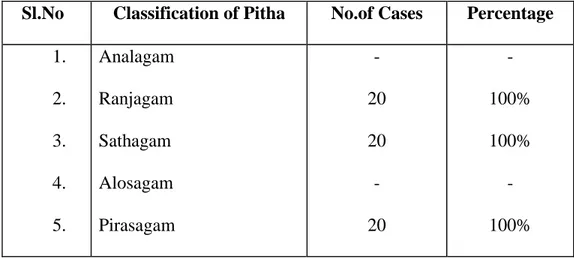
5. Prasaga pitham : Responsible for the complexion of skin In the case of Kalanjaha padai,
1. Ranjagam : Paleness of the skin, conjunctiva and tongue 2. Sathagam : Difficulty to do the routine works properly and
sluggishness
KABAM:
Stabilizes, maintains and lubricates all movements.
Kabam is found in samanan, semen, brain, head, tongue, nose, bones, bone marrow, fat, nerves, chest, blood, large intestine, eye, stomach and pancreas.
Kabam is classified in to 5 types, they are,
1. Avalambagan : Heart is the centre for avalambagam. It controls all other forms of kabam
2. Kiletham : Stomach is the centre for kiletham. It give moisture and softness to the ingested food, and helps for digestion.
3. Pothagam : Tongue is the centre for pothagam and it is responsible for the sense of taste 4. Tharpagam : Head is the centre for tharpagam. It gives
cooling effect to eyes
5. Santhigam : It lies in the joints and is responsible for
the locomotive action of movable bony
joints. In the case of Kalanjaha padai,
Tharpagam : Burning sensation in the eye
Santhigam : Pain in kneejoints, elbow and inter phallangeal joints.
UDAL VANMAI (Body Immunity)
The Udal Vanmai is classified into 3 types. They are, y Iyarkai Vanmai
y Seyarkai Vanmai y Kaala Vanmai
IYARKAI VANMAI
Natural immunity of the body itself by birth.
SEYARKAI VANMAI
Improving the health by intake of nutritious food materials, activities and medicines.
KAALA VANMAI
Development of immunity according to age and the environment.
When Udalvanmai is affected there may be a possibility of Kalanjaha Padai.
Line of treatment
In accordance with the Siddha system of medicine, certain basic principles are developed before starting the specific drug therapy. These procedures are initially followed to balance, the deranged kutrams ie. Vatham, pitham and kabam.They are purgation, vomiting and application of drugs on the eyes to balance the deranged vatham, pitham and kabam respectively. This can be understood by the following verbs,
“tpNurdj;jhy; thjk; jhOk; tkdj;jhy; gpj;jk; jhOk; mQ;rdj;jhy; Iak; jhOk;”
In addition to this following medications are practiced in the Siddha system. y Aha marunthugal (Internal medicines)
y Pura marunthugal (External medicines)
y Restriction regarding food habits and routine day to day life style. y Sirappu Maruthuvam - a special feature of Siddha medicine
like Pranayamam, Yoga.
y AHA MARUTHUVAM :
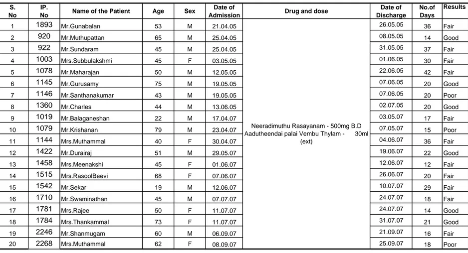
These are to administer rejuvenating drugs adopted for every one healthy life Neeradimuthu Rasayanam -500mgs two times a day with hot water.
y PURA MARUTHUVAM:
Aadutheendapalai vembu thylam - applied as an external application
Restriction Regarding Food and Habits 1. Avoid Karappan food Items
“ngUFQ; Nrhs kpWq;Fk; ngUk; fk;G tuF fhUld; thioapd; fhnahL
ciu nfhs; ghfw; nfspw;W kPd; cz;bby; tphptjha; fug;ghD kpFe;jNj”
2. Avoid bitter guard, guava, egg, fish, chicken 3. Obese must be restricted.
4. Avoid alcohol, smoking etc.
5. Should not be mentally stressed and strained
6. Since it is a chronic and not a life threatening disease, it should not be loaded with heavy drugs.
7. The medications should calm the mind just free from stress and strain
8. The patches should be washed with lukewarm water, to remove the scales everyday early morning. After the bath, external applications are to be applied there after.
SPECIAL NON - DRUG THERAPEUTICS
Several special medicaments of non-drug therapeutics like Yoga, Pranayamam, Asanas, Kalpa medicines are employed in Siddha system. These are employed during diseased state and for the prevention of diseases during healthy days.
In Kalanjaha padai, patients are also advised to follow Pranayamam, Yoga and Asanas for the early cure, in order to avoid the remissions and exacerbation of this disease.
PRANAYAMAM
It is a form of Kayakalpa method. By practicing this one can prevent any disease. This is explained in the following verse,
“Vw;wp ,wf;fp ,UfhYk; G+hpf;Fk; fhw;iwg; gpbf;Fk; fzf;fwp thhpy;iy fhw;iw gpbf;Fk; fzf;fwp thshf;F $w;iw cijf;Fk; FwpaJthNk”
YOGA:
Yoga is maintained by the body in a particular posture for a particular period of time. This is totally different from the ordinary exercise. Yoga vitilises, both physical body and the mental set-up unlike exercise which tones only the muscles.
The common benefits are,
y It tones the internal organs y It prevents obesity and disease
y It maintains normal circulation to all the organs of the body. y It is very safeguard for all the vital organs.
y It avoids laziness, enhances pure mind and cleverness and memory power. There will be no problems like psychomotive disturbances if practised daily.
ASANAS
Regarding skin disease the following Asanas can be adviced 1. gj;khrdk;
2. rh;thq;fhrdk;
1. gj;khrdk;
rkjsj;jpy; rk;kzkpl;L cl;fhh;e;J tyg;ghjj;ij ,lj; njhil kPJk;> ,lg;ghjj;ij tyJ njhil kPJk; Vw;wp ,uz;L iffisAk; Kd; Gwk; xd;wd; gpd; xd;whf Nfhh;j;J kyh;e;jpUf;FkhW ,Uj;jy;> ,jdhy; cly; eyKk; kd kfpo;r;rpAk; Vw;gLk;.
2. rh;thq;fhrdk;
ky;yhe;J gLj;J fhy;fis nkJthf xl;bagbNa NkNy J}f;fpg; gpd; Gl;l ghfj;ijAk;> ,Lg;Gg; ghfj;ijAk; NkNy J}f;fpf; iffshy; KJFg; Gwj;jpy; jhq;fp epw;wy;.
eiu> jpiu> %g;G khwp ,sikAz;lhFk;.
tPjdNfhsk; J}z;lg;gLfpwJ.
clypd; vy;yh cWg;GfSk; gyg;gLk;.
3. G+uz rtrhe;jp Mrdk;
ky;yhe;J gLj;Jf; fhy;fis NeuhfTk;> iffis clNyhL gf;fthl;bYk; itj;J Neuhfg; gLj;J ,Uj;jy;.
,J fisg;igg; Nghf;Fk;. Gj;Jzh;r;rpia cz;lhf;Fk;. PATHIYAM
Disease mainly occur due to wrong diet habits. Siddhars stressed this in every aspect of treatment and prevention from further occurrences. This view is well understood in this verse.
“kUe;njd Ntz;lhthk; ahf;iff;F mUe;jpaJaw;wJ Nghw;wp Azpd;”
During diseased states, diet restrictions or pathiyam are strictly to be followed. These are to be administered to normalize the deranged doshas and for the good manifestation of given medicines to be more effective. This is given in the verse,
“gj;jpaj;jpdhNy gyd; cz;lhFk; kUe;J
gj;jpaq;fs; Nghdhy; gyd; NghFk; - gj;jpaj;jpy; gj;jaNkntw;wp jUk; gz;bjh;f;F - Mjypdhy; gj;jpaNk cj;jpnad;W ghh;”.
- Njiuah; ntz;gh
So it is very essential to adhere pathiyam strictly for the early cure of the disease.
DIET
Food habits that reduce the vatha, pitha, kaba to the normal level has to be taken. Patients are strictly convinced to avoid all the non-vegetarian items except goat’s flesh. Avoidence of the following food items were also strictly advised.
y Agathi Keerai - Leaves of sesbania grandiflora y Seeni avaraikai - Cynampsis psoratoides
y Pagarkai - Bitter guard y Poosanikai - Great pumpkin y Perum payaru - Cow – gram
y Solam - Maize
y Kanam - Horsegram
y Motchai - Flac bean
MODERN ASPECT
SKIN
ANATOMY
The skin is a large organ of the human body. Forming a major interface between man and his environment. It covers an area of approximately 2m2 and weighs about 4kg. The structure of human skin is complex consisting of a number of layers and tissue components.
The superficial epithelial layer – EPIDERMIS
Underlying connective tissue layer – DERMIS or CORIUM Subcutaneous layer – HYPODERMIS
Epidermis
The epidermis is formed of non vascular stratified epithelium. Its usual thickness is between 0.07mm and 0.12mm. It is divisible into two main systems,
1. Keratinising or malphigian systems which forms the bulk. 2. Pigmentary system produces pigment.
Epidermis composed of the following layers from base to the surface.
1. Stratum Germinatum (Basal cell layer)
This is the deepest portion of the epidermis and consists of a single layer of keratinocytes.
2. Stratum malphigii (Prickle cell layer)
3. Stratum Granulosum (Granular cell layer)
This layer consists of 1 to 3 layers of flat cells containing keratohyaline basophilic granules which are PAS negative.
4. Stratum lucidum
It is a pale wavy looking layer, formed by many layers of flattened and closely packed cells devoid of nuclei.
5. Stratum corneum (Horny layer)
Most superficial layer, consists of many layers of non-nucleated flattened cornfield cells.
Dendritic cells of epidermis
These are melanocytes, langerhans cells and indeterminate cells.
Basement Membrane
The junction of the epidermis and dermis is formed by basement membrane. It allows movement of cells and nutrients between the dermis and epidermis.
Dermis
The dermis consist of 2 parts, 1. Superficial papillary dermis 2. Deeper reticular dermis
The dermis is composed of fibro collagenic tissue containing blood vessels, lymphatics and nerves. Besides these structures, dermis contain appendages or adnexal structure
DERMAL APPENDAGES 1. Sweat glands
These are of 2 types:
Eccrine and Apocrine. Eccrine glands
They are coiled tubular glands distributed all over the skin except on the beds of nails, margins of lips and the glans penis. But are most numerous in the palms, soles and axillae.
Apocrine glands
They are tubular glands with large lamina. They occur in the axillae, areola and nipples of breasts, umbilicus, the anus and the genitalia. Their secretion is odoriferous with a secondary sexual significance.
2. Sebaceous glands
They are scattered all over the integument except palms and soles.Meibomian gland, mammay glands and smegma glands of the penis are modified sebaceous glands.
3. Hair
Hair is found on every part of the body except on the palms and the soles, the dorsal surface of the terminal phalanges, lips, inner surface of the labia, inner
A hair is composed of a root, the part embedded in the skin and a shaft the portion projecting from the surface. Hairs differ in length, thickness and colour in different parts of the body and in different races.
4. Nails
The nails are thickness of the deeper part of the stratum corneum. The nail is composed of keratinised clear horny cells.
Blood vessels:
The blood supply of the skin originates from a large number of anterior forming anastamosis in the deepest part of the cortex. From here single vessels run upwards and from a second network in the upper cortex. Finally terminal arterioles ending in capillary loops, which drain into connecting venules. The blood is returned to the large veins in the subcutaneous tissue.
Lymphatics:
The skin contains a rich network of lymphatics, which drain into a few large vessels in the hypodermis.
Nerve Supply:
The nerve supply of the skin consists of a motor sympathetic portion derived from the sympathetic ganglia and sensory spinal portion arising from the dorsal route ganglia. The sympathetic fibers innervate the blood vessels, erector pilorum muscles and apocrine duct.
Physiological Functions of the Skin
Function Structure/cell involved
Protection against: Chemicals, particles Ultraviolet radiation Antigens, haptens Microbes Horny layer Melanocytes
Langerhan cells, lymphocytes, Mononuclear phagocytes, mast cells Preservation of a balanced internal
environment
Prevents loss of water, electrolytes and macromolecules
Horny layer
Shock absorber
Strong, yet elastic and compliant covering Dermis and subcutaneous fat
Sensation Specialist nerve endings
Calorie reserve Subcutaneous fat
Vitamin D synthesis Keratinocytes
Temperature regulation Blood vessels, eccrine sweat glands
Lubrication and waterproofing Stratum corneum
Protection and praising Nails
Hormonal
Testosterone synthesis from inactive precursors and testosterone conversion to other androgenic steroids
Hair follicles Sebaceous glands
Body odour (more important in animals) Apocrine sweat glands
Psychosocial Hair, nails, appearance and tactile quality
PSORIASIS
DefinitionPsoriasis is a common, genetically determined, inflammatory skin disorder of unknown cause which in its most usual form is characterized by well-demarcated raised red scaling patches that preferentially localize to the extensor surfaces.
PSORIASIS the word comes from ancient Greece and means “to itch”.
Red eruptions appear on the surface of the skin and begin to itch. These areas form plaques over the reddened lesions. The plaques resemble multilayered scales of skin. Psoriasis varies in intensity from a few random spots to a massive out break covering the entire body and requiring hospitalization. Psoriasis has a tendency to be genetically inherited.
Recently it has been classified as being an auto immune disorder (the body’s immune system turning on itself). This disorder can originate in juveniles or not be evident until adulthood, it has been reported to initiate as early as birth or not occur until very late in life.Once Psoriasis begins there are only remissions and relapses of varying degrees of intensity.
Historical Aspects of Psoriasis
The earliest descriptions of what appears to represent Psoriasis are given at the beginning of medicine in the Corpus Hippocratium. This work was edited in Alexandria 100 years after the death of Hippocrates (460 -377 B.C). Hippocrates used the terms Psora and Lepra for conditions that can be recognized as Psoriasis.
Later Celsus (25 B.C) who translated the writings of Tiberius Claudius Menekrates (the personal physician of Emperor Tibericus) in the Greek, described among 40 different dermatoses a form of impetigo that was interpreted by R.William (1757-1812) as being Psoriasis. William separated two diseases as Psoriasiform entities, a discoid lepra Greaconum and a polycyclic confluent Psora leprosa which later was called Psoriasis. In 1841 dermatologist Ferdinand Non Hebra (1816-1880) unequivocally showed that William's lepra Graeconum and Psora leprosa were one disease that had caused much confusion because of differences in the size distribution growth and involution of lesions.
Aetiology :
Exact cause of Psoriasis is unknown. Hypothesis of aetiology. y Hyperplastic epidermis
y Immunological defects y Biochemical abnormalities
y Increased epidermal proliferation / inflammatory component y Alteration in skin content.
y Infection (retro viruses)
Psoriasis a disease with Genetic Predisposition
Numerous studies over many years support the finding that genetic predisposition, an inherited tendency to develop the disease which has a major role in the pathogenesis of Psoriasis. Genetic predisposition does not mean a 100 percent guarantee that the disease will appear; other initiating factors such as
injury or infection may act together with the disease process. Supporting evidence for genetic predisposition includes,
1 There is a higher-than-average incidence of Psoriasis in relatives of people with Psoriasis, indicating “familial tendency” to develop the disease; however in some people with Psoriasis no family history is evident.
2 There is an increased incidence of Psoriasis in children when one or both parents have Psoriasis.
3 In studies of identical and non-identical twins, Psoriasis is much more likely to appear in both identical twins than in both non-identical twins a finding that also confirms that more than one gene must be inherited to establish genetic predisposition for Psoriasis.
4 There is higher than expected frequency of certain white cell antigens (class I human Leucocyte Antigens or HLA' S ) on cells of people with Psoriasis and their close relatives; this finding also supports Psoriasis inheritability and also suggests that the gene(s) involved in Psoriasis may be some chromosome that holds the genes for HLA. There are many types of HLA in the HLA complex and studies have shown that HLA type may be associated in some degree with timing of disease onset, type of Psoriasis and severity of disease.
Psoriasis Triggers:
A trigger is usually needed to make Psoriasis appear whether it is for the first time or the thirtieth.
Common Psoriasis Triggers are: (a) Infection:
• Candida albicans (thrush)
• Human Immuno deficiency virus (HIV)
• Staphylococcal skin infections (boils)
• Streptococcal pharyngitis (strep throat)
• Viral upper respiratory condition (b) Reaction to certain medications:
• Anti-malarial drugs.
• Beta-Blockers (used to treat high blood pressure)
• Cortico steroids(to treat Psoriasis – overuse/with drawal)
• Indomethacin (NSAID – to treat arthritis)
• Lithium (to treat manic depression / other psychiatric conditions). (c) Skin injury:
People with psoriasis often notice new lesions 10 to 14 days after the skin is cut, scratched , rubbed, or severely sun burned. This is called “KOEBNER'S
PHENOMENON” and is named after Dr. Koebner who in the 19th century
observed that a patient developed new lesions in areas where his horse bit him. A wide range of traumas and skin conditions are known to trigger “KOEBNER’S PHENOMENON”:
Skin trauma: • Acupuncture • Bites • Bruises • Burns • Chafing • Chemical irritation
• Cuts and scrapes
• Pressure against skin
• Shaving
• Sunburn and peeling
• Adhesive tape on the skin
• Tattoos • Vaccinations • Others Skin conditions: • Boils • Dermatitis • Herpes blisters • Lichen planus • Scabies • Vitiligo •
(d) Stress:
• Isolated from the society because of skin changes
• Difficulty in activities of daily living because of Psoriasis
• Time-consuming treatment
• Spending money for treatment
• Ineffective treatment
• Stressful event (etc). (e) Weather:
• Cold winter – trigger psoriasis
• Hot / sunny – help clear psoriasis
• Air conditioning – trigger psoriasis (f) other triggers:
• Hormones
• Smoking
• Heavy drinking Pathogenesis
Accelerated epidermopoiesis has been considered to be the fundamental pathological event in Psoriasis. The transit rate of Psoriatic keratinocyte is increased and the deoxyribo nucleic acid synthesis time is decreased. It has been suggested that it is the heightened proportion of epidermal cells participating in the proliferative process rather than the actual rate of epidermopoiesis, that is the
basic fault in Psoriatic lesions. The result in either case is greatly increased production of keratin.
The earliest histologic change was a inflammatory perivascular upper dermal infiltrate, with only epidermal acanthosis and parakeratosis after the transformation of the lesion into a scaly papule.
Polyamines are significantly increased in Psoriatic lesions. There is an increased production of the leucotrienes and 12-hydroxy eicosatetraenoic acid, both of which are chemotactic for polymorphonuclear leukocytes.
The Arachidonic acid cascade
The complexity of the pattern of inducers of inflammation is nowhere better shown than in the production of leucotrienes, prostaglandins and eicosatetraenoic acid from arachidonic acid. Arachidonic acid is obtained from dietary sources, membranes of red meat, green leafy vegetables, corn and safflower oil. Its products are central to the aggravation of Psoriasis by trauma and to the pathogenesis of Psoriatic lesions.
Psoriasis is associated with different HLA antigens. They are B13 or B17 has a five fold risk of developing Psoriasis. In Pustular Psoriasis HLA- B27 may be seen whereas B13 and B17 are increased in Guttate and Erythrodermic Psoriasis. In palmoplantar Psoriasis, there is an increased proportion of persons having HLA-B8, Bw 35. - Cw7, and -DR3.
Farber affirms an abnormal nucleoprotein metabolism in the incomplete keratinization process in Psoriasis. In the stratum corneum the free amino acids are low, with an accumulation of MPS and of free and esterified choline. DNA and RNA accumulate and pentose, purines, uracil and organic phosphates are also increased.
There is prominent neutrophil response in psoriatic lesions. How this relates to findings of increased plasminogen activator activity in psoriatic skin or to the enhanced physiologic functions. Both Psoriasis and Reiter's disease occur with increased frequency in patients with AIDS. Also interleukin-2 therapy for malignancy may induce Psoriasis.
(Reference - Text book of Dermatology-fitz patrik) Weddell showed that there is a profuse hyperplasia of nerve endings beneath and into the lesions of Psoriasis.
Histopathology:
The histopathology appearance of Psoriasis is distinctive but not specific.Main feature may be subdivided into
• Epidermal thickening
• The inflammatory component
• The vascular component a) Epidermal Thickening:
• Epidermis shows marked exaggeration of the rete pattern.
enlargement of their ends.
• Many mitotic figures can be seen
• Rate of epidermal cell production increased.
• The turn-over time of psoriatic epidermis and stratum corneum is very much shortened than normal epidermis.
b) The inflammatory component:
• Desiccated polymorpho nuclear leukocytes – Munro microabscesses.
• Epidermis is oedematous/infiltrated by inflammatory cells.
• Dermis also contains many inflammatory cells mostly lymphocytes.
• Leukotriene B4 – seen in psoriatic stratum corneum. c) The vascular component:
• Papillary capillaries are dilated and tortuous
• Larger gaps between the endothelial cells
• Abnormal capillaries.
Cell Systems involved in Pathogenesis of Psoriasis Keratinocytes
A characteristic feature of involved skin of Psoriatic subjects is hyper proliferation. There is two fold increase in proliferative cell population and 100 percent of the germinative cells of the epidermis appear to enter the growth fraction compared with 60 to 70 percent for normal subjects.
T cells
Numerous T cells are present in psoriatic lesions predominantly surrounding the vessels of the upper dermal plexus.
Granulocytes
Formation of spongiform microabscesses (Munromicro abscesses) filled with granulocyte is a hallmark of Psoriasis. The presence of these cells in psoriatic lesions variable and becomes more pronounced with disease activity (eg) in acute or pustular Psoriasis.
Endothelial cells
Changes in the dermal capillary endothelium have been implicates at the site of primary defect. Hyper proliferation of endothelial cells is most pronounced in the tortous dilated capillaries at the advancing edges of a lesion.
Mast cells
Mast cell densities are increased in lesional psoriatic skin compared with normal or uninvolved psoriatic skin.
Fibroblasts
Fibroblasts are potent producers of cytokines and lipid mediators that could affect the epidermis as well as the inflammatory reaction.
CLINICAL FEATURES
The lesions
• Well demarcated margin and is raised above the skin surface (plaque)
• Plaques vary enormously in size and shape
• They often start out discoid but end up polycyclic Sites Affected
• Extensor aspects of trunk and limbs preferentially. Sometimes flexor aspects also affected.
• Knees, elbows, scalp are frequently affected.
• Nails are often affected and may show – “thimble pitting”, separation of the nail plate from the nail bed (anycholysis) subungual debris, brownish – black discolourations and deformities of the nail plate.
• Major body folds in elderly who are over weight (flexor aspects)
• Groins, genitalia, axillae
• Inframammay folds in women.
• Skin of abdominal folds/ umbilicus.
• Scalp margin, paranasal folds, retro auricular folds. CLINICAL VARIANTS OF PSORIASIS
Guttate Psoriasis
• Guttate Psoriasis, named for its droplet –shaped lesions.
• Guttate lesions range in diameter from 0.1cm to 1 c.m
• They are predominate on the trunk and proximal areas of the extremities and are more likely to involve the face.
• Often it develops some two or four weeks after an episode of tonsillitis or phargngitis, mostly due to beta-haemolytic streptococci.
• Represent acute flare of pre-existing chronic plaque type Psoriasis. Plaque – type Psoriasis (Psoriasis vulgaris)
• Occuring in 75% to 80% of all psoriasis patients.
• Lesion is well demarcated, red – violet round or oval plaque.
• Leison is 1c.m or larger in diameter and surrounded by white silvery scales, which overlie bony prominences.
• In darkly pigmented patients, lesions are hyperpigmented with various shades of brown or black, which are accentuated by chronic rubbing or scratching.
• Symmetry of distribution of skin lesions is the rule in plaque type psoriasis.
• Face is frequently spared. Most commonly involved areas are elbows, knees, scalp, sacrum, umbilicus, intergluteal cleft and genitalia.
• 70% of patients complain of pruritis, skin pain, or burning, especially when the scalp is involved.
PUSTULAR PSORIASIS.
It is characterized by sterile pustules either generalized or localized to palms and soles.
(a) Localized Pustular Psoriasis (Palmoplantar Pustulosis)
• Yellowish white, sterile pustules on the central parts of the palm and soles.
• Older lesions take on a brownish appearance and later are shed in a scale at the surface
• Lesions are observed in all stages of development, including vesicles, vesicopustules, frank pustules and dried brown maculopapules
• The disorder is resistant to treatment and is subject to relapses and remission over many years
(b) Generalized pustular psoriasis (Von Zumbusch)
• Characterized by fiery – red, irregular patches with round, arcuate, serpiginous borders that are studded with myriad 1m.m to 2 m.m superficial pustules.
• Severe systemic upset, a swinging pyrexia, arthralgia and a high polymorpho nuclear leuco cytosis, hypocalcaemia, hypoalbuminaemia accompanying the skin disorder.
• The skin patches have a predilection for flexural or skin – fold areas such as armpits, groin, or under breasts but may occur anywhere.
• The tiny pustules coalesce into lakes of pus, desqumate, and form new pustules as the borders moves in waves every 24 to 72 hours.
(C) Other rare variants of pustular psoriasis 1. Acrodermatitis Continua (Dermatitis repens)
Recalcitrant pustular erosive disorder on the fingers and toes around the nails and occasionally elsewhere
2. Pustular bacteriod
Sterile pustules suddenly appear on the palms and soles after an infection. 3. Subcorneal pustular dermatosis
There is generalized eruption of sterile superficial pustules. ERYTHRODERMIC PSORIASIS
Plaque type Psoriasis progress to erthrodermic Psoriasis in which the plaque – like appearance disappears and the skin is universally red and scaly.
• Concomitant Psoriatic arthropathy is common
• Heat loss (because of increased blood supply to skin)
• Water loss leads to dehydration because of the disturbed barrier function of the abnormal stratum corneum.
• Hyperdynamic circulation
• Risk of high cardiac output failure
• Loss of protein, electrolytes, metabolites via the shed scale and exudates and may develop deficiency states
• Depressed because of malaise, pruritis and discomfort NAPKIN PSORIASIS
Infantile napkin dermatitis some times takes on Psoriatic lesions develop on the scalp and trunk.
NAIL PSORIASIS
• Finger nails are affected more than toe nails
• Pitting of nail plate. The pits tend to be large, deep and randomly dispersed on the nail plate.
• Small red spots in the lanula or yellow-brown spots (“oil droplet sign”) in the nailbed correspond to early guttate lesions of Psoriasis.
• The distal nail plate may separate from the nail bed (onycholysis)
• Chronic inflammation of the nail matrix may lead to scarring and permanent dystrophy, mimicking onychomycosis, especially when toe nails are affected.
Terms describing Morphologic features of Psoriasis Psoriasis ostracea
Old patches may be thickened and tough and covered with lamellar scales like outside of an oyster shell.
Psoriasis guttata
The lesions are the size of water drops. Psoriasis follicularis
Tiny scaly lesions are located at the orifices of the pilosebaceous follicles. Psoriasis figurate, Psoriasis annulata & psoriasis gyrate
The lesions have curved linear patterns produced by central involution. Psoriasis dicodiea
Psoriasis rupioides
In this type crustaceous lesions occur resembling syphilitic rupia. Psoriasis flexura
Better known as inverse Psoriasis and is found in intertriginous area. Volar Psoriasis
On palms and soles.
Systemic Association of Psoriasis Inflammatory Bowel Disease
The strong linkage of HLA-B 27 to ankylosing spondylitis and ulcerative colitis and the increased frequency of this haplotype in patients with psoriasis and arthritis (about six times normal) suggest that ulcerative colitis should be seen more frequently in patients with psoriasis. The frequency of psoriasis among patients with ulcerative colitis and crohn’s disease is respectively, 3.8 and 7.6 times normal.
Occlusive Vascular Disease
Investigators to propose an association between Psoriasis and large vessel disease. Current evidence suggests that patients with Psoriasis especially males have an increased incidence of occlusive vascular disease (Thrombophlebitis, Myocardial infarction, pulmonary and cerebral vascular accidents).
Systemic effects of Psoriasis
Generalized pustular Psoriasis described by Von Zumbusch, is the form associated with systemic findings. This form of Psoriasis appears as waves of sterile pustules on an erythematous skin, characteristically short episodes of fever 390 to 400C followed by another wave of new pustules. In addition to fever there are systemic signs of disease such as weight loss, muscle weakness, leucocytosis, hypocalcemia and an increased sedimentation rate. In patients with pustular Psoriasis, arthropathy is common as it the HLA – B27-haplotype.
Studies of approximately 500 patients suggest that patients with Psoriasis have a normal risk of systemic cancer and possibly increased risk of cutaneous cancer.
Dignosis of Psoriasis Is Based Upon
• Family History of Psoriasis
• Typical distribution of the lesion on scalp, elbows, knees, front of legs and nails
• Well defined non - indurated, dry erythematous areas with silvery layer
• The candle - grease sign
• Auspitz sign
• Koebner’s phenomenon
Severity of psoriasis
y Extensive plaque type psoriasis involving 20% or more of the body surface area (BSA)
y Psoriatic erythroderma
y Generalized pustular psoriasis
A broader definition of severe disease include
y Disabling plaque type Psoriasis involving the face, genitalia, hands and feet
y Psoriasis complicated by medication, such as rotational therapy or by a need for medication withdrawal, such as that caused by flare due to systemic corticosteroids.
y Disabling psoriatic arthritis with skin disease of any extent.
COMPLICATIONS OF PSORIASIS Complications are infrequent
y Psoriatic arthritis can cause disability y Exfoliative dermatitis
y Eczematous lesion caused by scratching and infection y Lichenification brought by scratching
PROGNOSIS
Psoriasis can be controlled satisfactorily. General health and longevity are unaffected. The clinical course of the lesion is chronic with various periods of remissions (weeks to years). The whole position should be explained to the
patient. He should be encouraged for persisting towards the treatment until the lesions have disappeared. Psoriasis does not leave scars. The nail gradually assumes the normal appearance. The palmar and nail psoriasis are more resistant to the treatment.
Common Laboratory Abnormalities in Psoriasis y Elevated Uric acid
y Mild anemia
y Negative nitrogen balance y Increased sedimentation rate y Increased-α2 Macro globulin
y Increased IgA levels and increased quantities of immune complexes.
Serum Uric acid
Serum uric acid is elevated in 30-50 percent of patients with Psoriasis. This is thought to be caused by the increased epidermal proliferation seen in Psoriasis and it is associated with break down of DNA. Elevated Uric acid levels increase the risk of gouty arthritis and there are reported cases of typical gouty arthritis with Psoriasis.
Haematologic findings Folate metabolism
It is not uncommon for patients with Psoriasis to present with mild anemia. Although the anemia is usually categorized as anemia of chronic disease, there is .evidence of folate and iron abnormalities.
Iron metabolism
Iron content in normal stratum corneum is 26 μg/g and the normal loss of stratum corneum per day approaches lg. In Psoriasis the iron loss approach 50 gms. The mean iron content of the shed stratum corneum of involved sites in patient's with Psoriasis is two times normal. These calculations suggest that up to 2.5 mg of iron can be last per day via desquamation.
Protein loss
Negative nitrogen balance, defined as protein loss exceeding nutritional requirements, may be reflected in serum albumin levels. Although the pathologic significance is unknown, hypoalbuminemia has been noted in patients with severe psoriasis.
Serum proteins
Patients with Psoriasis have increased levels of C- reactive protein and α2 macro globulin and generally their sedimentation rates are elevated. Recently it has been observed that serum IgA levels and IgA immune complexes are elevated in patients with Psoriasis.
DIFFERENTIAL DIAGNOSIS
Psoriasis must be differentiated from Seborrheic dermatitis, Pityriasis rosea, Lichen planus, Eczema, Psoriasiform syphilis, and Lupus erythematoses.
SEBORRHEIC DERMATITIS
In Psoriasis lesions are on the extensor surfaces, especially of elbows, knees and on the scalp, whereas in seborrheic dermatitis, scalp is involved there is a predilection for the eyebrows, nasolabial angle, the ears, the sternal region and the flexures. The scales in Psoriasis are dry, silvery and shiny, whereas those in seborrheic dermatitis are greasy and lusterless. On removal of the scales in Psoriasis there is a oozing of blood from the capillaries (Auspitz sign) whereas this does not occur in seborrheic dermatitis.
PITYRIASIS ROSEA
In pityriasis rosea, the eruption is located on the upper arms, trunk and thighs and the duration is a matter of weeks. There are oval, rose coloured patches that centrally show a crinkling of the epidermis and on almost perceptible scaling, often of colarette type. The onset with the herald patch and the tendency of the subsequent lesions to arrange themselves so that their long diameters are parallel to the direction of the rib, helps to distinguish between pityriasis rosea and psoriasis.
LICHEN PLANUS
Lichen planus affects chiefly the flexor surfaces of the forearms and wrists and the shins and ankles. The patches are pruritic and thickened. Often the violaceous colour is pronounced, but at other times the patches are a dirty brown
colour, only distinguished from psoriasis by close examination, which reveals that the scaling is not at all micaceous, but scanty and tightly adherent at the edge of the patch. The scalp is much less frequently involved, and the nails are not pitted as in psoriasis, but longitudinally ridged and thickened with pterygium a characteristic finding.
ATOPIC DERMATITIS
In atopic dermatitis the distribution is usually not on the extensor surfaces of elbows and knees and exudation and a slight greyish scaling, accompanied by severe itching are present.
PSORIASIFORM SYPHILIS
The psoriasiform syphilis, has infiltrated patches of copper coloured papules, often arranged in a configurate manner. The scales are brownish and sparse. Serologic tests for syphilis are positive; a general adenopathy and often mucuous patches, condylomas and other symptoms of late secondary syphilis are present. Itching is usually absent.
LUPUS ERYTHEMATOSUS
In lupus erythematosus, the lesions are discrete plaques, usually on the face, scalp associated with atrophy, scaling and alopecia. Rarely face is affected in psoriasis. The scales of lupus erythematosus are greyish and adherent. Removal of the scale the undersurface is seen to be papillous due to the projecting follicular plugs. There is a psoriasiform subset of subacute cutaneous lupus erythematosus that may be distinguished by its location on the upper trunk, arms, legs, face and by other signs of Lupus erythematosus such as photosensitivity.
PSORIATIC ARTHRITIS (Arthropathic psoriasis)
Psoriatic arthritis is a destructive arthropathy and enthesopathy (tendinitis,dactylitis and fascitis) with some clinical features in common with rheumatoid arthritis.It is an autoimmune disease.
Symptoms of Psoriatic arthritis
y Symmetrical involvement of the small joints of hands, feet, wrists, ankles in patients with psoriasis.
y No obvious skin findings.
y Minimal scaly red skin on the scalp, in the belly button, or between the buttocks.
y Nail abonormalities. y Conjunctivits
y Iritis
y Inflammation of muscles and tendons, especially in heel, sole of foot.
Patterns of Psoriatic arthritis
(a) Asymmetrical Oligoarticular arthritis
y Arthritis that involves a few joints but not necessarily the same joints on both sides of the body or other similar joints on the same side of the body. y Usually, fingers and toes are affected, first fingers have a “Sausage”
appearance (Called dactylitis).
(b) Symmetrical Polyarthritis
y Arthritis that involves the same joints on both sides of the body. y Hands, wrists, ankles and feet may be involved.
(c) Distal interphalangeal arthropathy
Arthritis in the joints at the ends of the fingers and toes.
(d) Arthritis mutilans
A long term psoriatic arthritis in which the joints are severely damaged and deformities can be seen/ especially in the hands and feet.
(e) Spondylitis
Inflammation of the vertebrae in the spine with or without inflammation of the sacroiliac joint and inflammation of the hip.
(f) Juvenile psoriatic arthritis