CagA and VacA Gene Expression in Helicobacter pylori
Infected Patients in Dr. Soetomo General Hospital
Burhana Mawarasti*, Herry Purbayu**, Lindawati Alimsardjono***
*Faculty of Medicine, Universitas Airlangga, Surabaya**Division of Gastroentero-hepatology, Department of Internal Medicine Universitas Airlangga/Dr. Soetomo General Hospital, Surabaya
***Department of Microbiology, Faculty of Medicine, Universitas Airlangga, Surabaya
Corresponding author:
Herry Purbayu. Division of Gastroentero-hepatology, Department of Internal Medicine, Dr. Soetomo General Hospital. Jl. Mayjend Prof Dr Moestopo No.6-8 Surabaya Indonesia. Phone/facsimile: +62-31-5501614. Email: [email protected]
ABSTRACT
Background: Helicobacter pylori (H. pylori) is known as the main pathogen which causes infection in human’s stomach. There are three strains of H. pylori, which are type I, intermediate, and type II strains of H. pylori. Strain type I has cytotoxin-associated gene A (cagA) and vacuolating cytotoxin gene A (vacA) while strain type II has only vacuolating cytotoxin gene A (vacA). The aim of this study is to determine whether the stool sample shows cagA gene and or vacA gene expression. The bene¿t of this study is to understand the strains of H. pylori in order to prevent misdiagnosis and recurrent infection.
Method: This study was conducted by using descriptive purposeful sampling method upon patients in Endoscopy Department of Internal Medicine in the Division of Hepatology Gastroentero Dr. Soetomo starting from 20 October to 25 November 2015. Total patients in this study were ten patients each was selected by random sampling. The aim of this study is to determine whether the stool sample shows cagA gene and or vacA gene expression. The data was processed by observing results of polymerase chain reaction (PCR) assays to look at the genes that were expressed by H. pylori. DNA was extracted from stool by using QIAamp (Qiagen) stool kit. Results: Results of the study showed only one patient was positive for vacA gene while cagA gene was found in none of those ten patients. DNA examinations with different concentrations and temperatures also showed similar results. One sample from the stool specimens showed positive for type II strain, indicating that it only had vacA gene. PCR examination through gastric biopsy has been known for its high speci¿city.
Conclusion: In polymerase chain reaction (PCR) examination of extracted gene from patients’ faecal specimen, cagA gene expression which was type I strain of H. pylori was not found. Meanwhile, vacA gene expression was found to be positive in one patient which indicated type II strain of H. pylori.
.H\ZRUGVHelicobacter pylori, cagA, vacA, polymerase chain reaction (PCR), stool specimen
ABSTRAK
CagA atau gen vacA. Manfaat dari penelitian ini adalah untuk mengetahui strain H. pylori agar dapat mencegah kesalahan diagnosis dan infeksi berulang.
Metode: Penelitian menggunakan metode deskriptif secara purposeful sampling pada pasien endoskopi di departemen penyakit dalam divisi gastroentero hepatologi yang dirawat di RSUD Dr. Soetomo terhitung sejak 20 Oktober hingga 25 November 2015. Total pasien adalah sepuluh yang mana kesemuanya dipilih secara acak. Pengolahan data dilakukan secara observasi melalui hasil pemeriksaan reaksi berantai polimerase assays dengan melihat gen yang diekspresikan oleh H. pylori. Ekstraksi DNA dari feses dilakukan dengan menggunakan QIAamp (Qiagen) stool kit.
Hasil: Hanya didapatkan satu pasien positif terhadap gen vacA sedangkan untuk gen cagA tidak didapatkan satu pun hasil yang positif dari sepuluh pasien. Pemeriksaan dengan konsentrasi DNA yang berbeda serta dengan suhu yang berbeda juga menunjukkan hasil yang sama. Satu sampel dari spesimen tinja menunjukkan positif untuk strain tipe II, yang hanya memiliki gen vacA. Pemeriksaan reaksi berantai polimerase melalui biopsi lambung dikenal memiliki spesi¿tas tinggi.
Simpulan: Pada pemeriksaan reaksi berantai polimerase melalui ekstraksi gen dari spesimen feses pasien tidak ditemukan gen cagA yang merupakan strain tipe I H. pylori sedangkan ditemukan gen vacA pada satu pasien yang merupakan strain tipe II H. pylori.
Kata kunci: Helicobacter pylori, cagA, vacA, reaksi berantai polimerase, spesimen feses
INTRODUCTION
Helicobacter pylori (H. pylori) is one of infectious disease bacteria agents which play role in a variety form of peptic ulcer disease. Increased production of acid which is secreted in the stomach or even ingested food in the stomach or decreased mucosal defence may provoke the presence of peptic acid disease. H. pylori is a pathogen which can be found in 50% of world population with higher incidence rate in poor countries with deprived sanitation facilities and poor personal hygiene.1 From national consensus data by the Indonesian Society of Gastroenterology, prevalence of
H. pylori infection in dyspepsia patients who underwent endoscopy in various medical education hospital in Indonesia (2003-2004) was 10.2%. Moderately high prevalence was found in Makassar in year 2011 (55%), Solo in year 2008 (51,8%), Yogyakarta (30.6%) and Surabaya in 2013 (23,5%), and lowest prevalence in Jakarta (8%).2
Through several studies which have been successfully developed, currently H. pylori is categorized into three different strain types, which are type I strain, intermediate strain and type II strain.3 This grouping is based on the antigen being e[pressed in each strain, wherein type I strain, vacuolating cytoto[in gene $ (9ac$) dan cytoto[in associated gene $ (&ag$) are found. In patients with lesion in their stomach, H. pylori type I strain infection was frequently found.4 Several forms of disease which can appear from H. pylori infection, such as gastric ulcer, acute erosive gastritis, chronic erosive gastritis, duodenal ulcer until
the presence of gastric cancer.1 This infection can be con¿rmed through gastric and duodenal biopsy. From the biopsy result, microbial culture can be performed to support histology e[amination. The presence of urease activity can support the diagnosis of H. pylori infection. Treatments which can be administered include antibiotic and proton pump inhibitor. $mo[icillin, metronidazole, and omeprazole are drugs commonly used due to its e[pected ef¿cacy.5 However, it is often reported that reactivation occurs due to therapy which does not eradicate H. pylori; thus, the recurrence rate from this peptic acid disease is quite high.1
It is still unclear why one form of acid peptic disease is present in a person; and not the other form. H. pylori can cause peptic acid disease through various mechanisms, including changing the transduction signal and decrease mucosal defence. This bacterium can also inÀuence apoptosis in the digestive tract.1
$dditionally, identi¿cation of this strain is important from its distribution globally, high number of individuals as carrier of this pathogen, and the vague yet various mechanisms of transmission.6 Later, the results of this study can be used as a reference in managing and administering the appropriate drug in order to prevent misdiagnosis and recurrent infection. It is also e[pected that patient can be cured totally without reactivation and carrier of H. pylori.
METHOD
PCR assays to patients’ faeces specimen who suffered from clinical manifestation of H. pylori infection. The population in this study was patients in Internal Medicine Department in Gastroenterohepatology Division who were hospitalized in Dr. Soetomo General Hospital from 20 October to 25 November 2015. Method of sample collection was purposeful sampling, which is sample collection in accordance with speci¿c quali¿cation and particular criteria.7 Faeces specimen was collected from 10 patients based on the determined criteria.
Patients were e[plained about e[amination of their faeces specimen before informed consent was taken. Patients who agreed to undergo the e[amination were further being e[plained regarding method of faecal collection as the specimen needs to be collected in a sterile condition and should not be mi[ed with urine. After specimen collection was performed, specimen must be brought to the laboratory as soon as possible using ice bo[ to maintain the temperature in 2-4oC and stored in the refrigerator with temperature of -80oC.8
The researcher performed DNA e[traction and PCR assays in Laboratory of Tropical Disease Centre, Airlangga University. To obtain DNA template, method which was used by researcher was DNA stool QIAamp (Qiagen) kit. Procedure was conducted as mentioned in the kit. Faecal specimen collection was taken with sterile spatula. It is important to prevent specimen e[change between patients; thus, microtube was labelled as a marker. Labels were written using number from one to ten. Previously, researcher sorted the numbering in the tube with data written in the researcher’s notebook. After puri¿ed DNA was obtained, sample was transferred to the new tube and was labelled according to the number and the word DNA was also written to avoid it to be mi[ed up with other samples. Puri¿ed DNA was stored in a small bo[ which has been labelled with researcher’s name. Sample was stored in refrigerator under the temperature of -20oC. It is essential to ensure that, in addition to
maintaining specimen and reagent always in sterile condition, researcher also needs to protect himself from germs which probably colonized in patients’ specimen. Therefore, it is critical to maintain aseptic actions by wearing laboratory gowns, hand gloves, and
disinfection through washing hands every time they are contaminated or changing hand gloves.
After DNA template was obtained, MQ solution 2.5 ȝl buffer; 0.5 ȝl dNTP mi[; 2.5 ȝl forward primer; 2.5 ȝl reverse primer; 5 ȝl DNA template and 0,2 ȝl DNA polymerase were added to the tube for further PCR e[amination. DNA ampli¿cation with PCR used two oligonucleotide primer for CagA and VacAgenes. Centrifugation was performed after lysis solution, purifying solution, and ethanol had been added. This was aimed to obtain a homogenous mi[ture; thus DNA ampli¿cation can be performed after pure DNA has been obtained.9
RESULTS
Below is the list of collected specimen.
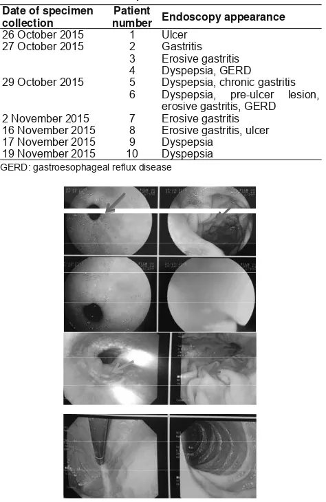
Table 2. List of collected specimens
Date of specimen collection
Patient
number Endoscopy appearance
26 October 2015 1 Ulcer
27 October 2015 2 Gastritis
3 Erosive gastritis
4 Dyspepsia, GERD
29 October 2015 5 Dyspepsia, chronic gastritis 6 Dyspepsia, pre-ulcer lesion,
erosive gastritis, GERD
2 November 2015 7 Erosive gastritis
16 November 2015 8 Erosive gastritis, ulcer
17 November 2015 9 Dyspepsia
19 November 2015 10 Dyspepsia
GERD gastroesophageal reÀu[ disease
Table 1. Primer used to amplify cagA and vacA gene allele s14,9
Gene Oligonucleotide Product size (bp) PCR conditions
cagA 5’-GATAACAGGCAAGCTTTTGAGG-3’
PCR: polymerase chain reaction; cagA: cytotoxin associated gene A; vacA: vacuolating cytotoxin gene A
Table 3. Examination results
GERD: gastroesophageal reÀux disease
From the study results, in PCR e[amination, we only obtained one patient positive for vacA gene, meanwhile for cagA gene, none was positive among all ten patients. Additional data, the results of patients’ biopsy, was obtained one week after PCR e[amination had been conducted. It is e[pected that additional data did not inÀuence researcher during performing the e[amination. Therefore, the results were e[pected to be objective.
Furthermore, researcher compared the results of e[amination with clinical manifestations suffered by patients. VacA gene is known to bring symptomatic characteristic in which symptoms suffered by the patient is more dominant and severe compared to H. pylori infection which did not have this particular gene. Nonetheless, from the e[amination, we found patient number 4 was positive for vacA but revealed negative cagA whose clinical manifestation was in the form of dyspepsia and GERD. If we compare patients in whom we found pre-ulcer, ulcer, and erosion lesions in their gaster endoscopy, they showed negative results for these two particular genes. E[amination with different DNA concentration and different temperature also showed the same results.
In the ¿rst patient, we found ulcer appearance. By the appearance of seemingly signi¿cant clinical manifestations, we e[pected that the results of PCR e[amination would reveal gene which brings tendency characteristic to cause quite severe infection. However, in this case, researcher has not obtained the results. Similarly, patient number eight had clinical manifestations of erosive gastritis and ulcer, however, e[pressions of cagA and vacA gene were not found. But, in patient number four whose clinical manifestations were not as severe as patient number one and eight, we found vacA gene and had not found cagA gene. Researcher considers the presence of other factors which caused severe infection without gaining cagA and vacA gene e[pression as e[pected.
The appearance of relatively severe clinical manifestations in several patients was possibly caused by the use of drugs that may increase gastric acid secretion, for e[ample NSAID. The consumption of drugs continuously without physicians’ supervision may lead to the appearance of lesion which was further aggravated by bacterial infection.
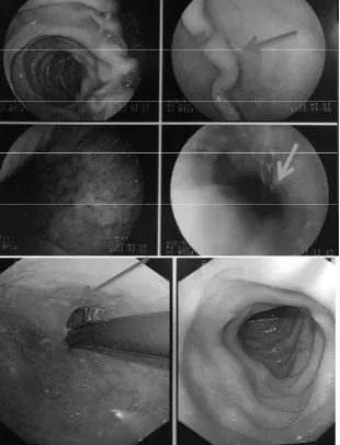
In addition, researcher evaluated patients’ medical records to obtain further information. From the results of patients’ biopsy, 8 patients revealed negative results for H. pylori e[amination using diff count method, and no data was available in 2 patients. It is )LJXUH (QGRVFRSLF DSSHDUDQFH UHYHDOHG SUHXOFHU OHVLRQ
hyperaemia, and erosion
)LJXUH (QGRVFRSLF DSSHDUDQFH VKRZHG SUHXOFHU OHVLRQ
erosion, and hyperaemia
presumed that these patients did not take the results of e[amination, thus the biopsy results were not reported in their medical records. Comparing biopsy results with PCR e[amination for cagA and vacA gene, we found discrepancy for patient number 4 who showed positive result for vacA gene, while the result of biopsy showed negative for H. pylori. Further study should be performed to identify if PCR e[amination of faecal specimen which tends to show high sensitivity allows for biopsy e[amination with false negative results. In this particular matter, another new question arose regarding the possibility of methods during biopsy sample collection, specific location where biopsy sample was taken, as well as the biopsy sample e[amination.
The subjects in this study performed self-sample collection. DNA e[traction was done using particular kit. QIAamp (Qiagen) stool kit is known to have high sensitivity, which is 98.8%. However, DNA e[traction from faeces has metagenome characteristic; thus, it has low speci¿city. Possible presence of other bacteria other than H. pylori is an obscuring factor to identify the gene which infects the patient.
CagA and vacA genes are very speci¿c; therefore, it is not impossible that the gene brought by the bacteria cannot be identi¿ed through PCR e[amination, particularly if specimen is obtained from patients’ faeces. Higher specificity will be obtained if e[amination was performed using biopsy specimen from the patients’ stomach.
DISCUSSION
This study was a descriptive study using PCR e[amination to determine cagA and vacA gene e[pression in patients who described clinical manifestations with suspicions of H. pylori infection. Researcher performed PCR e[amination to specimens from ten patients who underwent endoscopy e[amination in Dr. Soetomo General Hospital, Surabaya from 26 October to 19 November 2015.
From the results of study with study subjects of patients who underwent endoscopy in Dr. Soetomo General Hospital, Surabaya, we found one patient positive for vacA but negative for cagA and in the other nine patients, none of both gene e[pressions was found, either cagA or vacA. The patient who gave positive result for vacA gene e[pression was patient with clinical manifestation of dyspepsia and GERD which, if compared with other patient, had the tendency for milder degree of infection. Similar to
several patients with erosive gastritis, pre-ulcer and ulcer lesions, gene e[pressions of either cagA or even vacA was not found. In this study, cagA or even vacA gene could not show the severity of patient’s clinical manifestation, but might describe that those strains were present in dyspepsia patients.
A study conducted by Sulaksma et alin November 2011 - April 2012 in Makassar revealed that from 35 paediatric patients who were e[amined with PCR method, only 9 patients were positive for H. pylori; this meant that only 25,71% from total sample was found to have positive results. This percentage also described that if 10 patients’ samples were taken, at least 2 patients would show positive results for H. pylori. In this study, e[amination of glmM and cagA genes were performed. However, in this study, they did not state which criteria were being used to rule in positive result. In this case, it was not written if both genes appear or only one of them was enough to be used as a reference that the patients’ specimens were positive for H. pylori.10
Many factors support the struggle in obtaining minimal results of 2 positive samples for cagA or vacA gene from 10 e[amined samples. First, during specimen transport process. Based on the clinical laboratory guideline book, stated that fresh faeces only could stand for < 1 hour if stored in room temperature. Meanwhile, for transport > 1 hour, it need to be stored in 2-40C. Therefore, to support this transportation process, researcher prepared ice bo[ with dry ice with storing temperature of 20C. After sample collection, researcher immediately stored the samples in the refrigerator with temperature of -800 C.8 But, in this case, there were limitations, such as specimen collection time by patient which could not be predicted, thus faeces could not be stored immediately in temperature of -800 C. Outdoor temperature which could reach 380C also did not make it impossible that temperature change was present during temporary storage in the ice bo[.
Second, as H. pylori is not a bacteria located in the intestine, it has small possibility that DNA concentration can be found in faeces. Although PCR e[amination has quite high sensitivity and speci¿city. Based on the study performed by Falsa¿ T et alin 2009, the success rate to detect this bacterium from faeces varied, from 25% to 100%.11
results. In this study, researcher had not obtained the most possible annealing temperature, thus e[amination still required a gradient. Therefore, due to limited resources and cost, researcher could not determine precisely the annealing temperature to obtain positive results.11
In their study, Falsa¿ T et aladded that to obtain good results, a researcher must know the characteristic of transmission and severity of H. pylori in patients’ gaster. However, in this study, considering patients’ endoscopy appearance and results of PCR e[amination, there was no inclination that in more severe infection, both cagA and vacA genes were found to support the appearance of patients’ clinical manifestations. Falsa¿ et al also said the need of adding more than one primer to increase detection rate from the desired DNA.11
Several journals stated different nucleotide base for each gene. In the study conducted by C h a t t o p a d h y a y e t a l i n I n d i a , t h e y u s e d 5 ’ - AT G G A A ATA C A A C A A A C A C A C - 3 ’ f o r Forward vacA s1/s2 primer with 259 bp and 5’-GTTGATAACGCTGTCGCTTC-3’ for Forward cagA primer with 350 bp.12 Later, a study performed by Falsafi T et al used the same Forward vacA primer with the study done by Chattopadhyay et al, but used different Forward cagA primer, which was 5'AATACACCAACGCCTCCA3' with size of 400 bp.11 Nonetheless, in this study, researcher used the same Forward vacA primer, but used different Forward cagA primer. This was similar with the study done by Syaifudin et al in 2006. As this study was performed in Jakarta, researcher chose to adjust with the aforementioned study based on consideration of geographic location. For Forward vacA gene, similar with previous studies either in India or Iran correspond to the consideration of being in Asian continent.9
Results of study by Chattopadhyay et al in 2004 obtained 75.6% positive and study performed by Falsa¿ T et al in 2009 determined sensitivity of 62.5% and speci¿city 92.3%. Meanwhile, from study by Syaifudin et al in 2006 5.47% positive results were obtained, but was taken from biopsy specimen.9
Argyros et al in 2000 conducted a study to compare 5 methods being used for DNA e[traction. This study measured the sensitivity in CFU/mL and obtained that QIAamp kit method e[hibited high sensitivity result, which was 8.0 [ 102 CFU/mL for pure H. pylori and 7.0 [ 103 CFU/mL for e[tracted H. pylori from faecal specimen. In their study, they also stated that to attain positive results from faecal specimen, at least [ &)8P/ZDVQHHGHG VSHFLPHQ RU HYHQ IURP SDWLHQWV¶ IDHFDO VSHFLPHQ 7KHUHIRUHLWZDVQRWLPSRVVLEOHWKDWERWKH[DPLQDWLRQV JDYHGLIIHUHQWUHVXOWV
CONCLUSION
Based on the results of the study, we concluded that PCR e[amination through gene e[traction from patients’ faeces specimen did not ¿nd cagA gene which is a type I strain H. pylori, while vacA gene was found in one patient which is a type II strain H. pylori.
REFERENCES
1. Bunnet NW, Lingappa VR. Penyakit Gastrointestinal. In: McPhee SJ, Ganong WF, eds. Pato¿siologi penyakit pengantar menuju kedokteran klinis. 5th ed. Jakarta: Penerbit Buku Kedokteran EGC 2010.p.363-418.
2. Simadibrata M, Makmun D, Abdullah M, Syam AF, Fauzi A, Maulahela H, et al. Konsensus Dispepsia dan Helicobacter pylori 2014 [serial online] 2014 [cited 2015 September 2]. Available from: URL: http://pbpgi.or.id/wp-content/ uploads/2015/10/ Konsensus-Dispepsia-dan-Helicobater-Pylori-2014.pdf.
3. Enroth H, Kraaz W, Engstrand L, Nyrén O, Rohan T.
Helicobacter pylori strains types and risk of gastric cancer: a case control study [serial online] 2000 [cited 2016 January 17]. Available from: URL: http://cebp.aacrjournals.org/ content/9/9/981.full.
4. Kawahara T, Teshima S, Oka A, Sugiyama T, Kishi K, Rokutan K. Type I Helicobacter pylori Lipopolysaccharide stimulated toll-like receptor 4 and activates Mitogen O[idase 1 in gastric Pit cells [serial online] 2001;69:4382–9. [cited 2014 July 10]. Available from: URL: http://www.ncbi.nlm.nih.gov/pmc/ articles/PMC98510/.
5. Gillespie SH, Bamford KB. At a glance mikrobiologi medis dan infeksi. In: Astikawati R, Tania S, eds. 3rd ed. Jakarta: Penerbit Erlangga 2007.p.8-30.
6. Shahamat M, Alavi M, Watts JEM, Gonzales JM, Sowers KR, Maeder DW, et al. Development of two PCR-based techniques for detecting helical and coccoid forms of Helicobacter pylori
[serial online] 2004;42:8 [cited 2014 July 20]. Available from: URL: http://jcm.asm.org/content/42/8/3613.full.pdf+html. 7. Budiarto, Eko. In: Syahril S, Dayyana TM, eds. Metodologi
penelitian kedokteran sebuah pengantar. Jakarta: Penerbit Buku Kedokteran EGC 2003.p.28-57.
Dehydrogenase gene for detection of Helicobacter pylori in feces. [serial online] 2000 October [cited 2015 September 2]. Available from: URL: http://www.ncbi.nlm.nih.gov/pmc/ articles/PMC87470/.
9. Syaifudin M, Tetriana D, Nurhayati S, Darlina, Purnami, S, Ramadhani D. Aplikasi isotop dan radiasi untuk deteksi
Helicobacter pylori dan resistensi Mycobacterium tuberculosis
serta pengembangan vaksin malaria. [serial online] 2010 [cited 2014 July 18]. Available from: URL: http://digilib.batan.go.id/ ppin/katalog/¿le/MukhBSyaifudin.pdf.
10. Sulaksma W, Sukardi, Razak A, Mutaqqin Z. Deteksi
Helicobacter pylori pada anak menggunakan teknik PCR dan kultur feces. [serial online] 2014; Vol. 41 No.1 [cited 2015 November 10]. Available from: UL: http://www.kalbemed. com/Portals/ 6/10B212Deteksi%20Helicobacter%20 pylori%20pada%20Anak%20Menggunakan%20Teknik%20 PCR%20dan%20Kultur%20Feses.pdf.
11. Falsa¿ T, Favaedi R, Mahjoub F, Naja¿ M. Application of Stool-PCR test for diagnosis of Helicobacter pylori infection in children [serial online] 2009 Jan 28; 15:484–8 [cited 2015 September 2]. Available from: URL: http://www.ncbi.nlm. nih.gov/ pmc/articles/PMC2653372/.
12. Chattopadhyay S, Patra R, Ramamurthy R, Chowdury A, Santra A, Dhali GK, et al. Multiple[ PCR assay for rapid detection and genotyping of Helicobacter pylori directly from biopsy specimens. [serial online] 2004;42:2821–4 [cited 2015 September 2]. Available from: URL:http://www.ncbi.nlm.nih. gov/pmc/articles/PMC427847/.