ES
BIT
T
ET
A
L.:
A
ZE
N
D
O
H
SA
U
RU
S M
AD
AG
AS
KA
RE
N
SIS
A
M
N
H
BU
LL
ET
IN
3
98
20
15
BULLETIN OF THE AMERICAN MUSEUM OF NATURAL HISTORY
POSTCRANIAL
O
STEOLOGY OF
A
ZENDOHSAURUS
MADAGASKARENSIS
(?MIDDLE TO
U
PPER
TRIASSIC,
ISALO
G
ROUP, M
ADAGASCAR) AND I
TS
S
YSTEMATIC
POSITION AMONG
STEM
A
RCHOSAUR
R
EPTILES
STERLI N G J. N ESBIT T, J O H N J. FLYN N,
A DA M C . PRITCH A RD, J. M I CH A EL PA RRISH,
LOVA SOA R A N I VO H A RI M A N A N A, A N D A N D RÉ R . W YSS
American Museum NovitatesBulletin of the American Museum of Natural History
Anthropological Papers of the American Museum of Natural History
Publications Committee Robert S. Voss, Chair Board of Editors
Jin Meng, Paleontology
Lorenzo Prendini, Invertebrate Zoology Robert S. Voss, Vertebrate Zoology Peter M. Whiteley, Anthropology Managing Editor
Mary Knight
Submission procedures can be found at http://research.amnh.org/scipubs
O n t h e c o v e r :
Reconstruction of
Azendohsaurus
madagaskarensis
from the
?
Middle to Upper Triassic, Isalo
group, Madagascar. Drawing by Matth Celeskey.
All issues of Novitates and Bulletin are available on the web (http://digitallibrary.amnh. org/dspace). Order printed copies on the web from:
http://shop.amnh.org/a701/shop-by-category/books/scientific-publications.html or via standard mail from:
American Museum of Natural History—Scientific Publications Central Park West at 79th Street
New York, NY 10024
AZENDOHSAURUS MADAGASKARENSIS
(?MIDDLE TO UPPER TRIASSIC, ISALO GROUP,
MADAGASCAR) AND ITS SYSTEMATIC POSITION
AMONG STEM ARCHOSAUR REPTILES
STERLING J. NESBITT
Center for Integrative Research, Field Museum of Natural History;
Division of Paleontology, American Museum of Natural History;
Department of Geosciences, Virginia Polytechnic Institute and State University,
Blacksburg
JOHN J. FLYNN
Richard Gilder Graduate School and Division of Paleontology
American Museum of Natural History
ADAM C. PRITCHARD
Department of Anatomical Sciences, Stony Brook University,
Stony Brook, New York
J. MICHAEL PARRISH
College of Science, San Jose State University, San Jose, California
LOVASOA RANIVOHARIMANANA
De´partment de Pale´ontologie et d’Anthropologie Biologique,
Universite´ d’Antananarivo, Madagascar
ANDRE
´ R. WYSS
Department of Geological Sciences, University of California at Santa Barbara;
Division of Paleontology, American Museum of Natural History
BULLETIN OF THE AMERICAN MUSEUM OF NATURAL HISTORY
Number 398, 126 pp., 81 figures, 12 tables Issued December 7, 2015
Abstract . . . 4 Introduction . . . 4 Institutional acronyms . . . 5 Methods . . . 6 Geological context . . . 7 Systematic paleontology . . . 9 Archosauromorpha . . . 9 Allokotosauria . . . 9 Azendohsaurus . . . 10 Azendohsaurus madagaskarensis . . . 10 Holotype . . . 10 Paratypes . . . 10 Referred Material . . . 10 Locality . . . 10 Revised Diagnosis . . . 11 Description . . . 11 Axial Skeleton . . . 11
Vertebral column (general) . . . 11
Cervical vertebrae . . . 13 Trunk vertebrae . . . 21 Sacral vertebrae. . . 25 Caudal vertebrae . . . 27 Ribs . . . 31 Gastralia . . . 33 Chevrons . . . 33 Pectoral girdle . . . 35 Clavicle . . . 35 Interclavicle . . . 36 Scapula . . . 37 Coracoid . . . 39 Forelimb . . . 40 Humerus . . . 40 Radius . . . 42 Ulna . . . 43 Manus . . . 44 Radiale . . . 47 Intermedium . . . 48 Ulnare . . . 50 Lateral centrale . . . 51 Medial centrale . . . 51
First–fourth distal carpals. . . 51
Metacarpals . . . 53 Manual digits . . . 54 Terminal phalanges . . . 55 Pelvic girdle . . . 57 Ilium . . . 57 Pubis . . . 60 Ischium . . . 61 Hind limb . . . 62 Femur . . . 62 2
Pes. . . 66 Astragalus . . . 68 Calcaneum . . . 73 Distal tarsals . . . 75 Metatarsals . . . 76 Pedal digits . . . 78 Phylogenetic analysis . . . 79
Taxon and character sampling . . . 79
Tree search strategy . . . 81
Results and Discussion . . . 81
Allokotosauria: tree topology and clade support . . . 84
Allokotosauria+Archosauriformes. . . 84 Allokotosauria . . . 85 Azendohsauridae+Trilophosauridae . . . 86 Azendohsauridae . . . 86 Trilophosauridae . . . 87 Trilophosaurus. . . 88
Implications for the divergence of Archosauromorpha . . . 92
Convergence and the prevalence of herbivory in Triassic Archosauromorpha . . . 93
Conclusions and perspective . . . 96
Acknowledgments . . . 97
References . . . 97
Appendix 1. Referred material ofAzendohsaurus madagaskarensis. . . 106
Appendix 2. The manus ofTrilophosaurus buettneri. . . 108
Appendix 3. Extended description of newly added terminal taxa in the phylogenetic analysis . . . 111
Appendix 4. Phylogenetic characters . . . 113
Appendix 5. Character scores . . . 124
ABSTRACT
During the Triassic, archosauromorphs became one of the first groups of diapsid reptiles to diversify in terms of body size and morphological disparity in both terrestrial and marine ecosystems across Pangaea. This seemingly rapid divergence, and the numerous unique body plans stemming from it, concomitantly has confounded reconstructions of archosauromorph relationships. Teasing apart homology from homoplasy of anatomical characters in this broad suite of body types remains an enormous challenge with the current sample of taxa. Here, we present the postcranial anatomy ofAzendohsaurus madagaskarensis, an early archosauromorph from ?Middle to Upper Triassic strata of Madagascar.Azendohsaurus madagaskarensisis known from nearly the entire skeleton in an ontogenetically variable sample. The holotype locality consists of a monotypic bone bed; preservation ranges from complete but disarticulated bones to articulated sections of the skeleton. Azendohsaurus madagaskarensis embodies an aberrant constellation of archosauromorph features, including an elongated neck, a short, stocky tail, robust limbs, and unexpectedly short digits terminating in large recurved unguals on the manus and pes. Together with the cranium, the postcrania revealA. madagaskarensisto be another representative of a growing coterie of highly apomorphic and bizarre Triassic archosaur-omorphs. At the same time, recovery and description of the full anatomy ofA. madagaskarensis
helps to identify a monophyletic grouping of specialized taxa that includes the North American Late Triassic–aged archosauromorphs Trilophosaurus, Spinosuchus, and Teraterpeton, Indian
Pamelaria, and MoroccanAzendohsaurus laaroussii. Moreover, information derived from the skeleton of A. madagaskarensis solidifies the systematic position of these taxa among other archosauromorphs. Using the most comprehensively sampled phylogenetic analysis of early archosauromorphs, we found the clade encompassing the aforementioned taxa as the nearest outgroup of Prolacerta broomi + Archosauriformes. The newly recognized clade containing
Azendohsaurus, Trilophosaurus, Spinosuchus, Pamelaria, and Teraterpeton demonstrates high morphological disparity even within a closely related group of archosauromorphs, underscores the polyphyly of protorosaurs (5prolacertiforms), and suggests that most major divergences within this group occurred in the Triassic. Furthermore, our results indicate that craniodental character states ascribed to a herbivorous diet were much more pervasive across Triassic Archosauromorpha than previously conjectured.
INTRODUCTION
The initial diversification of Archosauria during the Triassic has been the focus of voluminous research (e.g., Romer, 1956; Charig, 1980; Benton, 1983; Gauthier, 1986; Benton and Clark, 1988; Sereno, 1991; Parrish, 1993; Juul, 1994; Nesbitt, 2003, 2011; Nesbitt et al., 2011; Brusatte et al., 2008; 2011; Butler et al., 2011) because this clade: (1) includes two important extant groups, crocodylians and birds; (2) is a classic example of an adaptive radiation; and (3) has recently been the focus of many ground-breaking macroevolutionary studies. Arch-osauria is just one component of a larger radiation of Archosauromorpha (stem arch-osaurs and the crown clade Archosauria) that arose either just prior to the end of the Permian or soon after the end-Permian extinction (Ezcurra et al., 2014). Stem archo-saur interrelationships have received some attention (Benton, 1985; Gauthier et al.,
1988a, 1988b; Dilkes, 1998; Muller, 2004; Ezcurra et al., 2014; Pritchard et al., 2015), but a consensus is elusive. Disagreement about relationships partially reflects the tremendous divergence among taxa (e.g.,Trilophosaurus,
Tanystropheus, long-necked “protorosaurs”) with extremely long ghost lineages. Moreover, the membership of Archosauromorpha re-mains fluid. Currently, it is unclear whether turtles (Cao et al., 2000; Bhullar and Bever, 2009), drepanosaurids (Dilkes, 1998), saurop-terygians (Caldwell, 1996), or other extinct clades (e.g., Ichthyosauria; Caldwell, 1996) are archosauromorphs. Placement of these clades at the base of Archosauromorpha would have a profound influence on our understanding of character evolution and macroevolutionary patterns within the group. Even relationships among the clades that are clearly part of Archosauromorpha, such as archosauriforms, rhynchosaurs, and tany-stropheids, are challenging because these
clades appear “fully derived” at their first appearance in the fossil record, providing few clues to assist in teasing out plesiomorphy from homoplasy and derived character states. Additionally, bizarre taxa such as Trilopho-saurusandTeraterpetondo not clearly fit into any of the monophyletic components of the clade. The problem of the phylogenetic place-ment of early archosauromorph taxa often owes less to incomplete skeletal records of these taxa (although that is sometime a major factor) than to a lack of understanding about which character states are plesiomorphic for Archosauromorpha.
To help resolve uncertainties involving character transformations among early arch-osauromorphs, we continue our morpho-logical description of the early archosau-romorph Azendohsaurus madagaskarensis
(fig. 1) from the ?Middle to Upper Triassic Isalo Group of Madagascar. Our most recent work (Flynn et al., 2010) onA. madagaskar-ensis focused on naming the taxon, a de-scription of its cranial anatomy, and a re-jection of the taxon’s previously accepted dinosaurian affinities (Dutuit, 1972; Gauffre, 1993; Flynn et al., 1999). The cranial morphology of A. madagaskarensis revealed an enigmatic taxon combining a patchwork of seemingly dinosaurian dental characters and various plesiomorphic characters indica-tive of an earlier-diverging position within Archosauromorpha (Flynn, et al., 2010). Herein we describe and thoroughly illustrate the known, almost entire, postcranial skele-ton of A. madagaskarensis. We provide a complete diagnosis for the taxon, compare it to other early archosauromorphs, and determine its affinities based on the most
comprehensive phylogenetic analysis focused on Triassic noncrown archosauromorphs undertaken to date. Finally, we discuss the implications of the phylogenetic placement of
A. madagaskarensison our understanding of the relationships of other aberrant archo-sauromorphs. We also use the well-preserved material ofA. madagaskarensisto reinterpret portions of the skeletal anatomy of several other archosauromorphs.
In the description below, we compare
Azendohsaurus madagaskarensis to many archosauromorphs, but preferentially focus our comparisons on the Triassic archosaur-omorphs with the most complete skeletal records, such as Trilophosaurus buettneri,
Mesosuchus browni, Proterosuchus fergusi,
Protorosaurus speneri, and Tanystropheus longobardicus.
INSTITUTIONAL ACRONYMS AMNH American Museum of Natural
His-tory, New York
BP Evolutionary Studies Institute (for-merly Bernard Price Institute for Palaeontological Research), Univer-sity of the Witswatersrand, Johan-nesburg, South Africa
FMNH Field Museum of Natural History, Chicago, Illinois
GPIT Pala¨ontologische Sammlung der Universita¨t Tu¨ bingen, Tu¨ bingen, Germany
GR Ruth Hall Museum of Paleontolo-gy, Ghost Ranch, New Mexico ISIR Geological Studies Unit of the
Indian Statistical Institute, Kolkata, India
IVPP Institute of Vertebrate Paleontology and Paleoanthropology, Beijing, China
MCSN Museo Civico di Storia Naturale di Milano, Milan, Italy
MCSNB Museo Civico di Storia Naturale Enrico Caffi, Bergamo, Italy MCZ Museum of Comparative Zoology,
Cambridge, Massachusetts
MFSN Museo Friulano di Storia Naturale, Udine, Italy
MNHN (and MTD)
Muse´um National d’Histoire Natu relle, Paris, France
NHMUK Natural History Museum, London, United Kingdom
NMK Naturkundmuseum im Ottoneum, Kassel, Germany
NMMNHS
New Mexico Museum of Natural History and Science, Albuquerque, New Mexico
NMQR National Museum, Blomfontein, South Africa
NMT National Museum of Tanzania, Dar es Salaam, Tanzania
NSM Nova Scotia Museum of Natural History in Halifax, Nova Scotia, Canada
PIMUZ Pala¨ ontologisches Institut un Muse-um, Zu¨ rich, Switzerland
PIN Paleontological Institute of the Rus-sian Academy of Sciences, Moscow, Russia
PVSJ Divisio´ n de Paleontologı´a de Ver-tebrados del Museo de Ciencias Naturales y Universidad Nacional de San Juan, San Juan, Argentina SAM Iziko South African Museum, Cape
Town, South Africa
SMNS Staatliches Museum fu¨r Natur-kunde, Stuttgart, Germany
TTU-P Museum of Texas Tech University, Lubbock, Texas
UA Universite´ d’Antannanarivo, An-tannanarivo, Madagascar
UCMP University of California Museum of Paleontology, Berkeley, California UFRGS Instituto de Geocieˆncias,
Universi-dade Federal do Rio Grande do Sul, Porto Alegre, Brazil
UMMP University of Michigan Museum of Paleontology, Ann Arbor, Michi-gan
USNM National Museum of Natural His-tory, Washington, DC
VMNH Virginia Museum of Natural Histo-ry, Martinsville, Virginia
WMsN Westfa¨ liches Museum fu¨ r Natur-kunde, Mu¨ nster, Germany
METHODS PERMITS
Fieldwork was conducted under permit from Ministe`re des Mines de Madagascar and in collaboration with the Ministe`re de l’Enseignement Supe´rieur et de la Recherche Scientifique de Madagascar.
FIELDWORK ANDPREPARATION
Field crews from the Field Museum of Natural History, University of California Santa Barbara, and University of Antana-narivo (see Acknowledgments) excavated the type locality ofA. madagaskarensisin south-western Madagascar from 1997 until 1999 (fig. 2). Specimens were excavated with hand tools (e.g., small shovels, rock hammers) using standard paleontological techniques and consolidated with cyanoacrylate glues; they were wrapped in locally obtained tissue paper and then secured by tape, or encapsu-lated in metal cans or plaster jackets when necessary for transport. Specimens were shipped to the Field Museum of Natural History for preparation by staff and volun-teer preparators (see Acknowledgments). The red clay matrix and thin calcium carbonate veneer adhering to the surfaces of fossils were removed with microjacks (www.paleotools. com) and pin-vices holding carbide needles; fine-scale preparation was undertaken under magnifiers or stereomicroscopes. Surface coats of thin B-72 were applied to protect the surfaces of many of the bones. Cyanoac-rylate glues and epoxies (e.g., 2-ton) were used to secure fragile areas or bond breaks.
The material of Azendohsaurus madagas-karensis is permanently housed at the Uni-versity of Antananarivo (including the holo-type and other material) and the Field Museum of Natural History. Most specimens
are referred to throughout the text by their original field collection number (see appendix 1) and these specimen identification numbers will always be kept with the specimens at both institutions. Most specimens perma-nently housed at the University of Antana-narivo were molded, and casts are deposited at the Field Museum of Natural History. Preparation volunteers and staff molded and cast specimens at the FMNH. Elements were embedded in plastaline modeling clay and encased in room temperature vulcanizing tin-cured silicone rubber, typically in two or three part molds. Surfaces were cleaned with acetone applied by brush.
GEOLOGICAL CONTEXT
The geological and geographic provenance of the Azendohsaurus madagaskarensis sam-ple is detailed in Flynn et al. (2010), and consequently just a few of the more pertinent details are highlighted here. The monospecific sample derives from a single, geographically tightly circumscribed locality (M-28) within tens of meters of the east (right) bank of the Malio River immediately west of the western boundary of Isalo National Park, which itself lies between the cities of Ranohira and Sakaraha (prior to the fairly recent discovery of sapphires in Quaternary gravels near
Ilakaka and the ensuing mining boom, Ranohira and Sakaraha were the largest centers of habitation in the region). Fossils ofA. madagaskarensis were recovered along
,100 m of strike of a less than 1 m thick stratigraphic interval. The fossiliferous unit, which dips nearly horizontally, is exposed at midelevation along a,15 m high portion of an uplifted river terrace. As a result, the fossiliferous horizon projects into a steep slope of outcroup exposures. We thus were able to excavate specimens over a swath of only a few meters wide into this slope.
Fossils of A. madagaskarensis have been recovered exclusively from a lens of red, fine-grained sequence of overbank deposits in the limited area just described (M-28) (fig. 3). Reduced (green) ovoid granules and flakes,
#1 cm in diameter, occur sporadically through the fossiliferous mudstone. Several aspects of this occurrence are curious. First, numerous other terrestrial vertebrate taxa (see Flynn et al., 2000, for summary) have been recovered from the same formation from many locations within a 20–30 km radius of the A. madagaskarensis site. Al-though many of these other sites are litho-logically indistinguishable from M-28, none have yielded A. madagaskarensis. Similarly, none of the dozen or so reptile and synapsid taxa (see below) occurring at these other locations are recorded at M-28. Many of the best-preserved specimens from other loca-tions, particularly rhynchosaurs and traver-sodontids, are derived from medium-grained, well-sorted channel sands. Whether the cur-rent restriction of A. madagaskarensis to a single monotypic bone bed, reflects pro-found habitat partitioning between it and other components of the assemblage, a taph-onomic/depositional bias (A. madagaskaren-sis being the largest-bodied taxon in the entire assemblage), behavioral factors, or some other cause, remains to be established.
Azendohsaurus madagaskarensis and other elements of the Triassic assemblage from the Malio River drainage are derived from basal levels of a lithostratigraphical unit that is classically termed the Isalo II (Besairie, 1936; 1972)—of the Isalo Group, or the Makay Formation (Razafimbelo, 1987). The age of the fossiliferous unit is best constrained by vertebrate biostratigraphy of a single cynodont
Fig. 2. Map of the location of the holotype locality ofAzendohsaurus madagaskarensis.
and the absence of certain Late Triassic archosauriforms otherwise found throughout Pangaea. The shared presence of Menadon besairiei in the Makay Formation and the Ladinian Santacruzodon Assemblage Zone (AZ) of the Brazilian Santa Maria Formation suggests that the Makay Formation is also Ladinian or late Middle Triassic in age (Melo et al., 2010; Schultz and Langer, 2010). Moreover, theSantacruzodonAZ is biostrati-graphically correlated with the Chan˜ ares Formation of northwestern Argentina. The latter unit has been recently considered to be late Middle Triassic–early Late Triassic in age by Desojo et al. (2011), a hypothesis further bolstered by radioisotopic dating (Irmis et al., 2013). The possible extension of these South American units into the early Late Triassic and the shared presence of the Massetog-nathus-Santacruzodon clade with the Makay Formation provide further evidence for a late Middle Triassic–early Late Triassic age for the Makay Formation. Additionally, we note that dinosaurs and other archosauriform taxa (e.g., phytosaurs, aetosaurs) typically found in Upper Triassic deposits of neighboring areas (e.g., Maleri Formation of India; Chatterjee, 1978) are absent thus far in the Makay Formation. The Makay Formation thus is almost certainly older (by an unknown span) than units producing the earliest dino-saurs, including the Ischigualasto Formation of Argentina and Santa Maria Series of Brazil (see Flynn et al., 1999; 2000).
As mentioned, the remains of A. mada-gaskarensis have been recovered solely from a monotypic bone bed. Of the hundreds of elements removed from the small quarry, none obviously pertain to any other taxon. Specimens range from incomplete isolated elements to articulated partial skeletons, sometimes preserved in life position (e.g., the upright manus in fig. 3C). As discussed more fully below in conjunction with the
r
Fig. 3. The holotype locality ofAzendohsaurus madagaskarensisnear the Malio River in southern Madagascar (A) during the second year of excavation in 1998, (B) during the third year of excavation in 1999, and (C) a photograph of the articulated right manus (FMNH PR 3820) in situ. All photographs by William Simpson.
description of particular elements, the sample exhibits a size range of roughly 25%from the smallest to the largest specimens, reflecting ontogenetic variation, intraspecific variation, and/or sexual dimorphism; these factors cannot be differentiated from one another in the context of the given sample.
The quality of preservation of the A. madagaskarensis sample varies considerably but is generally excellent. Some postmortem disturbance (by other organisms) and de-formation (by geological processes) is evident in the sample, possibly the result of soft-sediment deformation or trampling. Given the multiple duplicates of most elements in the assemblage, little ambiguity about origi-nal morphology exists. The surfaces of the bones vary from heavily cracked (weathering stage 3 or 4 of Behrensmeyer, 1978; fig. 4A)
to pristine (fig. 4C) suggesting that some were exposed at the surface for many years before burial, whereas others were rapidly and completely buried. In general, the bones with heavily cracked surfaces tend to be isolated, whereas the articulated sections, for the most part, tend to be better preserved. Diagenetic factors may have been responsible for cracking in some specimens (e.g., fig. 4B). The cracking in this specimen (UA 7-12-99-564) is similar to the bones from the Triassic Santa Maria Formation that were cracked and displaced as a consequence of calcite cementation during or after initial fossiliza-tion (see Holz and Schultz, 1998).
SYSTEMATIC PALEONTOLOGY Archosauromorpha Huene, 1946 (sensu
Benton, 1985) Allokotosauria (new)
DEFINITION: The least-inclusive clade
con-taining Azendohsaurus madagaskarensis, Flynn et al., 2010, and Trilophosaurus buett-neri, Case, 1928a, but not Tanystropheus longobardicus, Bassani, 1886, Proterosuchus fergusi, Broom, 1903, Protorosaurus speneri
von Meyer, 1830, or Rhynchosaurus articeps
Owen, 1842 (fig. 5).
DIAGNOSIS: Lateral surface of orbital margin of frontal rugose (char. 237, state 1, abbreviated to 237-1; this simplification is followed throughout the text); posterior side of quadrate head expanded and hooked (207-1); and prominent tubercle developed superi-or to glenoid fossa of scapula (146-0). These unambiguous character states most conspic-uously diagnose Allokotosauria, but are only a subset of the full unambiguous character state set that characterizes the clade (see Results and Discussion).
Fig. 4. The various states of preservation of bones from theAzendohsaurus madagaskarensis. A poorly preserved humerus (UA 8-28-97-143) (A) in ventral view and close-up (right); a heavily cracked cervical vertebra (UA 7-12-99-564) (B) in ventral view and close-up (right); and an exquisitely preserved pedal ungual (FMNH PR 3815) (C) in lateral view and close-up (right). Scales51 cm.
Fig. 5. Illustration of the phylogenetic use of Allokotosauria, Azendohsauridae, and Trilopho-sauridae for this paper. Curved lines mark stem groups.
REMARKS: Here we recognize a clade
containingAzendohsaurus-like taxa (in Azen-dohsauridae) paired with Trilophosaurus-like taxa (in Trilophosauridae). Allokotosauria, meaning “strange reptiles” in Greek, contains a bizarre suite of Triassic-aged archosaur-omorph taxa with a high disparity of craniodental features typically associated with herbivory. Given the previous difficulty with finding a well-supported position of
Trilophosaurus and kin within Archosauro-morpha (Dilkes, 1998), and the currently fluid state of understanding both the inter-relationships of early Archosauromorpha and their placement within Diapsida, we err on the side of caution in naming this clade. As defined, the stem-based clade name Allokotosauria is used to refer to Azendoh-sauridae and TrilophoAzendoh-sauridae and their closest relatives exclusive of other archosaur-omorphs. If Azendohsauridae and Trilopho-sauridae are found to be more distantly related in future analyses, Allokotosauria would thus serve no purpose and should summarily be abandoned. Azendohsauridae and Trilophosauridae currently appear to share a number of apomorphies exclusive of other archosauromorph groups, supporting the monophyly and value of naming this new clade (see below).
Azendohsauridae (new)
DEFINITION: The most inclusive clade containing Azendohsaurus madagaskarensis, Flynn et al., 2010, but not Trilophosaurus buettneri, Case, 1928a, Tanystropheus long-obardicus (Bassani, 1886),Proterosuchus fer-gusi, Broom, 1903,Protorosaurus spenerivon Meyer, 1830, Rhynchosaurus articeps Owen, 1842, or Passer domesticus Linnaeus, 1758 (fig. 5).
DIAGNOSIS: A prominent
anteroposter-iorly oriented ridge is present on the medial surface of the maxilla (201-1); the dorsal apex of the maxilla is a separate, distinct process with a posteriorly concave margin (202-1); the crowns of the upper dentition are lower (i.e., apicobasally shorter) than those of the lower dentition (211-1).
REMARKS: Here we use Azendohsauridae
to refer to Azendohsaurus madagaskarensis -like taxa from the Triassic. This stem-based
definition specifies the most completely known taxon, Azendohsaurus madagaskaren-sis, to distinguish members of Archosauro-morpha that are more closely related to A. madagaskarensis than to any other clade within Archosauromorpha (e.g., Trilopho-sauridae, Rhynchosauria) and thus the name would be applied regardless of whether the phylogenetic position of Azendohsaurus ma-dagaskarensis would change in the future relative to various basal archosauromorphs.
AzendohsaurusDutuit, 1972
Azendohsaurus madagaskarensisFlynn et al., 2010
HOLOTYPE: UA 7-20-99-653 (field number
7-20-99-653) (figs. 3–12), a nearly complete skull with associated vertebrae (fig. 6).
PARATYPES: FMNH PR 2751 (field
num-ber 8-30-98-376), nearly complete disarticu-lated skull (associated with postcranial speci-mens, FMNH PR 2788, FMNH PR 2789, FMNH PR 2792 and possibly FMNH PR 2796).
REFERREDMATERIAL: See appendix 1.
LOCALITY: Basal Isalo IIof Besairie (1972),
termed the Makay Formation by Razafim-belo (1987); drainage of the Malio River, Morondava Basin, southwestern Madagascar (fig. 2). Locality details are on file at the AMNH and FMNH. Cynodonts (Dadadon isaloi, Flynn et al., 2000; Menadon besairiei, Flynn et al., 2000; Chiniquodon kalanoro, Kammerer et al., 2010), rhynchosaurs ( Isa-lorhynchus genovefaeBuffetaut, 1983, What-ley, 2005), a kannemeyeriifrom dicynodont (Flynn et al., 1999), an enigmatic archosaur (Nesbitt et al., unpublished data) and dino-sauromorphs (Kammerer et al., unpublished data) occur in the Makay Formation within a 10 km radius of the Azendohsaurus pro-ducing locality. The stratigraphic relation-ships among these localities are not well
Fig. 6. Elements present in the holotype of
understood because most outcrops are iso-lated and difficult to trace.
REVISEDDIAGNOSIS(see also Flynn et al., 2010): Azendohsaurus madagaskarensis is a medium-sized (2–3 m in length), early-diverging archosauromorph with large pala-tal teeth, a pineal foramen, and lanceolate, or leaf-shaped, teeth. Based solely on the holotype, Azendohsaurus madagaskarensis
differs from all other archosauromorphs in possessing a unique combination of character states, including: ventral curvature of the anterior portion of the dentary; a robust dorsal process of the maxilla, the base of which occurs on the anterior third of the bone; a concave anterior margin of the dorsal process of the maxilla; lanceolate teeth with denticles; a series of small nutrient foramina on the medial surface of the maxilla; elongat-ed cervical vertebrae with small epipophyses dorsal to the postzygapophyses (unknown in
A. laaroussii); and a posteriorly expanded, T-shaped interclavicle (unknown in A. laar-oussii).
Azendohsaurus madagaskarensis is distin-guished from Azendohsaurus laaroussii by a lower maxillary tooth count (11–13, vs. 15–16 for the Moroccan form); apicobasally longer teeth in both maxillae and dentaries; more densely packed serrations in both maxillary and dentary teeth (fig. 7), the absence of a mediolateral swelling at the base of the dentary teeth (fig. 7); and a prominent longitudinal keel on the medial surface of the maxilla present only on the posterior half of the maxilla, as opposed to occurrence along its entire length in A. laaroussii.
Furthermore, we have identified seven additional possible autapomorphies present in the postcranial elements now referred to
Azendohsaurus madagaskarensis (see figures in the description). These include: (1) a small tuber located on the ventrolateral surface of the prezygapophyseal stalk in the middle to posterior cervical vertebrae; (2) a well-defined fossa at the base of the neural spine, just posterior to the prezygapophyses in the second sacral vertebra; (3) the lateral side of the calcaneal tuber expanded laterally and ventrally, with the ventral expansion being clearly visible in proximal view; (4) deep fossae between well-developed laminae in the
posterior cervical vertebrae (autapomorphy among non-archosaurian archosauromorphs, but these structures are found in some crown-group archosaurs andAenigmastropheus par-rintoni [Ezcurra et al., 2014]); (5) hypos-phene-hypantra intervertebral articulations in the posterior cervical, anterior trunk, and sacral vertebrae (autapomorphy among non-archosaurian archosauromorphs, but these structures are found in some crown-group archosaurs); (6) proximal projection on the proximal surface of metatarsal IV; and (7) an oval and proximodistally oriented tuber on the lateral surface of the scapula that nearly contacts the edge of the glenoid fossa.
DESCRIPTION AXIALSKELETON
VERTEBRAL COLUMN: A complete verte-bral column is not available from any single individual of Azendohsaurus madagaskaren-sis. Instead, we present a composite axial column (fig. 8) assembled from short, artic-ulated sections of vertebrae, associated (but not articulated) vertebrae from several in-dividual specimens, and isolated vertebrae found throughout the monospecific bone bed. For example, FMNH PR 2751 and UA 7-20-99-653 both preserve the anterior cervical series, including the atlas and axis— in addition to nearly complete skulls (Flynn et al., 2010).
Full descriptions of the intracolumnar variation of the vertebrae are presented below, but a few general introductory com-ments about the vertebral column of Azen-dohsaurus madagaskarensis are warranted. The neck of A. madagaskarensis is long, as in “protorosaurs” (e.g., Macrocnemus bassa-nii,Prolacerta broomi,Protorosaurus speneri) and in sauropodomorph dinosaurs (e.g.,
Plateosaurus engelhardti), poposauroids (e.g., Xilousuchus sapingensis), and early neotheropods (Coelophysis bauri). The cervi-cal vertebrae immediately posterior to the axis are the longest, maintaining a similar length from the third through fifth cervical elements. Vertebrae become progressively shorter posteriorly, with this trend continu-ing to the pelvis. The anterior articular facets of the anterior cervical vertebrae are elevated
dorsally relative to their corresponding pos-terior articular facets, indicating that the head and neck were raised above the level of the trunk vertebrae.
The shape and height of the neural spines ofAzendohsaurus madagaskarensisare highly variable throughout the axial vertebral col-umn. The neural spines of the anterior
Fig. 7. Comparison between the tooth-bearing elements and teeth ofAzendohsaurus madagaskarensis
(A–B,E–G,J) andAzendohsaurus laaroussii(C–D,H–I). Right maxilla ofA. madagaskarensis(FMNH PR 2756) in (A) lateral and (B) medial views. Left maxilla ofA. laaroussii(MNHN-ALM 364) in (C) medial view with a close up of a posterior maxillary tooth in (D) medial view. Middle to posterior maxillary teeth of A. madagaskarensis (FMNH PR 2751) in (E) lateral view. Left dentary of A. madagaskarensis
(unnumbered) in (F) lateral and (G) medial views. Left dentary ofA. laaroussii(MNHN-ALM 351) in (H) lateral view with a close-up of a posterior dentary teeth in I, lateral view. Middle dentary teeth ofA. madagaskarensis(FMNH PR 2751) inJ, lateral view. Scales51 cm inA–C,E–H,I; scales55 mm inD,I. Arrows indicate anterior direction. Abbreviations:ad,anteroventral deflection;dp,dorsal process;h,heel;
cervical vertebrae are anteroposteriorly long and low, whereas they are anteroposteriorly short and tall on the posterior cervical vertebrae (presacral vertebrae 7–9). Neural spines of the posterior trunk vertebrae (5 dorsals) are inclined anterodorsally, whereas they slant posterodorsally on the anterior caudal vertebrae (fig. 8). All other neural spines are nearly vertically oriented. All centra are amphicoelous. Amphicoely is common among early archosauromorphs (e.g., Gottmann-Quesada and Sander, 2009; Gow, 1975), with procoely occurring in
Trilophosaurus buettneri (TMM 31025-140) and a subclade of Tanystropheidae (Olsen, 1979; Pritchard et al., 2015).
The number of vertebrae in each region of the axial column is difficult to estimate because of gaps in the articulated series of vertebrae throughout the column. We esti-mate 24 presacral vertebrae based on gradual transitions of centrum length, positions of diapophyses and parapophyses, and the sizes and shapes of the corresponding prezygapo-physes and postzygapoprezygapo-physes. Two sacral vertebrae are present, based on the morphol-ogy of the sacral ribs and the two sacral rib scars on the medial side of the ilium (see below). The number of caudal vertebrae is the most uncertain. Nevertheless, the tail of
Azendohsaurus madagaskarensis appears short compared to those of other early archosauromorphs with preserved caudal series, such as Langobardisaurus pandolfii,
Protorosaurus speneri, and Trilophosaurus buettneri. We estimate the caudal vertebral count to be between 45 and 55, with centra decreasing in length distally.
Postaxial vertebral intercentra do not occur in Azendohsaurus madagaskarensis,
based on three lines of evidence. First, no intercentra have been found between any of the articulated segments of presacral verte-brae. Second, the ventral edges of the anterior and posterior articular faces of the
presacral centra are not beveled to any degree, leaving no room for intercentra. Third, no isolated postaxial intercentra have been recovered from the extensive, multi-individual, monospecific bone bed. Postaxial intercentra also are absent in Tanystrophei-dae (see Pritchard et al., 2015) and Arch-osauria (Nesbitt, 2011).
Vertebrae of A. madagaskarensis vary in size by roughly 25%across individuals in the sample. In all instances, however, the neural arch is completely fused to the centrum and no suture between the two vertebral compo-nents is apparent. It should be noted that neurocentral fusion occurs early in ontogeny in some other early archosauromorphs (see Nosotti, 2007). In some cases, a slightly raised rim demarks the contact between the neural arch and the centrum, but, even in those vertebrae, the suture is completely obliterated. Thus, given the size variation, in spite of fusion without evidence of a suture,
A. madagaskarensis continued to grow after the neural arch and centrum had fused (see below).
CERVICAL VERTEBRAE: The complete atlas and axis are best preserved as disarti-culated elements in the holotype UA 7-20-99-653 (fig. 9A–D) and as partial specimens in FMNH PR 3823 (fig. 9E–F) and FMNH PR 3818. Originally in articulation, the atlas and axis of UA 7-20-99-653 were partially dis-articulated for study, whereas in FMNH PR 3823 these elements remain articulated, but are missing the atlantal neural arch and proatlas. The atlas of A. madagaskarensis
consists of six elements: paired proatlantes, paired neural arches, the atlantal centrum (fused to the axial intercentrum), and the atlantal intercentrum. The left proatlas lies on the dorsal surface of the neural arch of the atlas whereas the right element lies within the foramen magnum of the skull of specimen UA 7-20-99-653 (labeled “pa” in Flynn et al., 2010: fig. 2). The dorsoventrally compressed,
oval proatlas is similar to that in other archosauromorphs, such as Trilophosaurus buettneri (TMM 31025-140) and the croco-dylomorph Hesperosuchus agilis (Colbert, 1952). Proatlas elements are present in a variety of diapsids as either paired elements that lack bone-to-bone articulations (e.g.,
Sphenodon) or as fused bones (e.g., crocody-lians) (Romer, 1956), but these are rarely preserved. The left atlantal neural arch of UA 7-20-99-653 appears complete, and in-cludes a prominent, winglike posterior ex-pansion at its dorsal end. In articulation, this posterior process lies on the anterolateral surface of the neural arch of the atlas, a condition widespread in reptiles (e.g., Reisz, 1981; Broili and Schroeder, 1934). A thin ridge lies on the dorsal surface of the posterior process and terminates in a point posteriorly. Medially the atlantal neural arches are dorsoventrally thin, failing to meet at the midline in UA 7-20-99-653. The atlantal intercentrum, a prominent crescent-shaped structure in anterior view, articulates with the ventral surfaces of the two neural arches, and posteriorly with the atlantal centrum. The anterior surfaces of the atlantal intercentrum and the two neural spine elements form a hemispherical concavity for articulation with the occipital condyle. The convex posterior surface of the atlantal intercentrum mirrors a concave surface of the axial centrum just ventral to the odontoid process. No rib attachments occur on the lateral or ventral surfaces of the intercentrum of the atlas, but the presence of ribs attaching elsewhere cannot be ruled out (see below).
The anteroposteriorly foreshortened atlan-tal centrum is atlan-tall and almost completely fused to the intercentrum of the axis (fig. 10), as in Trilophosaurus buettneri(TMM 31025-140). Unfused odontoid complexes occur in
Mesosuchus browni (Dilkes, 1998), Protero-suchus fergusi (Broili and Schro¨ der, 1934), and Tanystropheus longobardicus (Wild, 1973). The anterior surface of the bone in
A. madagaskarensis is complex, with a med-iolaterally expanded crescent-shaped odon-toid process at its dorsal half overlying a crescent-shaped concavity that articulates with the atlantal intercentrum. The odontoid process of A. madagaskarensis is relatively wider than that of Trilophosaurus buettneri
(TMM 31025-140). The posterior face of the atlantal centrum ofA. madagaskarensisforms an ovoid concavity that is taller than wide, articulating with the convex anterior surface of the centrum of the axis. The presence of a rib fragment ventrolateral to the atlantal centrum on the right side suggests that an
Fig. 9. Atlas and axis ofAzendohsaurus mada-gaskarensis. Atlas (UA 7-20-99-653) in (A) anterior and (B) left lateral view. Odontoid process and axis intercentrum (UA 7-20-99-653) in (C) anterior and (D) posterior views. Articulated odontoid, axis intercentrum, and axis (FMNH PR 3823) in (E) left lateral and (F) ventral views. Scales 51 cm. Arrows indicate anterior direction. Abbreviations:
a., articulates with; ain, axis intercentrum; ana,
atlas neural arch;ati,atlas intercentrum;ax,axis;
dp,diapophysis;epi,epipophysis;nc,neural canal;
atlantal rib was present, despite the lack of a clear rib facet on the centrum.
The axis is preserved in the holotype, UA 7-20-99-653, as part of an articulated series (fig. 11), and in FMNH PR 3823 (fig. 9E–F) and FMNH PR 3818. All three axes preserve the axis intercentrum, which contacts the anteroventral surface of the axial centrum. This wedge-shaped element is partially fused to the atlas centrum in UA 7-20-99-653 and FMNH PR 3823, but completely fused in FMNH PR 3818 without any remnants of a suture (fig. 10). In all three specimens, the axis intercentrum remains independent of the centrum. The dorsal extent of the axis intercentrum reaches the ventral extent of the axial diapophysis. The axis parapophysis lies entirely on the axis intercentrum, an arrangement best seen in FMNH PR 3818 (fig. 12). The facets of the parapophysis are directed posteriorly at the ventral edge of the lateral sides of the axis intercentrum.
The broad subrectangular neural spine of the main body of the axis is just over twice as long anteroposteriorly as it is tall. The neural spine inclines slightly posterodorsally and the posterior termination is expanded sligh-tly transversely. The dorsal margin of the axial neural spine is inclined posterodorsally in Proterosuchus alexanderi (NMQR 1484; Broili and Schro¨ der, 1934),Protorosaurus spe-neri (Gottmann-Quesada and Sander, 2009), and Trilophosaurus buettneri (TMM 31025-140). It is inclined anterodorsally in tanystrop-heids (e.g.,Macrocnemus bassanii[MCSN V 457],Tanystropheus longobardicus[PIMUZ T/ 2819)]).
The anterior aspect of the axial neural spine differs in the three Azendohsaurus
specimens, but the anterior end overhangs the rest of the axis in all of them; in UA 7-20-99-653, the anterior end tapers to a point dorsally, whereas in FMNH PR 3823 and FMNH PR 3818 the anterior edge is rounded (fig. 12). The posterior edge of the neural spine also differs among the three specimens; the posterior surface exhibits a midline groove in UA 7-20-99-653 and FMNH PR 3818, whereas in FMNH PR 3823 a thin vertical ridge occurs on the midline.
The anteriorly short, semicircular prezyga-pophyses face dorsolaterally, articulating with the neural arches of the atlas. A slight ridge connecting the lateral edge of the prezygapophysis with the anterior extent of the postzygapophysis (5 interzygapophyseal lamina sensu Wilson, 1999) occurs in all three specimens, but is most prominent in the largest (FMNH PR 3823). The flat articular surfaces of the postzygapophyses are de-flected dorsally about 20u from horizontal in posterior view. Laminae extending from near the dorsal surface of the neural spine to the dorsal margin of the postzygapophyses, and a thinner one between the postzygapo-physes, frame a deep, triangular, and poster-iorly facing fossa at the midline of the neural arch. Epipophyses lie on the dorsal surface of the postzygapophyses but do not extend posteriorly to the articular surface of the postzygapophyses. Similar small projections occur in a variety of early archosauromorphs, including Mesosuchus browni (SAM-PK-5882), tanystropheids (e.g., Wild, 1973; Pritchard et al., 2015),Teraterpeton hrynewi-chorum(Sues, 2003),Trilophosaurus buettneri
(TMM 31025-140), andSpinosuchus caseanus
(Spielmann et al., 2009). The postzygapo-physes extend about 30u laterally from the midline whereas the postzygapophyses of
T. buettneri (TMM 31025-140) project 45u
laterally from the midline. The neural canal is circular in both anterior and posterior views.
The gently convex anterior surface of the axis centrum is subtriangular and pointed ventrally in anterior view. The slightly ventrally directed anterior articular surface of the centrum is convex in UA 7-20-99-653, whereas the ventral half of the anterior face is
Fig. 10. The fused odontoid and axis intercen-trum ofAzendohsaurus madagaskarensis (FMNH PR 3818) in (A) anterior, (B) posterior, and (C) left lateral views. Scale 5 1 cm. Abbreviations: a.,
articulates with; ain, axis intercentrum; ati, atlas intercentrum; ax, axis; nc, neural canal; od,
concave in FMNH PR 3818. In lateral view, the axis centrum is parallelogram shaped, with the anterior articular surface elevated dorsally relative to the posterior articulation, suggesting a curved neck in life. A similar parallelogram-shaped axis is present in Tri-lophosaurus buettneri(Gregory, 1945) and, to a lesser degree, in other archosauromorphs with elongated necks (e.g., Macrocnemus bassanii, MCSN V 457; Prolacerta broomi, BP/1 2675, UCMP 37151; Gow, 1975). In
Azendohsaurus madagaskarensis, the diapo-physis is poorly developed and anteriorly bordered by the atlas centrum. A fossa is located ventral to the diapophysis, but this fossa shallows posteriorly anterior to the anteroposterior midpoint of the centrum. The posterior articular surface of the centrum is deeply concave, to a degree unmatched throughout the rest of the vertebral column. In contrast, this surface is convex in Trilo-phosaurus buettneri (Gregory, 1945) and slightly concave in a South American rhynch-osaur (MCZ 1529). The ventral surface of the centrum bears a distinct ventral keel at the midline in all three preservedAzendohsaurus
axes. In UA 7-20-99-653 and FMNH PR 3818 the keel extends along the entire length of the centrum, whereas in FMNH PR 3823 it is highly incomplete. The ventral keel of FMNH PR 3818 is much more ventrally expanded than in the other specimens (fig. 12).
The anterior cervical vertebrae of A. madagaskarensis occur in articulation in the
holotype (UA 7-20-99-653; fig. 11) and in one larger individual (FMNH PR 2788); they are also known from disarticulated examples throughout the quarry (e.g., FMNH PR 2791; fig. 13). The anterior cervical vertebrae are elongated relative to the mid- to posterior dorsal vertebrae (fig. 8). The axis of UA 7-20-99-653 is considerably shorter than the third through fifth cervical vertebrae, which are all similar in length. A similar pattern occurs in Macrocnemus bassanii (MCSN V 457),Prolacerta broomi(UCMP 37151), and
Trilophosaurus buettneri (TMM 31025-140). In Tanystropheus longobardicus, vertebrae lengthen from the axis to the eighth or ninth cervical vertebra (MCSN BES 1018; Wild, 1973), before shortening to the ultimate (13th) cervical vertebra (Rieppel et al., 2010). The anterior cervical neural spines of A. madagaskarensis are about twice as long (anteroposteriorly) as tall (dorsoventrally). Similarly low anterior cervical neural spines occur inLangobardisaurus pandolfii(MCSNB 2883), Macrocnemus fuyuanensis (MCSN V 457; Jiang et al., 2011), andProlacerta broomi
(UCMP 37151; Gow, 1975). InA. madagas-karensis, the anterior cervical neural spines are transversely compressed into thin blades, resembling those in Prolacerta broomi
(UCMP 37151) andTrilophosaurus buettneri
(TMM 31025-140). The anterodorsal portion of the neural spine overhangs the base of the neural spine in some anterior cervical verte-brae (e.g., the fourth cervical of UA 7-20-99-653; fig. 11) whereas the anterior edge of the
Fig. 11. Articulated cervical vertebrae ofAzendohsaurus madagaskarensis(UA 7-20-99-653) in lateral view. Scale51 cm. Arrow indicates anterior direction. Abbreviations:ain,axis intercentrum;at,atlas;ax,
neural spine is vertical in a much larger anterior cervical vertebra (FMNH PR 2791; fig. 13). The orientation of the posterior edge of the neural spine also varies; the poster-odorsal corner of the neural spine in the fourth cervical vertebra of UA 7-20-99-653 over-hangs the base of the neural spine, whereas the posterior edge of the neural spine is vertical in the fifth cervical vertebra in this specimen and in the larger FMNH PR 2791. The neural spines of the anterior cervicals consistently overhang the anterior margins of their bases in tanystropheids (Peyer, 1937; Wild, 1973; Dilkes, 1998; Pritchard et al., 2015) and in
Prolacerta broomi(BP/1/2675).
The neural arches of the anterior cervical vertebrae bear well developed pre- and postzygapophyses that extend horizontally in lateral view; the articular facets are angled 30u (medially and laterally in the transverse plane, respectively). Shallow fossae occur at the base of the neural spine, between the prezygapophyses anteriorly and the postzy-gapophyses posteriorly. Similarly positioned but substantially deeper pits are known in some tanystropheids (Wild, 1973; Pritchard et al., 2015), and likely represent attachment sites for the interspinous ligaments. In dorsal view, the pre- and postzygapophyses project about 30u (medially and laterally in the transverse plane, respectively), but do not diverge anterolaterally to the degree seen in
Spinosuchus caseanus(Spielmann et al., 2009) and Trilophosaurus buettneri (TMM 31885-140).
In the fourth cervical vertebra of UA 7-20-99-653, a small knob is located on the ventrolateral surface of the prezygapophyseal stalk; this feature is more prominently expressed in FMNH PR 2791 (fig. 13). Knobs are inconsistently present on the anterior cervical vertebrae. For example, the third and fifth cervical elements of the same specimen (UA 7-20-99-653) lack this struc-ture. The knob is also present in other cervical vertebrae (see below) and may represent an autapomorphy of A. madagas-karensis. A ventrally deflected shelf (with respect to the midline) occurs between the postzygapophyses of the third cervical verte-bra of UA 7-20-99-653. This structure is distinct from the well-developed, mediolater-ally horizontal shelf linking the medial
Fig. 12. Variation in the axis ofAzendohsaurus madagaskarensis. Axis of the (A) holotype (UA 7-20-99-653) in left lateral view, (B) FMNH PR 3818 in left lateral view, and (C) FMNH PR 3823 in left lateral view. The 1arrows highlight variation of the anterior portion of neural spine. The2arrows highlight variation of the posterior edge of the neural spine. The 3arrows highlight variation of the ventral keel. Scale51 cm. Abbreviations:ain,
margins of the postzygapophyses in the anterior cervical vertebrae of T. buettneri
(TMM 31885-140) andSpinosuchus caseanus
(Spielmann et al., 2009)—a possible synapo-morphy of those two taxa (Spielmann et al., 2009; see below). Epipophyses occur dorsal to the postzygapophyses but fail to extend posteriorly beyond the articular surface of the postzygapophyses, resembling the condi-tions inLangobardisaurus pandolfii(MCSNB 2883),Macrocnemus bassanii(MCSN V 457), and in tanystropheid cervical vertebrae from the Hayden Quarry (GR 269). A slight extension of epipophyses beyond the level of the articular facet occurs inTrilophosaurus buettneri (TMM 31025-140), and an even greater extension occurs in species of Tany-stropheus(MCSN BES SC 1018; Wild, 1973). A small ridge occurs medial to the post-zygapophysis in FMNH PR 2791. Just anterior and ventral to the articular surface of the postzygapophysis in the anterior cervical of UA 7-20-99-653 lies a small notch, prominently seen in FMNH PR 2791 (fig. 13). A small ridge that projects poster-iorly beyond the lateral wall of the neural canal frames the notch ventrally. Hypo-sphene-hypantrum intervertebral articula-tions are absent in the anterior cervical vertebrae. The neural canal is circular in both anterior and posterior views.
The centra of the third through fifth cervical vertebrae of A. madagaskarensis are three times longer than high (fig. 11), as in
Protorosaurus speneri (Gottmann-Quesada and Sander, 2009) andTrilophosaurus buett-neri (TMM 31025-140). Cervical centra are proportionally more elongate in Tanystro-pheidae (e.g., Wild, 1973; Pritchard et al., 2015), and proportionally shorter in both late-diverging rhynchosaurs (e.g., Montefel-tro et al., 2013) andErythrosuchus africanus
(NHMUK R 3592; Gower, 2003). As with the axis, the cervical centra in A. madagas-karensisare parallelogram shaped, where the anterior articular surface is positioned dorsal to the posterior articular surface in lateral view. The anterior and posterior articular
Fig. 13. Anterior cervical vertebra of Azendoh-saurus madagaskarensis (FMNH PR 2791) in (A) right lateral, (B) ventral, (C) dorsal, (D) anterior, and (E) posterior views. Scale 5 1 cm. Arrow indicates anterior direction. Abbreviations: dp,
diapophysis;epi, epipophysis;kn,knob;nc,neural
r
canal;ns,neural spine;poz,postzygapophysis;pp,
surfaces of the cervical vertebrae are circular and concave. The diapophyses and parapo-physes are situated on pedicles distinct from but located near the anterior rim of the centrum, as in most early archosauromorphs (e.g., Gow, 1975; Gregory, 1945). These articular surfaces are anteroposteriorly elon-gate and concave inA.madagaskarensis. The diapophyses and parapophyses very nearly contact each other in the anteriormost cervicals, but quickly diverge posteriorly in UA 7-20-99-653, as in archosauromorphs basal to Erythrosuchus (Gower, 2003). The diapophysis projects ventrolaterally and the parapophysis laterally. A dorsally convex crescentic fossa indents the centra laterally, originating between the diapophyses and parapophyses and arching posteriorly. The length and relative depth of these fossae increase posteriorly within the vertebral column, as seen in the articulated series of the holotype (UA 7-20-99-653). A weak midline keel occurs ventrally on the anterior cervicals, as in most early archosauromorphs (Pritchard et al., 2015).
Amidcervical vertebra, possibly the fifth or sixth presacral vertebra, of A. madagaskar-ensis is represented in specimen UA 8-28-9-141 (fig. 14). This element is shorter than more anterior vertebrae, but longer than the more posterior cervicals, a pattern similar to
Trilophosaurus buettneri(Gregory, 1945) and
Spinosuchus caseanus(Spielmann et al., 2009). This contrasts with Protorosaurus speneri
(Gottmann-Quesada and Sander, 2009) and Tanystropheidae (Peyer, 1937; Wild, 1973), wherein the midcervical centra are propor-tionally the longest.
InA. madagaskarensis, the neural spines of the midcervicals are shorter anteroposteriorly than those of the anterior cervicals. The neural spines are oval in cross section—the major axis of which is oriented anteroposter-iorly—and their tips are slightly expanded transversely. They are canted slightly anteri-orly, as in some of the more posterior cervical vertebrae (e.g., FMNH PR 3818). The anterior edge of the neural spine is flat, whereas a thin vertical ridge occurs along the midline posteriorly.
Deep, dorsally opening fossae are present at the base of the neural spine between the prezygapophyses and postzygapophyses.
Similar fossae are seen in some archosaur-omorphs (e.g., Erythrosuchus africanus, Gower, 2001;Prolacerta broomi, BP/1/2675). A shallow fossa occurs between the base of the neural spine and the prezygapophyses, and a much deeper one between the base of the neural spine and the postzygapophyses. The articular facets are more inclined in the midcervical than in the anterior cervical vertebrae; the prezygapophyses and postzy-gapophyses are banked 45uto the horizontal plane. A knob similar to that seen in the anterior cervical vertebrae occurs on the lateral side of the prezygapophyses (fig. 14). Deep, anteriorly opening fossae are present lateral to the neural canal, just ventral to the articular facets of the prezygapophyses (fig. 14). Fossae of this kind have not been reported in any non-archosaurian archosaur-omorph to date, but do occur in theropod dinosaurs such as coelophysoids (e.g., AMNH FR 2701, cervical vertebra).
A distinct gap occurs between the pre-zygapophyses of the midcervicals of A. madagaskarensis (fig. 14), resembling the hypantra of saurischian dinosaurs and those of a variety of pseudosuchians (see Nesbitt, 2011). In support of the view that this feature represents a true hypantrum inA. madagas-karensis, a ventrally elongated lamina of bone resembling the hyposphene of sauris-chian dinosaurs is present between the post-zygapophyses in the same midcervical verte-bra. The A. madagaskarensis hypantrum clearly extends ventral to the articular facets of the postzygapophyses and its ventral sur-face is flush with the dorsal border of the neural canal. A hyposphene-hypantrum in-tervertebral articulation in A. madagaskar-ensis, restricted to the midcervical vertebrae, represents the first occurrence of this feature among non-archosauriform archosauromor-phs. Epipophyses adorn the dorsal surface of the postzygapophyses but do not extend posterior to the articular surfaces. A rounded ridge separates the postzygapophyseal artic-ular surface from a shallow, anteroventral fossa.
The centra of the midcervical vertebra in
A. madagaskarensis are less parallelogram shaped than those of the anterior cervical vertebrae. The surface area of the anterior articular surface is larger than the posterior;
both are round. Shallow fossae ventral to the posterior diapophyseal laminae span the length of the centrum laterally. A fine, anterolaterally oriented lamina subdivides this fossa on one side (left) of UA 8-28-97-141. Similar laminae occur in a number of archosauriforms (Butler et al., 2012). The diapophysis projects ventrolaterally. Its con-cave articular surface is smaller than that of the parapophysis. The articular surface of the parapophysis lies on the lateral edge of the anterior articular surface of the centrum. The ventral surface of the centrum bears a midline keel that fails to meet the anterior margin of the centrum.
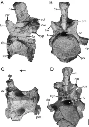
Two isolated posterior cervical vertebrae
(UA 8-30-98-349; fig. 15; FMNH PR 3818; fig. 16) that likely correspond to the eight or
ninth presacral vertebrae of A. madagaskar-ensis are well preserved. The neural spines, positioned on the posterior half of the centrum, are much more transversely ex-panded than those of the anterior cervical vertebrae, resulting in a subcircular cross section. The vertically oriented neural spine flares laterally at its dorsal end but does not form a distinct “spine table” as occurs in phytosaurs (e.g., Butler et al., 2012: fig. 7) and aetosaurs (e.g.,Desmatosuchus spurensis, MNA V9300; Parker, 2008). The edges of the dorsal surface of the neural spine are dis-tinctly convex. A midline ridge is present post-eriorly, whereas the neural spine is concave anteriorly.
As on the midcervicals, the pre- and postzygapophyses of UA 8-30-98-349 are angled about 45u from horizontal (medially and laterally in the transverse plane, re-spectively) and, thus, are comparatively more steeply oriented than in the anterior cervical vertebrae. The pre- and postzygapophyses meet their counterparts at the midline at an acute angle. The dorsal tips of the articular surface of the pre- and postzygapophyses taper to a point; distinct epipophyses occur on the dorsal margin of the postzygapo-physes. Rounded knobs lateral to the pre-zygapophyses are larger than those in more anterior elements of the vertebral column.
A complicated set of laminae (figs. 15, 16) connects the prezygapophyses, postzygapo-physes, diapopostzygapo-physes, and parapophyses of the posterior cervical vertebrae, structures more typical of archosaurs than of basal saurians. Although laminae have been descri-bed in non-archosaurian archosauromorphs (Ezcurra et al., 2014), they are not present among all archosauromorphs. Three deep fossae (anterior, ventral, and posterior) occur at the base of the diapophysis. Employing the terminology of Wilson (1999), a paradiapo-physeal lamina that joins the ventral surface of the diapophyses with the parapophysis together with a vertical centroprezygapophy-seal lamina that connects the ventral portion of the prezygapophysis with the centrum frame a deep anterior fossa just lateral to the neural canal (fig. 16). The ventral fossa, the deepest of the three, is bordered anteriorly by the paradiapophyseal lamina and posteriorly by the centrodiapophyseal lamina that
con-Fig. 14. Midcervical vertebra ofAzendohsaurus madagaskarensis (UA 8-28-97-141) in (A) left lateral, (B) anterior, (C) ventral, and (D) posterior views. Scale 5 1 cm. Arrow indicates anterior direction. Abbreviations: dp, diapophysis; epi,
epipophysis;hyp, hyposphene;kn,knob;nc,neural canal;ns,neural spine;poz,postzygapophysis;pp,
nects the ventral surface of the diapophyses with the posterior portion of the centrum. The fossa posterior to the diapophysis is roofed by the postzygodiapophyseal lamina, which extends from the posterior side of the diapophysis to the postzygapophysis. Simi-lar, but less pronoumced laminae occur in
Trilophosaurus buettneri and Spinosuchus caseanus (Spielmann et al., 2009). In A. madagaskarensis, the diapophyses, which are deflected ventrolaterally at their distal tips, expand laterally, making them club shaped. The articular surfaces are distinctly concave. The parapophysis arises from the anterior margin of the centrum and the major axis of its oval articular surface is vertically oriented.
The centrum is rectangular, lacking any vertical offset between the anterior and posterior surfaces. The anterior and posterior articular surfaces are round, the former much larger than the latter. No ventral beveling is present, in contrast to the condition in
archosauromorphs with vertebral intercen-tra (e.g., Proterosuchus alexanderi [NMQR 1484],Trilophosaurus buettneri[TMM 31025-140]). A shallow fossa occurs laterally near the contact between the neural arch and the centrum. A ventral keel is absent.
TRUNK VERTEBRAE: Trunk vertebrae (5
dorsals; presacral vertebrae ,11–24) of A. madagaskarensis are well represented in the Malio River bone bed, either in articulated series (e.g., FMNH PR 2789, fig. 17) or as isolated elements (e.g., FMNH PR 2779, fig. 18; UA 8-26-98-250, fig. 19).
The anterior trunk vertebrae are best represented by the spectacularly preserved FMNH PR 2779 (fig.18). The neural spine is mediolaterally compressed; its ventral base is about as long (anteroposteriorly) as the centrum, whereas its dorsal margin is one-third shorter, resulting in the anterior margin of the neural spine angled posterodorsally. The dorsal surface of the neural spine is unexpanded and dorsally convex in lateral
Fig. 15. Posterior cervical vertebra ofAzendohsaurus madagaskarensis(UA 8-30-98-349) in (A) right lateral, (B) anterior, (C) posterior, (D) ventral, and (E) dorsal views. Scale 51 cm. Arrows indicate anterior direction. Abbreviations:dp,diapophysis;epi,epipophysis;kn,knob;nc,neural canal;ns,neural spine;poz,postzygapophysis;pp,parapophysis;prz,prezygapophyses.
view. Posteriorly, the neural spine bifurcates ventrally into thin laminae that terminate at the dorsal margin of the postzygapophyses; these laminae and the postzygapophyses together frame a posteriorly oriented inter-spinous fossa at the midline. A similarly positioned, but substantially deeper fossa is known in some tanystropheids (Pritchard et al., 2015). No corresponding fossae occur at the bases of the neural arches between the prezygapophyses in the anterior trunk verte-brae—in contrast to the cervical vertebrae, where one is present. A ventrally deep fossa with mediolaterally oriented striations is present lateral to the base of the neural spine, immediately medial to the articular surface of the diapophysis. This structure is divided by a rounded ridge of bone from a second, more anterior fossa that sits lateral to the base of the neural spine (fig. 18E).
The articular surfaces of the pre- and postzygapophyses are deflected about 45u
to the horizontal (medially and laterally in the transverse plane, respectively), as in the mid- and posterior cervical regions. Weakly developed hyposphene-hypantrum interver-tebral articulations are present in FMNH PR
2779 (fig. 18). The hypantrum (fig. 18) exists as a small gap between the prezygapophyses; it is located roughly half the distance between the anterior ends of the prezygapophyses and the base of the neural spine. The hyposphene arises from a vertically oriented lamina at the midline between the postzygapophyses. This lamina reaches the posterior end of the postzygapophyses and roofs the neural canal. The well-developed laminae between the diapophyses, parapophyses, neural arches, and centra of the anterior trunk vertebrae of Azendohsaurus madagaskarensis resemble those of its posterior cervical vertebrae, as well as the anterior trunk vertebrae of
Erythrosuchus africanus (Gower, 2003), Ae-nigmastropheus parrintoni (Ezcurra et al., 2014), and certain archosaurs (e.g., paracro-codylomorphs, saurischians). The diapophy-sis and prezygapophydiapophy-sis are connected by the prezygadiapophyseal lamina, the parapophy-sis by the paradiapophyseal lamina, the posterior portion of the centrum by the posterior centrodiapophyseal lamina, and the postzygapophysis by the postzygapodia-pophyseal lamina (fig. 18). As in the poste-rior cervical vertebrae, three well-defined
Fig. 16. Posterior cervical vertebra of Azendohsaurus madagaskarensis (FMNH PR 3818) in (A) anterior, (B) ventrolateral, (C) anteroventral, and (D) posterolateral views. Scale51 cm. Arrow indicates anterior directions. Abbreviations: acdl, anterior centrodiapophyseal lamina; dp, diapophysis; hyp,
hyposphene; nc, neural canal; ns, neural spine; pcdl, posterior centrodiapophyseal lamina; podl,
postzygadiapophyseal lamina;poz,postzygapophysis;pp,parapophysis;prdl,prezygadiapophyseal lamina;
fossae are present among laminae radiating from the diapophysis, with the one ventral to the diapophysis the deepest. This fossa is clearly separated from the more ventrally located lateral centrum fossa. Smaller, shal-low fossae lie within a fossa formed by the postzygapodiapophyseal and posterior centrodiapophyseal laminae. Compared to the trunk vertebrae of Spinosuchus caseanus
(UMMP 7507), material referred to Tany-stropheus conspicuus(Wild, 1973), and Trilo-phosaurus buettneri (TMM 31025-173 but now cataloged as FMNH PR 259), the verte-bral laminae ofA. madagaskarensisare better developed and, consequently, the fossae fra-med by them are much deeper. InS. caseanus
and T. buettneri, the prezygadiapophyseal and postzygapodiapophyseal laminae are present, but given that the parapophysis is either absent or fused with the diapophysis, the paradiapophyseal and posterior centro-diapophyseal laminae are absent.
In A. madagaskarensis, the concave artic-ular facets of the diapophysis and parapo-physis are about the same size in the anterior trunk vertebrae. The diapophysis projects laterally and is inclined slightly dorsally, whereas the parapophysis projects directly laterally. The diapophysis sits ventral to the dorsal margin of the prezygapophysis.
In lateral view, the centra of the anterior trunk vertebrae are rectangular, their anterior and posterior articular facets are vertical, and the ventral margins are slightly concave. The circular anterior and posterior articular facets are both concave. A distinct lateral fossa is centered on the centrum below the neural arch. This feature is absent in the trunk vertebrae of
Trilophosaurus buettneri (TMM 31025-173, now cataloged as FMNH PR 259).
Specimen UA 8-26-98-250 (fig. 19) repre-sents a midtrunk vertebra from a much
smaller individual than that represented by FMNH PR 2779. In general, the former element resembles the anterior trunk verte-brae, sharing the same set of laminae, fossae, and hyposphene-hypantrum intervertebral articulations. The midtrunk vertebrae (UA 8-26-98-250) differ from the anterior trunk vertebrae in having mediolaterally thicker neural spines and in lacking lateral fossae on the centra. The lack of lateral fossae in UA
Fig. 17. Articulated trunk vertebrae of Azen-dohsaurus madagaskarensis(FMNH PR 2789) in lateral view. Scale 5 1 cm. Arrow indicates anterior direction.
Fig. 18. Trunk vertebra of Azendohsaurus madagaskarensis (FMNH PR 2779) in (A) left lateral, (B) anterior, (C) ventral, (D) posterior, and (E) anterolateral views. Scale 5 1 cm. Arrows indicate anterior direction. Abbreviations: dp,
diapophysis;fo,fossa;hyp,hyposphene;nc,neural canal;ns,neural spine; prdl,prezygadiapophyseal lamina; poz,postzygapophysis; pp,parapophysis;
ppdl, paradiapophyseal lamina; prz, prezygapo-physes.
8-26-98-250 may simply reflect the small size of the specimen, given the presence of this feature in the midtrunk vertebrae of larger specimens of A. madagaskarensis (e.g., FMNH PR 2789).
The vertebral series of FMNH PR 2789 spans the midtrunk to posterior-trunk verte-bral transition (fig. 19). Preservation of FMNH PR 2789 is poorer than that of FMNH PR 2779 (fig. 18), but most general features are nonetheless discernable through-out the series in the former specimen. A few incremental changes occur posteriorly. The diapophysis and parapophysis converge pos-teriorly in the trunk region; consequently the paradiapophyseal lamina shortens and even-tually disappears as the diapophysis and the parapophysis merge into a single articular facet (fig. 20). A similar convergence of costal facets occurs in the trunk series of tanystropheids (Wild, 1973; Pritchard et al.,
2015) and in early archosauriforms (Hughes, 1963; Gower, 2003), although the position within the column where this convergence occurs varies among those taxa. InA. mada-gaskarensis, the deep fossa framed by the paradiapophyseal and posterior centrodiapo-physeal diminishes (and then disappears) posteriorly. Centra become less transversely “waisted” and shorter posteriorly (table 1). Because of preservation, it is unclear whether hyposphene-hypantrum intervertebral articu-lations occur in the middle to posterior trunk vertebrae.
The last trunk vertebra (FMNH PR 2780, fig. 21; FMNH PR 3822; fig. 22) was re-covered in association with the first sacral vertebra. The former differs strikingly from other trunk vertebrae. The neural spine, which sits over the posterior half of the centrum, is oval in cross section, with an anteroposteriorly oriented major axis; its dorsal portion is expanded laterally. The pre- and postzygapophyses are slanted ,45u to the horizontal (medially and laterally in the transverse plane, respectively); no hypo-sphene-hypantrum intervertebral articula-tions are present. All vertebral laminae and nearly all the fossae surrounding the diapo-physis are absent, apart from a shallow fossa ventral to the confluent diapophysis-parapo-physis. The articular surfaces of the diapo-physis-parapophysis are nearly vertical, with a slight posterodorsal cant; these surfaces are greatly reduced in area relative to their counterparts in the midtrunk vertebrae. In FMNH PR 2780 (fig. 21), a rib is partially fused to the diapophysis-parapophysis on both sides of the element. Fusion of ribs to their respective posterior trunk vertebrae creates a distinct lumbar region in Langobar-disaurus tonelloi(MFSN 1921),Proterosuchus fergusi(Cruickshank, 1972), and Tanytrache-los ahynis(VMNH 120015).
The centrum of FMNH PR 2780 is the shortest within the presacral vertebral series. Its concave anterior and posterior articular facets are larger in surface area than in any other presacral vertebrae. The posterior artic-ular facet of the centrum matches the anterior articular facet of the first sacral vertebra in size. The lateral edges of the anterior articular facet of the first sacral vertebra articulate against the laterally deflected edges of the
Fig. 19. Trunk vertebra of Azendohsaurus madagaskarensis (UA 8-26-98-250) in (A) right lateral, (B) anterior, (C) ventral, and (D) posterior views. Scale 5 1 cm. Arrow indicates anterior direction. Abbreviations: dp, diapophysis; nc,
neural canal;ns,neural spine;poz, postzygapophy-sis;pp,parapophysis;prz,prezygapophyses.