Sequence variations of mitochondrial DNA D-loop region are associated with familial
nasopharyngeal carcinoma
Zheng Peng, Congying Xie, Qiuyan Wan, Li Zhang, Wenfeng Li, Shixiu Wu
⁎
Department of Radiation Oncology, 1st hospital Wenzhou Medical College
a b s t r a c t
a r t i c l e
i n f o
Article history: Received 7 July 2010
Received in revised form 20 October 2010 Accepted 3 December 2010
Available online 16 December 2010
Keywords: D-loop
Familial nasopharyngeal carcinoma Mitochondrial DNA
Microsatellite instability Mutation
Mitochondrial DNA (mtDNA) D-loop has been identified as a frequent hot spot of mutations in various tumors. The aim here was to investigate the sequence variations of mitochondrial D-loop region in familial nasopharyngeal carcinoma (FNPC) and their possible associations with cancer risk. 29 subjects from 4 Chinese NPC families and 20 sporadic NPC as well as 122 cases of normal control were recruited. mtDNA extracted from peripheral blood was examined by PCR-based assay for D-loop sequence variations, followed by sequencing analysis. Compared with normal control, four high variations and 6 unrepoted novel polymorphisms were found. Particularly, the np16362 and 16519T to C variants show significantly higher (100%, 81.8%) and lower (0, 22.7%) frequencies in FNPC and unaffected pedigree members, respectively. The occurrence of mitochondrial microsatellite instability (mtMSI) at D310 in experimental groups was statistically significantly higher than in normal control (53.3%). Likewise, in Base Variation Rate consistent with the result, there was a statistically significant difference compared with NC (6.05%). Our results indicated that mtDNA exhibited a high rate of sequence variants in patients with NPC and pedigree members and the mtDNA np16362, np16519 variants and mtMSI at D310 are associated with an increased risk of familial nasopharyngeal carcinoma in pedigree members from families with NPC, which might be involved in the NPC carcinogenesis.
© 2010 Elsevier B.V. and Mitochondria Research Society. All rights reserved.
1. Introduction
Nasopharyngeal carcinoma (NPC) has a remarkable geographic and ethnic distribution. The incidence rates are low throughout most of the world, usuallyb1 per 100,000 population (Parkin and Muir, 1992). But it is common in Southern China, Southeast Asian, and in some North African countries (Yu and Yuan, 2002). However, in Southern China incidence of NPC is as high as 25 to 30 per 100,000 (Lo et al., 2004). Nasopharyngeal carcinoma represents a superb model of gene–environment–virus interaction in the pathogenesis of cancer (Jiang et al., 2006). Epidemiological studies have shown that familial clustering of NPC in Southern China is significantly higher than that of low-risk areas. Approximately 5–10% of NPC patients have a familial history in this endemic region (Zeng and Jia, 2002).The incidence of NPC is still high in the South Chinese population even after emigration from China. Offspring of admixture of southern-origin Chinese with non-Chinese groups showed an intermediate incidence (McDermott
et al., 2001). The high rate of familial clustering of this cancer strongly suggests that genetic components contribute to the high risk for this disease. However, how and what genes contribute to nasopharyngeal carcinoma-related genetic susceptibility still remain to be fully clarified. Several candidate chromosomal regions for susceptibility have been studied (Lu et al., 1990; Feng et al., 2002; Xiong et al., 2004; Hu et al., 2008). These studies, so far, have not arrived at consistent conclusions. Moreover, all of these studies focused on nuclear DNA. The relationships of genetic susceptibility of familial nasopharyngeal carcinoma (FNPC) and mitochondrial genomic alterations remain unknown.
Mitochondria are ubiquitous organelles in eukaryotic cells whose primary role is to generate energy supplies in the form of ATP through oxidative phosphorylation (Van Tuyle and McPherson, 1979). Mito-chondrial DNA is more susceptible to damage than nuclear DNA. Mitochondrial DNA (mtDNA) mutations in coding and non-coding regions have been reported in a variety of human cancers. Unlike nuclear DNA, mtDNA structure is not maintained in histone-like DNA bound protein scaffolds and has a weak nucleotide excision repair system (Anderson et al., 1981). In human tumors, multiple types of mitochondrial genomic alterations have been found, including point mutations, deletions and insertions. The human mitochondrial genome is a 16.6 kb circular double-stranded circular DNA of ~16 500 nucleotides (Attardi and Schatz, 1988). It contains 37 genes encoding 13 peptides for the oxidative phosphorylation apparatus, as
–
Abbreviations: FNPC, familial nasopharyngeal carcinoma; UAPM, unaffected pedigree member; SNPC, sporadic nasopharyngeal carcinoma; NC, normal control; mtMSI, mitochondrial microsatellite instability.
⁎Corresponding author. Department of Radiation Oncology. The First Affiliated Hospital of Wenzhou Medical College, No.2 Fuxue Lane, WenZhou City, ZheJiang 325000, China. Tel.: +46 8 52486285; fax: +46 8 319470.
E-mail address:[email protected](S. Wu).
1567-7249/$–see front matter © 2010 Elsevier B.V. and Mitochondria Research Society. All rights reserved. doi:10.1016/j.mito.2010.12.008
Contents lists available atScienceDirect
Mitochondrion
well as 22 tRNAs and 2 rRNAs essential for protein synthesis within mitochondria. In addition, mtDNA contains a non-coding region that includes a unique displacement loop (D-loop), which controls replication and transcription of mtDNA (Van Tuyle and McPherson, 1979). The control region (D loop) of mtDNA is a polymorphic region that accumulates point mutations (Corral-Debrinski et al., 1992). The majority of mtDNA mutations are reported to occur in the D-loop region in human malignancies (Penta et al., 2001; Polyak et al., 1998; Carew et al., 2003). D loop mutations are correlated with undifferen-tiated hepatocellular carcinomas (HCC) (Tamori et al, 2004) and with stage progression and unfavorable prognosis in non small cell lung cancers (Matsuyama et al, 2003). Specifically, it was suggested that in the D-loop region, a poly-C stretch (C-tract), termed the D310 region, is more susceptible to oxidative damage and electrophilic attack compared with the other regions of mtDNA (Mambo et al., 2003a) and has been identified recently as a frequent hot spot of deletion/ insertion mutations in tumors (Parrella et al., 2003; Sanchez-Cespedes et al., 2001).
With regard to familial nasopharyngeal carcinoma, little research has investigated associations between NPC and accumulation of mtDNA mution in D-Loop. Therefore, given the association of tumor with mtDNA alternations, we hypothesized that the alternations of mtDNA may contribute to the increased FNPC risk by affecting susceptibility. To test the hypothesis, we carried out studies on 4 families with NPC and 20 sporadic NPC from Zhejiang area located in the middle part of China. In particular, we analyzed the frequent occurrence of sequence variations of mitochondrial DNA D-loop region between experimental groups and normal control. To our knowledge, the correlations between mtDNA mutations and hered-itary factor in familial nasopharyngeal carcinoma have not been fully addressed. The results would uncover a possible link between mtDNA Sequence variants and familial nasopharyngeal carcinoma and may serve as a molecular marker that may have use in evaluating the tumorigenic potential of NPC.
2. Materials and methods
2.1. Patients and tumor samples
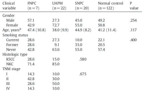
Briefly, a total of 29 subjects (7 affected and 22 unaffected pedigree members) from 4 families with NPC and 20 sporadic NPC as well as 122 cases of normal control were recruited from Zhejiang Han population, China. Concerning the patients' clinical characteristics, no significant differences were found in gender, age, smoking status, histologic types of tumors, or disease stages among all four groups of samples (Table 1). The histologic types and stages of nasopharyngeal carcinoma were classified according to the World Health Organization criteria. These families are from earlier epidemiological studies or from medical records in the First Affiliated Hospital of Wenzhou Medical College and Taizhou Hospital, which have at least two affected individuals with NPC. The familial history of these subjects was shown inFig. 1. All patients were diagnosed having NPC based on clinical pathological examinations as well as clinical records by local doctors. Except for NPC, other cancers were also found in one of these families such as liver cancer, pulmonary cancer. Five-to-10 ml of peripheral blood was collected from each subject before any chemotherapy, radiotherapy or pharmacotherapy, with fully informed consent. The study was approved by Wenzhou Medical College Ethic Committee.
2.2. DNA extraction
mtDNA and nuclear DNA from peripheral blood frozen at−70 °C was extracted respectively using the Blood Genomic/Mitochondrial DNA Extraction Kit (GenMed Scientifics, Inc. Arlington, MA, USA) according to the instructions of the manufacturer. Thefinal DNA pellet
was dissolved in 20 ul GENMED buffer solution and stored at−20 °C until use.
2.3. mtDNA D-loop PCR analysis
mtDNA fragments from the D loop were amplified using PCR primer pairs, which were designed according to MITOMAP Human mtDNA Cambridge Sequence data (www.mitomap.org) by using the Primer Express Software. The sequences of the primer are as follows: R: 5′-GAA TCG GAG GAC AAC CAG TA-3′S: 5′-TGA TGT GAG CCC GTC TAA AC-3′. PCR amplification was performed in 25 ulfinal volume consisting of 2.5 ul 10× PCR buffer, 200 uM of each dNTP, 1 U HoTaq polymerase (Nustar, America), 10 umol/l of each primer and 100 ng DNA template. The reactions were performed with a Bio-Rad thermocycler (Applied Biosystems Inc., USA) under the following conditions: 94 °C for 5 min, and 35 cycles of 94 °C for 35 s, 62.5 °C for 35 s, and 72 °C for 60 s, and a final extension step at 72 °C for 10 min. The PCR product amplified from D-loop mtDNA was detected by electrophoresis on a 1% agarose gel at 120 V and 60 mA for 60 min and under UV transillumination after ethidium bromide staining, and photographed.
2.4. Nucleotide sequence analysis
After purification with the AxyPrep™ PCR Cleanup Kit (Axygen Biosciences, America), all of the PCR products were sequenced in both directions using the same primers as PCR with the Applied Biosystems (ABI) 3730XL DNA Analyzer and BigDye® Terminator v3.1 Cycle Sequencing Kit (Applied Biosystems, America) under the following conditions: 25 cycles at 96 °C for 10 s, 51 °C for 10 s and 60 °C for 3 min. After nucleotide sequencing, sequence variations were deter-mined by comparison with revision of Cambridge reference sequence using CodonCode Aligner 2.0.6 software. Those not recorded in the database were categorized as novel mtDNA polymorphisms.
3. Statistical analysis
The clinicopathologic characteristics and prevalence of sequence variations between familial nasopharyngeal carcinoma (FNPC), unaffected pedigree member (UAPM), sporadic nasopharyngeal carcinoma (SNPC) group and normal control (NC) were compared using the one-way ANOVA and Chi-square test or Fisher's exact test. All statistical analyses were performed with the SPSS software Table 1
The clinical characteristics for the samplesa.
Clinical
Male 57.1 27.3 45.0 49.2 .254
Female 42.9 72.7 55.0 50.8
Age, yearsb 47.4 (10.8) 38.0 (9.9) 44.9 (8.2) 41.2 (11.4) .117 Smoking status
Current 28.6 27.3 10.0 22.1 .400
Former 28.6 9.1 35.0 20.5
Never 42.8 63.6 55.0 57.4
Histologic type
Abbreviations: FNPC, familial nasopharyngeal arcinoma; UAPM, unaffected pedigree member; SNPC, sporadic nasopharyngeal carcinoma; KSCC, keratinizing squamous cell carcinoma; NKC, non-keratinizing carcinoma.
a
Data are given as percentage of each group unless otherwise indicated. b
package 12.0 (SPSS Inc., Chicago, IL, USA). Hazard ratios are presented with their 95% confidence intervals (95% CI). p values less than 0.05 were considered significant. When the multiple experimental groups compared with the same control group, size of a test (α= 0.05) would be adjusted by α′=α/2(k−1). Related association results were adjusted for multiple testing.
4. Results
4.1. Sequence variants in four NPC families and SNPC
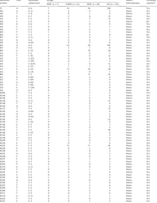
A total of 74 and 129 sequence variants were observed in 49 individuals from the experimental groups and 122 normal cases, respectively. Among these nucleotide positions, the overall numbers of variants were 67, 197, 186 and 832 in FNPC, UAPM, SNPC and NC, respectively (Table 5; Supplementary Information, Table S1). A significantly higher overall number of variants in FNPC, UAPM and SNPC compared with normal control were obtained (Pb0.05) (Table 2). Representative mtDNA sequence variation chromatograms are shown inFig. 2. Of the 74 detected sequence variants, 90.5% were homoplasmic, while 9.5% (7/74) of the mutations were heteroplasmic. There were 6 novel polymorphisms detected in this investigation. These include np186C→G, np206T–G, np310T→CCT, np314C–CCins (n), np556A→T and np16526G→T transition. Variations of the sites, transitions, and frequencies in familial NPC patients are summarized inTable 5. Moreover, there exist four high variations at np73A–G, np263A–G, np489T–C and np16223C–T. Unfortunately, except for np16362T–C and np16519T–C, all these alterations have no statistical significance compared with normal control.
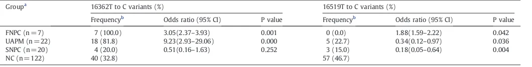
4.2. Frequency of T to C variants at np16362 and np16519 between groups
In this study, the study subjects were categorized into 4 subgroups (Table 3). The frequencies of np16362T–C were 100%, 81.8%, 20.0%, and 32.8% in FNPC, UAPM, SNPC and NC, respectively (Table 3). In contrast, we have observed a significantly higher frequency of np16362 variant in FNPC and UAPM(100%, 81.8%) compared with the other two groups (Pb0.05). Its frequency in SNPC (20.0%, 4/20) was found to be similar to that in NC (32.8%, 40/122), there was no statistically significant difference between SNPC and NC. At np16519T–C, the frequencies of variants were 0, 22.7%, 15%, and 46.7% in FNPC, UAPM, SNPC, and NC, respectively (Table 3). A significantly lower frequency of np16519 variant in FNPC, UAPM and SNPC compared with normal control were obtained (Pb0.05). Furthermore, there seems to appear a negative correlation between them. Thus, these results suggest that carriers of the T to C polymorphisms at 16362 might also be more susceptible to the development of familial nasopharyngeal carcinoma. With this reduction of frequency of np16519 variant, however, the risk of suffering from nasopharyngeal carcinoma more likely increase.
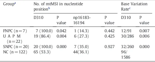
4.3. Microsatellite instability (MSI) in the D310 and np16183–16194 regions
The region between np303 and 315, designated as D310, is a highly homopolymeric C stretch in the D-loop region recognized as a mutational hot spot in several primary tumors. The other region at np16183–16194 also contains similar poly (C)-T-poly (C) sequences (ACCCCCTCCCCA). Thus, along with D310 were investigated in this study. As indicated inTable 4, totally 46 and 14 instances of mtMSI were detected in the D310 and np16183–16194 regions, which represent the most frequent mtMSI in experimental groups, respec-tively. The occurrence of mtMSI at D310 in FNPC, UAPM and SNPC groups was statistically significantly higher (Pb0.05) than those in NC (53.3%). Then, Base Variation Rate was calculated in the D310 regions. Consistent with the frequency of mtMSI at D310, there were statistically significant differences (Pb0.05) compared with NC (6.05%). Moreover, compared between FNPC, UAPM and SNPC, there was no statistically significant difference. However, no statistically significant difference (PN0.05) between frequency of variants at np16183–16194 regions and NC(36.1%) was detected. Overall, these results show that the individuals with family NPC and patients with nasopharyngeal carcinoma harbor Fig. 1.Four Chinese pedigrees with familial nasopharyngeal carcinoma. Patients with nasopharyngeal carcinoma are indicated byfilled symbols. Arrow denotes proband. Asterisk indicates the 7 affected and 22 unaffected pedigree members.
Table 2
Statistical analysis of total Base Variation Rate between groups.
Groupa Overall number of variants Base Variation Rateb P value
FNPC (n = 7) 67 67/7 1120 0.007
UAPM (n = 22) 197 197/22 1120 0.001
SNPC (n = 20) 186 188/20 1120 0.000
NC (n = 122) 832 832/122 1120
a
The number of cases analyzed is shown in parentheses for each group. b
more microsatellite instability (MSI) in the D310 regions than healthy cancer-free people, suggesting that these nucleotide changes or at least some of them might be related to nasopharyngeal carcinoma predis-position or family clustering.
5. Discussion
Warburg' (1956) proposal that cancer originated from a non-neoplastic cell that adopted anaerobic metabolism as a means of survival after injury to its respiratory system led to the notion that tumors were initiated by persistent damage to the mitochondria. Consequently, changes in the number, shape and function of mitochon-dria have been reported in various cancers (Pedersen, 1978).
Although mutations occur throughout the entire mitochondrial genome, most mutations are in the D-loop region. Because of its unique triple-stranded DNA structure that is more sensitive to oxidative damage due to the exposure of the displaced single strand in this arrested replication intermediate, the mutation rate in the D-loop region can reach 100- to 200-fold higher than that for nuclear DNA (Baldinu et al., 2002). Furthermore, the previous study (Wallace, 2005) has identified that the age is an important factor in mtDNA mutations, both of them appear positively correlated, that is, the accumulation of mtDNA mutations will gradually increase with the growth of the age. The age of subjects between study groups was well balanced, no significant differences were observed in age and gender between experimental groups and control subjects. Because of limited sample size the reviewer mentioned, we employed linkage/association tech-nology strategy (Risch and Merikangas, 1996; Strachan and Read, 1999) to extract or create the linkage or association conclusion in principle.
A total of 74 and 143 sequence variants were observed in 49 individuals from the experimental groups and 122 normal cases,
respectively. Among these, the overall number of variants in FNPC, UAPM and SNPC showed a positive significant difference, compared to normal control. This revealed that patients with NPC and pedigree members are enriched for sequence variants. There is a lower frequency of heteroplasmic mutation in 74 detected sequence variants (9.5%) than whatPang et al. (2008) reported (11.9%). It may be due to our current sequence analyses techniques, which are significantly better than in the past, do not detect heteroplasmies present at levels less than 20%. Furthermore,Sekiguchi et al. (2003)
demonstrated that mtDNA heteroplasmy from different individuals to tissue (hair, blood, orfinger nails) variation within individuals will show different proportions of the heteroplasmy.
In contrast to normal control, the frequent occurrence of these variations shows no significant difference by statistical analysis (PN0.05). Similarly, four high variations (np73A–G, np263A–G, np489T–C and np16223C–T) also display no significant difference (PN0.05). Interestingly, consistent with a previous study (Pang et al.,
2008), all subjects had these A→G transitions at sites 73 and 263 in mtDNA. These two variations may also play a role in distinguishing thefive major European mtDNA clusters (Wilkinson-Herbots et al., 1996; Zupanic Pajnic et al., 2004). The data support the concept that these haplotypes may represent a common mtDNA sequence type of a human being.
Another importantfinding in this study is the frequencies of T to C variants at np16362 and np16519 in different groups. Compared to normal control, the np16362 and 16519T to C variants were significantly higher and lower frequencies in FNPC and UAPM, respectively (Table 3). Though np16519 variant displays significantly lower frequency (Pb0.05), after this association results were adjusted for multiple testing (α′= 0.0083), there was no statistically significant difference in FNPC and UAPM. It means that larger samples to demonstrate the Fig. 2.Partial sequence chromatograms of D-loop sequence variation from experiment subjects. An arrow indicates the location of the base changes. A: np16362T→C fromIII7a, B: np16519T→C fromII5c, C: mtMSI at D310 from IV1a, D: mtMSI at np16183–16194 from III2a. a: pedigree1 b pedigree2 c: pedigree3 d: pedigree4.
Table 3
Statistical analysis of frequency of T to C variants at np16362 and np16519 between groups.
Groupa
16362T to C variants (%) 16519T to C variants (%)
Frequencyb Odds ratio (95% CI) P value Frequencyb Odds ratio (95% CI) P value
FNPC (n = 7) 7 (100.0) 3.05(2.37–3.93) 0.001 0 (0.0) 1.88(1.59–2.22) 0.042
UAPM (n = 22) 18 (81.8) 9.23(2.93–29.06) 0.000 5 (22.7) 0.34(0.12–0.97) 0.036
SNPC (n = 20) 4 (20.0) 0.51(0.16–1.63) 0.252 3 (15.0) 0.18(0.05–0.64) 0.004
NC (n = 122) 40 (32.8) 57 (46.7)
a
The number of cases analyzed is shown in parentheses for each group. b
association are necessary in follow-up experiments. Thus, ourfinding indicated the np16362 and 16519T to C variants may be associated with genetic susceptibility of familial nasopharyngeal carcinoma. The two variants are associated with an increased risk of familial nasopharyngeal carcinoma. We propose that these specific alterations, if confirmed, probably could be considered as promising markers in clinical diagnostic applications for familial nasopharyngeal carcinoma. Up to date, however, the possible biological significance and mechanism of action of the np16362 and 16519T to C variants are unclear.Zhou et al., 2007 consider that cancer-specific mitochondrial mutations may contribute to the development of a malignant phenotype by direct genotoxic effects from increased reactive oxygen species production as well as induction of aerobic glycolysis and growth promotion.
Similar frequencies of deletions and insertions at D310 region have been reported in other tumors (Sanchez-Cespedes et al., 2001), microsatellite instability (MSI) in the D310 appears to be good markers to determine nasopharyngeal carcinoma. The occurrence of mtMSI at D310 in FNPC, UAPM and SNPC groups was statistically significantly higher (Pb0.05) than in NC (53.3%,Table 4). Likewise, Base Variation Rate at D310 had statistically significant difference (Pb0.05) compared with NC (6.05%). Whereas MSI of np16183–16194 containing similar poly (C)-T-poly (C) sequences (Table 4) displayed no significant difference (PN0.05). This reinforces the notion that the D-loop has a selectively high mutation rate. The significantly higher frequency of mtMSI in nasopharyngeal carcinoma and pedigree members as compared to normal control could reflect a potentially significant role for mtMSI in the development and progression of cancer, in particular. At present,Sekiguchi et al. (2003) considered that automated DNA sequencing using either the ABI373 or 310 sequencers and either Dye Terminator (ABI373), dRhodamine Terminator (ABI 310) or BigDye-Terminator (ABI310) chemistries allow the unambiguous detection of the heteroplasmy present at the 30% level and these results show that PCR and sequencing are the gold standard for detecting single nucleotide polymorphisms (SNPs) and mutations. In view of this, we first applied the PCR-sequencing technique. But in follow-up experi-ments, we will use other methods to support.
Although mtMSI hot spots have been identified, it remains a challenge to address its function. In addition, the association between mtMSI and nuclear DNA has not yet been elucidated. Two recent studies (Harn et al., 2002; Trimeche et al., 2008), in Taiwan and Tunisia, have described a relative high frequency of nuclear MSI (47% and 31.2%) in NPC cases, respectively. The possibility that there may be a common initial triggering mechanism of mtMSI and nuclear MSI warrants further investigation. Some evidence indicates that mtMSI may be due to inefficient repair mechanisms (Mambo et al., 2003b). Another potential factor favoring mtMSI is DNA polymeraseγ, an enzyme responsible for mtDNA synthesis but with relatively weak proof-reading efficiency, therefore likely to be involved in the generation of mtDNA alterations.
Longley et al. (2001) have suggested that the homopolymorphic
nucleotide tracts in mtDNA were error prone because of the low frameshiftfidelity of the DNA polymeraseγthat carries out the mtDNA replication. Furthermore, DNA polymeraseγitself was also a target of oxidative damage (Graziewicz et al., 2002) and, when so damaged, may generate extensive errors during mtDNA replication and repair. Recent investigation showed that mtMSI at D310 seems to be associated as an early event in tumorigenesis and is increased with the severity of dysplasia in premalignant lesions of the head and neck (Ha et al., 2002). In addition, our experimental results were based on blood and not from the cancerous tissue. The mutation is one of hereditary factors, which is molecular basis for this disorder, while other mutations shared between tissue types are likely to have originated early in embryogenesis and may not increase risk, except that acquired somatic mutations are accumulated. But Pang et al. (2008) have reported that somatic mutations in the mtDNA D-loop region that shared concomitantly in blood and cancerous tissues are formed in NPC patients.
The study failed to detect a significant bias towards maternal inheritance in familial NPC, adding additional uncertainty regarding the role of inherited mtDNA variants in NPC. Mitochondrial genes are passed on from mothers to offspring but not from fathers to offspring. Therefore, to the degree that heritable mitochondrial genetic factors influence the risk of NPC, there should be a greater risk of NPC among offspring of women with NPC compared to offspring of men with NPC. However, this model has recently been challenged. Low levels of paternal transmission of mtDNA have been observed in crosses between mouse species, but not within species (Gyllensten et al., 1991), although further studies showed that this paternal mtDNA was not transmitted to the subsequent generation (Shitara et al., 1998). Studies of a patient with mitochondrial myopathy—who carried a novel, 2-bp pathogenic deletion in the NADH (reduced nicotinamide adenine dinucleotide) dehydrogenase subunit 2 (ND2) gene in his muscle mtDNA—have also challenged the maternal inheritance of mtDNA (Schwartz and Vissing, 2002). Another consideration is that other factors, possibly including somatic mtDNA mutations, virus and environment, may account for it. Because nasopharyngeal carcinoma is a complex disease with a proven genetic component. Similar to other solid tumors, NPC carcinogenesis likely occurs through a multistep process involving the interaction between environmental exposure, viral infection and somatic genetic alteration. However, the precise genetic component in this disease has remained largely elusive.
In summary, our data show that mtDNA exhibited a high rate of sequence changes in familial nasopharyngeal carcinoma and pedigree members, particularly those in np16362, np16519 and D310 seg-ments, might be involved in the NPC carcinogenesis and could be included in a panel of molecular biomarkers for cancer susceptibility early-detection strategy.
Supplementary materials related to this article can be found online atdoi: 10.1016/j.mito.2010.12.008
Conflict of interest statement
The authors declare no any potential conflict of interest.
Contributors
The following authors contributed to this work: conception and design: Shixiu Wu and Zheng Peng; collection and assembly of data: Zheng Peng, Qiuyan Wan, and Li Zhang; data analysis and interpre-tation: Zheng Peng; manuscript writing: Zheng Peng;final approval of manuscript: Zheng Peng, Congying Xie, Qiuyan Wan, Li Zhang, Wenfeng Li, and Shixiu Wu.
Acknowledgments
We sincerely thank the families for participating in this study and also we thank Dr. Wei Hu, Dr. Jianhua Wang for providing information about Table 4
Distribution of microsatellite instability (MSI) in the D310 and np16183–16194 regions.
Groupa
No. of mtMSI in nucleotide positionb
19 (86.4) 0.004 6 (27.3) 0.425 30/286 0.006
SNPC (n = 20) 20 (100.0) 0.000 7 (35.0) 0.927 32/260 0.000 NC (n = 122) 65 (53.3) 44(36.1) 96/
1586
a
The number of cases analyzed is shown in parentheses for each group. b The incidence (% of relevant samples tested) is shown in parentheses.
Table 5
Sequence variants of mtDNA D-loop region in all groups.
Nucleotide position
rCRSa Nucleotide replacement
Groupsb Homo-/
heteroplasmya
Previously reportedc FNPC (n = 7) UAPM (n = 22) SNPC (n = 20) NC (n = 122)
73 A A–G 7 22 20 108 Homo Yes
103 G G–A 1 2 0 2 Homo Yes
146 T T–A 0 0 1 0 Hetero Yes
150 C C–T 2 2 4 12 Homo Yes
152 T T–C 2 5 5 13 Homo Yes
186 C C–G 0 1 1 0 Hetero No
189 A A–G 1 2 0 6 Homo Yes
195 T T–C 0 0 1 1 Homo Yes
199 T T–C 0 0 2 1 Homo Yes
204 T T–C 1 3 4 7 Homo Yes
206 T T–G 1 1 0 0 Hetero No
207 G G–A 1 3 0 7 Homo Yes
235 A A–G 0 2 0 5 Homo Yes
249 A A-del 0 0 3 6 Homo Yes
263 A A–G 7 22 20 105 Homo Yes
297 A A–G 0 1 1 0 Homo Yes
309 C C–CC 4 5 10 22 Homo Yes
310 T T–C 0 0 0 7 Homo Yes
310 T T–TC 0 0 0 12 Homo Yes
310 T T–CCT 0 5 4 9 Homo No
310 T T–CTC 0 0 1 5 Homo Yes
310 T T–CCTC 2 2 0 11 Homo Yes
314 C C–CC 1 1 1 0 Homo No
315 C C–CC 5 17 16 30 Homo Yes
316 G G–CA 0 1 0 1 Homo Yes
489 T T–C 4 7 11 41 Homo Yes
521 A A-del 0 0 2 0 Homo Yes
522 C C-del 0 0 2 0 Homo Yes
523 A A-del 1 3 3 6 Homo Yes
524 C C-del 1 3 3 6 Homo Yes
524 C C–CAC 0 0 1 0 Homo Yes
556 A A–T 0 0 1 0 Homo No
16093 T T–C 1 4 3 6 Homo Yes
16104 C C–T 0 0 1 0 Hetero Yes
16126 T T–C 0 1 0 0 Hetero Yes
16129 G G–A 2 5 6 15 Homo Yes
16140 T T–C 1 3 0 4 Homo Yes
16164 A A–G 0 0 2 4 Homo Yes
16172 T T–C 2 2 5 13 Homo Yes
16182 A A-del 0 0 1 0 Homo Yes
16182 A A–C 0 0 1 2 Homo Yes
16183 A A-del 0 0 1 0 Homo Yes
16183 A A–C 1 6 2 23 Homo Yes
16184 C C–CC 0 0 1 0 Homo Yes
16184 C C–T 0 0 1 7 Homo Yes
16185 C C–T 0 0 0 2 Homo Yes
16189 T T–C 1 6 5 30 Homo Yes
16190 C C–CC 0 0 1 0 Homo Yes
16192 C C–T 0 0 0 2 Homo Yes
16194 A A–C 0 1 0 0 Homo Yes
16209 T T–C 1 2 0 2 Homo Yes
16218 C C–T 0 0 1 0 Homo Yes
16223 C C–T 4 12 11 43 Homo Yes
16228 C C–T 0 0 1 0 Homo Yes
16234 C C–T 0 2 0 3 Homo Yes
16235 A A–G 0 2 0 2 Homo Yes
16249 T T–C 2 3 0 7 Homo Yes
16257 C C–A 0 0 1 4 Homo Yes
16261 C C–T 0 0 1 1 Homo Yes
16263 T T–C 0 2 0 2 Homo Yes
16274 G G–A 1 2 0 3 Homo Yes
16278 C C–T 0 0 1 2 Homo Yes
16287 C C–T 0 0 1 0 Hetero Yes
16290 C C–T 0 2 0 4 Homo Yes
16293 A A–G 1 0 1 3 Homo Yes
16293 A A–C 0 2 0 2 Homo Yes
16296 C C–T 0 0 1 0 Hetero Yes
16297 T T–C 0 0 2 4 Homo Yes
16298 T T–C 0 0 2 5 Homo Yes
16304 T T–C 0 0 2 5 Homo Yes
16311 T T–C 0 0 3 6 Homo Yes
16319 G G–A 0 2 1 1 Homo Yes
16325 T T–C 0 2 0 5 Homo Yes
families with NPC. This work was supported by the National Natural Science Foundation of China (No. 30670621), Science and Technology Department of ZheJiang Province P.R. China: Major Program of High Incidence Disease Prevention and Control (No. 2007C13054) and Major program of WenZhou Science and Technology Bureau (No. S20070026).
References
Anderson, S., Bankier, A.T., Barrell, B.G., Debruijn, M.H.L., Coulson, A.R., Drouin, J., Eperon, I.C., Nierlich, D.P., Roe, B.A., Sanger, F., Schreier, P.H., Smith, A.J.H., Staden, R., Young, I.G., 1981. Sequence and organization of the human mitochondrial genome. Nature 290, 457–465. Attardi, G., Schatz, G., 1988. Biogenesis of mitochondria. Annu. Rev. Cell Biol. 4, 289–333. Baldinu, P., Cossu, A., Manca, A., Satta, M.P., Pisano, M., Casula, M., Dessole, S., Pintus, A., Tanda, F., Palmieri, G., 2002. Microsatellite instability and mutation analysis of candidate genes in unselected Sardinian patients with endometrial carcinoma. Cancer 94, 3157–3168.
Carew, J.S., Zhou, Y., Albitar, M., Carew, J.D., Keating, M.J., Huang, P., 2003. Mitochondrial DNA mutations in primary leukemia cells after chemotherapy: clinical significance and therapeutic implications. Leukemia 17, 1437–1447.
Corral-Debrinski, M., Horton, T., Lott, M.T., Shoffner, J.M., Beal, M.F., Wallace, D.C., 1992. Mitochondrial DNA deletions in human brain: regional variability and increase with advanced age. Nat. Genet. 2, 324–329.
Feng, B.J., Huang, W., Shugart, Y.Y., Lee, M.K., Zhang, F., Xia, J.C., Wang, H.Y., Huang, T.B., Jian, S.W., Huang, P., Feng, Q.S., Huang, L.X., Yu, X.J., Li, D., Chen, L.Z., Jia, W.H., Fang, Y., Huang, H.M., Zhu, J.L., Liu, X.M., Zhao, Y., Liu, W.Q., Deng, M.Q., Hu, W.H., Wu, S.X., Mo, H.Y., Hong, M.F., King, M.C., Chen, Z., Zeng, Y.X., 2002. Genome-wide scan for familial nasopharyngeal carcinoma reveals evidence of linkage to chromosome 4. Nat. Genet. 31, 395–399.
Graziewicz, M.A., Day, B.J., Copeland, W.C., 2002. The mitochondrial DNA polymerase as a target of oxidative damage. Nucleic Acids Res. 30, 2817–2824.
Gyllensten, U., Wharton, D., Josefsson, A., Wilson, A.C., 1991. Paternal inheritance of mitochondrial DNA in mice. Nature 352, 255–257.
Ha, P.K., Tong, B.C., Westra, W.H., Sanchez-Cespedes, M., Parrella, P., Zahurak, M., Sidransky, D., Califano, J.A., 2002. Mitochondrial C-tract alteration in premalignant lesions of the head and neck: a marker for progression and clonal proliferation. Clin. Cancer Res. 8, 2260–2265.
Harn, H.J., Fan, H.C., Chen, C.J., Tsai, N.M., Yen, C.Y., Huang, S.C., 2002. Microsatellite alteration at chromosome 11 in primary human nasopharyngeal carcinoma in Taiwan. Oral Oncol. 38, 23–29.
Hu, L.F., Qiu, Q.H., Fu, S.M., Sun, Di., Magnusson, Kristinn, He, B., Lindblom, A., Ernberg, I., 2008. A genome-wide scan suggests a susceptibility locus on 5p13 for nasopha-ryngeal carcinoma. Eur. J. Hum. Genet. 16, 343–349.
Jiang, R.C., Qin, H.D., Zeng, M.S., Huang, W., Feng, B.J., Zhang, F., Chen, H.K., Jia, W.H., Chen, L.Z., Feng, Q.S., Zhang, R.H., Yu, X.J., Zheng, M.Z., Zeng, Y.X., 2006. A functional variant in the transcriptional regulatory region of gene LOC344967 cosegregates with disease phenotype in familial nasopharyngeal carcinoma. Cancer Res. 66, 693–700.
Lo, K.W., To, K.F., Huang, D.P., 2004. Focus on nasopharyngeal carcinoma. Cancer Cell 5, 423–428.
Longley, M.J., Nguyen, D., Kunkel, T.A., Copeland, W.C., 2001. Thefidelity of human DNA polymerase gamma with and without exonucleolytic proofreading and the p55 accessory subunit. J. Biol. Chem. 276, 38555–38562.
Lu, S.J., Day, N.E., Degos, L., Lepage, V., Wang, P.C., Chan, S.H., Simons, M., Mcknight, B., Easton, D., Zeng, Y., Guy, D.T., 1990. Linkage of a nasopharyngeal carcinoma susceptibility locus to the HLA region. Nature 346, 470–471.
Mambo, E., Gao, X., Cohen, Y., Guo, Z., Talalay, P., Sidransky, D., 2003a. Electrophile and oxidant damage of mitochondrial DNA leading to rapid evolution of homoplasmic mutations. Proc. Natl Acad. Sci. USA 100, 838–1843.
Mambo, E., Gao, X., Cohen, Y., Guo, Z.M., Talalay, P., Sidransky, D., 2003b. Electrophile and oxidant damage of mitochondrial DNA leading to rapid evolution of homoplasmic mutations. Proc. Natl Acad. Sci. USA 100, 1838–1843.
Matsuyama, W., Nakagawa, M., Wakimoto, J., Hirotsu, Y., Kawabata, M., Osame, M., 2003. Mitochondrial DNA mutation correlates with stage progression and prognosis in nonsmall cell lung cancer. Hum. Mutat. 21, 441–443.
McDermott, A.L., Dutt, S.N., Watkinson, J.C., 2001. The aetiology of nasopharyngeal carcinoma. Clin. Otolaryngol. Allied Sci. 26, 82–92.
Pang, L.J., Shao, J.Y., Liang, X.M., Xia, Y.F., Zeng, Y.X., 2008. Mitochondrial DNA somatic mutations are frequent in nasopharyngeal carcinoma. Cancer Biol. Ther. 7, 198–207. Parkin, D.M., Muir, C.S., 1992. Cancer incidence infive continents. Comparability and
quality of data. IARC Sci. Publ. 120, 45–173.
Parrella, P., Seripa, D., Matera, M.G., Rabitti, C., Rinaldi, M., Mazzarelli, P., Gravina, C., Gallucci, M., Altomare, V., Flammia, G., Casalino, B., Benedetti-Panici, P.L., Fazio, V.M., 2003. Mutations of the D310 mitochondrial mononucleotide repeat in primary tumors and cytological specimens. Cancer Lett. 190, 73–77.
Pedersen, P.L., 1978. Tumor mitochondria and the bioenergetics of cancer cells. Prog. Exp. Tumor Res. 22, 190–274.
Penta, J.S., Johnson, F.M., Wachsman, J.T., Copeland, W.C., 2001. Mitochondrial DNA in human malignancy. Mutat. Res. 488, 119–133.
Polyak, K., Li, Y., Zhu, H., Lengauer, C., Willson, J.K.V., Markowitz, S.D., Trush, M.A., Kinzler, K.W., Vogelstein, B., 1998. Somatic mutations of the mitochondrial genome in human colorectal tumours. Nat. Genet. 20, 291–293.
Risch, N., Merikangas, K., 1996. The future of genetic studies of complex human diseases. Science 273, 1516–1517.
Sanchez-Cespedes, M., Parrella, P., Nomoto, S., Cohen, D., Xiao, Y., Esteller, M., Jeronimo, C., Jordan, R.C., Nicol, T., Koch, W.M., Schoenberg, M., Mazzarelli, P., Fazio, V.M., Sidransky, D., 2001. Identification of a mononucleotide repeat as a major target for mitochondrial DNA alterations in human tumors. Cancer Res. 61, 7015–7019. Schwartz, M., Vissing, J., 2002. Paternal inheritance of mitochondrial DNA. N. Engl. J.
Med. 347, 576–580.
Sekiguchi, K., Kasai, K., Levin, B.C., 2003. Inter- and intragenerational transmission of a human mitochondrial DNA heteroplasmy among 13 maternally-related individuals and differences between and within tissues in two family members. Mitochondrion 2, 401–414.
Shitara, H., Hayashi, J.I., Takahama, S., Kanedab, H., Yonekawab, H., 1998. Maternal inheritance of mouse mtDNA in interspecific hybrids: segregation of the leaked paternal mtDNA followed by the prevention of subsequent paternal leakage. Genetics 148, 851–857.
Strachan, T., Read, A.P., 1999. Human molecular genetics, Introduction to the Electronic Age, Second ed. Wiley-Liss. E-Publishing Inc., New York, pp. 283–294.
Tamori, A., Nishiguchi, S., Nishikawa, M., Kubo, S., Koh, N., Hirohashi, K., Shiomi, S., Inoue, M., 2004. Correlation between clinical characteristics and mitochondrial D-loop DNA mutations in hepatocel-lular carcinoma. J. Gastroenterol. 39, 1063–1068.
Trimeche, M., Braham, H., Ziadi, S., Amara, K., Hachana, M., Korbi, S., 2008. Investigation of allelic imbalances on chromosome 3p in nasopharyngeal carcinoma in Tunisia: high frequency of microsatellite instability in patients with early-onset of the disease. Oral Oncol. 44, 775–783.
Van Tuyle, G.C., McPherson, M.L., 1979. A compact form of rat liver mitochondrial DNA stabilized by bound proteins. J. Biol. Chem. 254, 6044–6053.
Wallace, D.C., 2005. A mitochondrial paradigm of metabolic and degenerative diseases, aging, and cancer: a dawn for evolutionary medicine. Annu. Rev. Genet. 39, 359–407. Warburg, O., 1956. On the origin of cancer cell. Science 123, 309–314.
Wilkinson-Herbots, H.M., Richards, M.B., Forster, P., Sykes, B.C., 1996. Site 73 in hypervariable region II of the human mitochondrial genome and the origin of European populations. Ann. Hum. Genet. 60, 499–508.
Xiong, W., Zeng, Z.Y., Xia, J.H., Xia, K., Shen, S.R., Li, X.L., Hu, D.X., Tan, C., Xiang, J.J., Zhou, J., Deng, H., Fan, S.Q., Li, W.F., Wang, R., Zhou, M., Zhu, S.G., Lu, H.B., Qian, Jun, Zhang, B.C., Wang, J.R., Ma, J., Xiao, B.Y., Huang, He., Zhang, Q.H., Zhou, Y.H., Luo, X.M., Zhou, H.D., Yang, Y.X., Dai, H.P., Feng, G.Y., Pan, Q., Wu, L.Q., He, L., Li, G.Y., 2004. A susceptibility locus at chromosome 3p21 linked to familial nasopharyngeal carcinoma. Cancer Res. 64, 1972–1974.
Yu, M.C., Yuan, J.M., 2002. Epidemiology of nasopharyngeal carcinoma. Semin. Cancer Biol. 12, 421–429.
Zeng, Y.X., Jia, W.H., 2002. Familial nasopharyngeal carcinoma. Semin. Cancer Biol. 12, 443–450.
Zhou, S.y., Kachhap, S., Sun, W., Wu, G.j., Chuang, A., Poeta, L., Grumbine, L., Mithani, S.K., Chatterjee, A., Koch, W., Westra, W.H., Maitra, A., Glazer, C., Carducci, M., Sidransky, D., McFate, T., Verma, A., Califano, J.A., 2007. Frequency and phenotypic implications of mitochondrial DNA mutations in human squamous cell cancers of the head and neck. PNAS 104, 7540–7545.
Zupanic Pajnic, I., Balazic, J., Komel, R., 2004. Sequence polymorphism of the mitochondrial DNA control region in the Slovenian population. Int. J. Leg. Med. 118, 1–4.
Abbreviations: FNPC,familial nasopharyngeal arcinoma; UAPM, unaffected pedigree member; SNPC, sporadic nasopharyngeal carcinoma. a
Revision of Cambridge reference sequence. b