Rare Distal Anterior Choroidal Artery Aneurysm
Introduction
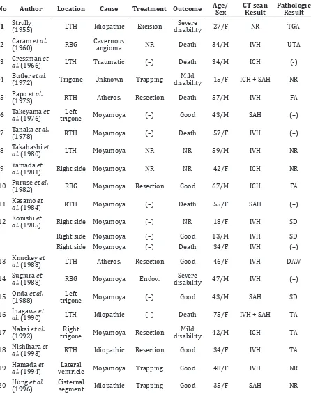
Reports on aneurysm of the distal anterior choroidal artery (AChA) are very rare. There are only 49 cases are reported in the literature. Twenty cases were associated with moyamoya disease; 10 cases with unknown causes; 8 cases are idiopathic; 3 cases were each associated with atherosclerosis and middle cerebral artery (MCA) occlusion; 2 cases with arteriovenous malformation (AVM), 1 case each for posterior cerebral artery (PCA) occlusion, cavernous angioma and head trauma (Table 1).
Case
A 56-year old female experienced a sudden onset of severe headache and vomiting. She
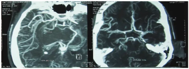
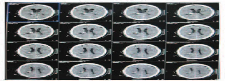
was brought to a local hospital near her house and was hospitalized for 2 days. She was later referred to our hospital, the Siloam Hospital because her symptoms did not disappear. She still complained of severe headache but was fully alert without any neurological deficits. Results from chest x-ray, laboratory examinations and electrocardiogram were normal. Head CT-scan revealed that there was intraventricular hemorrhage with most of it was found in the right lateral ventricle; the left lateral, 3rd and 4th ventricles were slightly enlarged; and an arachnoid cyst in the magna cistern (sized 3 x 2 x 2 cm). Angiography revealed the presence of a round, ruptured vascular lesion sized 5 x 5 mm at the wall of posterior cornu of right lateral ventricle. Digital substraction angiography examination presented a small aneurysm at the right distal AChA and total obstruction at the left MCA, with collaterals observed. Occlusion of the Abstract Objective: To describe a rare patient with ruptur aneurysm case of distal
anterior choroidal artery (AChA) and intraventricular hemorrhage. A 56-year old female came to our hospital with chief complaint sudden onset of severe headache and vomiting.
Methods: Head computed tomography (CT)-scan and angiography on the lesion was performed at the Department of Radiology, Siloam Hospital, Tangerang, Indonesia.
Results: Head CT-scan imaging revealed an intraventricular hemorrhage, primarily in the right lateral ventricle, with slight enlargement of both lateral, 3rd and 4th ventricles. Angiography examination revealed a round vascular
lesion at the wall of the posterior cornu of the lateral ventricle and an occlusion of the M1 base segment of the left middle cerebral artery.
Conclusions: The lesion, distal AChA aneurysm, at the posterior cornu was reached using an infratemporal lobe approach with the help of neuronavigation. Microsurgical clipping was successfully performed.
Keywords: Aneurysm, distal anterior choroidal artery, neuronavigation [IJIHS. 2016;4(2):86–92]
pISSN: 2302-1381; eISSN: 2338-4506; http://dx.doi.org/10.15850/ijihs.v4n2.837
Received:
Muhammad Zafrullah Arifin,1 Julius July,2 Bilzardy Ferry,1 Ahmad Faried1
1Department of Neurosurgery, Faculty of Medicine, Universitas Padjdajaran-Dr. Hasan Sadikin General Hospital 2Department of Neurosurgery, Faculty of Medicine, Pelita Harapan University–Siloam Hospital, Tangerang, Indonesia
Preoperative-axial Head CT-scan Showing an Intraventricular Hemorrhage on
Both Side Fig. 1
Preoperative-axial CT-angiography Shows an Aneurysm at the Posterior Cornu
Wall of Right Lateral Ventricle (with Diameter ±5.6 mm) Fig. 2
decreasing density of the aneurysm, meaning that thrombosis had already occurred. The patient underwent surgery on the next day. A right temporooccipital craniotomy was conducted and the inferior temporal gyrus was accessed through neuronavigation. A saccular aneurysm with 5 x 5 mm in the right lateral ventricle at the cornu posterior wall was found. The lesion was succesfully clipped, and an intraventricular drain was placed. One day after the operation, the patient’s complaints were completely resolved and patient was discharged with no neurological deficit.
Discussion
Preoperative CT Angiography Showing an Aneurysm at the Distal Anterior
Choroidal Artery Fig. 3
Positioning of the Patient and Marking of the Operation Area for Neuronavigation
No Author Location Cause Treatment Outcome Age/
Sex CT-scan Result Pathologic Result
1 Strully (1955) LTH Idiopathic Excision disabilitySevere 27/F NR TGA
2 Caram (1960)et al. RBG Cavernous angioma NR Death 34/M IVH UTA
3 Cressman al et
. (1966) LTH Traumatic (–) Death 34/M ICH (-)
4 Butler et al.
(1972) Trigone Unknown Trapping
Mild
disability 15/F ICH + SAH NR
5 Papo (1973)et al. RTH Atheros. Resection Death 57/M IVH FA
6 Takeyama al. (1976)et trigoneLeft Moyamoya (–) Good 43/M SAH (–)
7 Tanaka et al.
(1978) RTH Moyamoya (–) Death 57/F IVH (–)
8 Takahashi al et
. (1980) LTH Moyamoya NR NR 59/M IVH NR
9 Yamada al. (1981)et Right side Moyamoya NR NR 42/F ICH NR
10 Furuse (1982)et al. RBG Moyamoya Resection Good 67/M ICH FA
11 Kasamo al et
. (1984) RTH Moyamoya (–) Death 55/F SAH (–)
12 Konishi al et
. (1985) Right side Moyamoya (–) NR 18/F IVH SD
Right side Moyamoya (–) Good 13/M IVH SD
Right side Moyamoya (–) Death 34/F IVH (–)
13 Knuckey al et
. (1988) LTH Atheros. Resection Good 46/F IVH DAW
14 Sugiura al et
. (1988) RBG Moyamoya Endov.
Severe
disability 47/M IVH (–)
15 Onda (1988)et al. trigoneLeft Moyamoya (–) Good 43/M SAH SD
16 Inagawa al. (1990)et LTH Idiopathic (–) Death 75/F IVH + SAH TA
17 Nakai et al. (1992)
Right
trigone Moyamoya Resection
Mild
disability 42/M ICH TA
18 Nishihara al et
. (1993) RTH Idiopathic Resection Good 34/F IVH TA
19 Hamada al. (1994)et ventricleLateral Moyamoya Trapping Good 48/F IVH NR
20 Hung (1996)et al. Cisternal segment Idiopathic Trapping Good 35/F SAH NR
Table 1 List of Authors Reporting Patients with Distal Anterior Choroidal Artery
. (1996) horn Symptoms
22 Kawai (1997)et al. RTH Moyamoya (–) disabilityMild 19/M IVH TA
23 Yoneoka al et . (1998)
Right
side Unknown Clipping NR 69/M IVH TA
24 Yanaka et al. (2000)
Right
side AVM Resection Good 8/F IVH TA
25 Matsuura al. (2000)et Cisternal segment Idiopathic Conservative Good 42/M SymptomsIschemic NR
26 Lee (2001)et al. trigoneRight Moyamoya Resection Good 48/M ICH + IVH TA
27 Kuroda (2001)et al. NR Unknown Rev. Good F IVH NR
28 Wong et al. (2003)
Temporal
horn Moyamoya Clipping Good 62/F ICH + IVH NR
29 Ali et al.
(2004) NR Unknown Clipping Good 26/M ICH + IVH TA
30 Nishio (2004)et al. NR Unknown Embolization NR 47/F SAH NP
31 Ahn (2006)et al. Left side Unknown (–) Death 60/F NR NR
32 Inci et al. (2007)
Temporal
horn Idiopathic Resection Good 19/F ICH + IVH NR
Temporal
horn Idiopathic Resection Death 37/F ICH + SAH NR
33 Gandhi (2008)et al. NR Unknown Clipping NR M SAH NP
34 Yurt (2009)et al. Right side Unknown Clipping Good NR ICH + IVH NP
35 Kim et al.
(2009) NR Moyamoya
Vegetative
state NR 43/F IVH NR
36 Yang et al.
(2010) NR Moyamoya Endov. Good 56/F IVH NP
NR Moyamoya Endov. Good 38/F IVH NP
37 Choulakian et al. (2010) NR Moyamoya Endov. Good NR IVH NP
38 Nishida et al.
(2011) RTH
MCA
Occlusion Endov.
Mild
disability 84/F IVH NP
39 Leveque al et
. (2011) Left side Moyamoya Endov. Good 50/F IVH NP
40 Dolati (2012)et al. NR OcclusionPCA Endov. Good 55/M IVH NP
41 He (2013)et al. Left side Unknown Clipping Good M IVH NP
Right
42 Shimizu al. (2013)et posteriorLateral Atheros. Endov. Good 43/F ICH NP
NR AVM Endov. Good 6/F ICH NP
43 Oishi et al.
(2013) RTL
MCA
occlusion Endov.
Mild
disability 75/F ICH NP
44 Our case
(2014) RTH
MCA
occlusion Clipping Good 56/F IVH NP
No differences between gender were found. In terms of cause, most of the cases, i.e. 20 cases , were caused by moya-moya disease.5
In the case presented here, on the opposite side from the location of aneurysm, an occlusion of M1 segment of left MCA and narrowing or stenotic of P2 segment of left PCA were found; hence, it can be concluded that the cause of the aneurysm was MCA occlusion. In this case, the location of aneurysm is on the right lateral intraventricular at the temporal horn, known as plexal segment, while almost all patients had the aneurysm located in temporal horn (Table 1) eventhough most authors did not mention the location of the aneurysm in their case report. In this report, our patient was discharged from the hospital uneventfully. It is so unfortunate that most studies did not mention the outcome of their case.
Direct micosurgical intervention through
a transtemporal or ventricular approach is one of the options for managing distal AchA aneurysm with somehow additional damage to the brain and its collateral circulation may not be avoidable.6 Several reported cases of the distal AChA show that the cases were successfully treated using coils and n-butyl cyanoacrylate (nBCA) liquid embolization, arguing that the endovascular technique is a promising modality for this rare case.2–9
In conclusion, in the present case, aneurysm of distal choroidal anterior artery can be managed. Timing of surgery with great caution may be an advantage to improve the prognosis of the patient. The choice of treatment depends on the available expertise and equipment; the latest report has argued that the endovascular technique is a promising modality for this rare case.
1. Strully KJ. Successful removal of intraventricular aneurysm of the choroidal artery. J Neurosurg. 1955;12(3):317–21.
2. Oishi H, Suga Y, Nonaka S. Endovascular therapy of ruptured distal anterior choroidal artery aneurysm associated with moyamoya pattern collateralization secondary to middle cerebral artery occlusion. Indian J Neurosurg. 2013;2(3):278–81.
3. Shimizu T, Naito I, Fujita K, Miyamoto N, Sato K, Aishima K, et al. Aneurysm of ruptured
distal chroroidal artery embolized using n-butyl cyanoacrylate: case report. JNET.
2013;7(3):179–85.
4. Nishida A, Tokunaga K, Hisihikawa T, Sugiu K, Date I. Endovascular coil embolization of a ruptured diatal anterior choroidal artery aneurysm associated with ipsilateral middle cerebral artery occlusion: case report. Neurol Med Chir (Tokyo). 2011;51(10):716–9.
5. He K, Zhu W, Chen L, Mao Y. Management of distal choroidal artery aneurysm in patients with moyamoya disease: Report of three cases and review of the literature. World J Surg Oncol. 2013;11(2):187–93.
6. Inci S, Arat A, Ozgen T. Distal anterior choroidal Notes:
Atheros.: Atherosclerotic DAW: Degenerated artery wall Endov.: Endovascularization LTH: Left temporal horn
RBG: Right basal ganglia RTH: Right temporal horn RTL: Right temporal lobe Rev.: Revascularization
SAH: Subarachnoid haemorrhage SD: Spontaneus disappearance TGA: Thrombosed giant aneurysm TA: True aneurysm
UTA: Unruptured true aneurysm
7. Yang S, Yu JL, Wang HL, Wang B, Lou Q. Endovascular embolization of distal anterior choroidal artery aneurysm associated with moyamoya disease: a report of two cases and a literature review. Interv Neuroradiol. 2010;16(5):433–41.
8. Leveque M, McLaughlin N, Laroche M,
J Neurosurg. 2011;114(1):116–9.