Imaging Science
inDentistry
Volnme 41, Number 4, December 2011
-
zyxwvutsrqponmlkjihgfedcbaZYXWVUTSRQPONMLKJIHGFEDCBA-•
Sylviana Hardanti, Azhuri, Fahmi OscandarzyxwvutsrqponmlkjihgfedcbaZYXWVUTSRQPONMLKJIHGFEDCBA
Veparlm C !1I1ojzyxwvutsrqponmlkjihgfedcbaZYXWVUTSRQPONMLKJIHGFEDCBAD C"IO "ta ."IlJ (~f(1cj(l1N (Jdi(I/08Y.F uculty ofD entlstr ». P adjadjaran U II;\I(u':rit,y. /Jolldu'I$, indonesia
Description ohnandibular
bone quality based.on measurements of
cortical thickness using Mentallndex of male and female patients
-L51-zyxwvutsrqponmlkjihgfedcbaZYXWVUTSRQPONMLKJIHGFEDCBA Copyrighl®2011 b)' Korean Academy ofOrillluMiMtlXillo(ucrlllRadiology
.asOpen A«c.'''\.Ij1kkdhldtll~ UlI\!cr11a:Icntl8ltr!hcCn:a!j'1:CIlI~nllll1.\ALlribud!lllNlln·Cl)mllxtdllllktl!~(htlll!'lIcftlUi''eCIl(lIm'''''A.lrStlll'l!11._!br·lldl.Qj
whkhflCtll'lh.\I(!f9!IICINI nutt-tu,mncn:!alll)('. diMributiun.andn:l!lllllu(ooninlillymcdiam.proviiled II',,:~gillnl wDfkie prnf.Cr!yC(ted, rnfngiog.Sei<;ilCe tilDClntslrY: plSSN 2233·1822 cJSSN 2233-7810
ause 01'its capability 10obtain comprehensive image' of the maxillofacial structure.Y Although dentists mainly focus on teeth .and jaw anomalies, it is considered as an obligation for medical practitioners 10 recognize panora-mic images which shows the overall systepanora-mic health con-dition of thepatient.6 One of the systemic ,diseases that
reveal themselvesas a-specific image on panoramic radio-graph is osteoporosis. The characteristics of
this
diseasearc cortical thirining and more radiolucent trabecular areas.'
Quantil,y qnd-qualiry of the jaw bone have important roles inthe success Ofdenial care,S
Osteoporosis is diagnosed'by examining the BMD score.
Unfortunately. BMD tests arc very expensive. especially in developing countries such as Indonesia, thus usfunction
>ISan early detection 1001 for fracture risks is seldom used. Researches suggested that panoramic radiograph could be.
a
useful identification LOo)in female with low'B)vlD,scores."By examining panoramic. radiographs •.thequality and quantity of bone can be determined.
The purpose of thisstud), was to obtain the description on the mandibular bone quality of male and female between Q,,:_-oporo~is is on~ of the degenerative diseases.that can
~ ~fh problem in Indonesia recently as a result from
A..=.<e in life expectancy and life style alteration. Two
fivEIndonesian women have risk from osteoporosis.' ~ of OSICOP(lfO$isis essential todetect fracture risk. cs:>odally in high risk group. However, in Indonesla as a ,JC".dOping,COUHlry, the expenses (If the examination 'still
.:o::omea.major problem. Panoramic radiograph used
wide-•illdentistry is ayntlable (or early detection (If osreopo-..,.,.,. wilh.1()\\,er W<pense.One of the techniquesto detect
~'1pcltOS1s iscortical thickness measurement.
9 ..5ozmphy is utilized-as adiagn0stic tool forevaluation
~ ._..hand jaw anomalies indental practice.23 Panoramic
-:a.;...;;nphy isoneof the most comon radiographies
bee-Introduction
ll\"'II'ORDS : Panoramic 'Radiography; Mandible; Osteoporosis
~: The purpose of this study was to obtain the cescrlptiou of-me mandibular bone quality of male- and female
--"""lL<between 40·6.9 years old andtheir differences based On mandibular conical bone ihickness measured USing
~dl'J(
(~lli.. .
.
.
\&:teriaIs andMethods ;Forty digital panoramic n)aiographs \ wbich consisted of twenty male-and twenty female
~DlS, 40·60 yearsold, were observed. Mandibular conical bone thickness was measured USing
ivu:
on both sidestilemandible.The average MI scorcofrwc groups were ihenassessed usingt-sarnple independent test.
Results: There were-significant differences-of mandibular bone quality based on mandibular cortical bone thickness
_rement usiJ)!\Ml between'mille and femalepatienls(p<0.05).
Condusion.: Mandibular bone quality based
on
cortj.t;a)bon."thickness-measurement usinglInof male ana female~., indicated ~ xignificant.difference,zyxwvutsrqponmlkjihgfedcbaZYXWVUTSRQPONMLKJIHGFEDCBA(Tma gi1lCSciDelli 20J J ; 41 ;LSl·3)
•
~
Hardanti.
Azhan.FabmlOscandar
zyxwvutsrqponmlkjihgfedcbaZYXWVUTSRQPONMLKJIHGFEDCBAD e lllQ lll(L ,· {/{()j(l(;.lp lR< ,d/ology.F (l(;uIJ yoj Dentistr y,P (ulja dja r (ln Unlver sity, Ba ndllng.lndonesia
t:b..~
ofmanwhlliar
bone quality based on measurements of cortical thickness using
....qaf
Indexof
male and female patients between 40-60 years old
(hteoporoil
cytokieeszyxwvutsrqponmlkjihgfedcbaZYXWVUTSRQPONMLKJIHGFEDCBAi will [n.::u2
hol'UlO"'.e •
152Genetic factors. particularly gender. affected bone -Sexual hormone between mule and female are diner-However, both ofthem affectedbone growth. TesID'~ plays a role in male, while estrogen in female10 CDC
bone growth," Bone mass increases constaruly at1i.l
the peak bone mass at the uge of 40 in male BOO ageof30-35 in female.'
Different from male, rhe aging process in fem:zo.:
earlier, The reproductive age in female is o\~_
of 45· 50 when the menstruation cycle e~
hormone rapidly decreases. well known:!Sm<2::;=C::
Estrogendeficiency plays (Ivery important-r .:zyxwvutsrqponmlkjihgfedcbaZYXWVUTSRQPONMLKJIHGFEDCBA
pause as a cause of bone mass decr¢~lse.'E.s:t:""-=
Discussion
T obie 2 .zyxwvutsrqponmlkjihgfedcbaZYXWVUTSRQPONMLKJIHGFEDCBATIle meanM l valueH ndexurniuationresult ofboth grour
G ender N M1zyxwvutsrqponmlkjihgfedcbaZYXWVUTSRQPONMLKJIHGFEDCBAS O t-coum Hable p.v.l", remille 20 3.155 0,531
-4.164 2.02 0.000
Male 20 3.928 O.63R 1(5%) 19(95%)
6(30'lf. 14(10" <3.oolI\m
;a:3.00mm
Gender gruup
M ule Femnle
MI value
T able 1. D istribution or patients according togender nnd M Iv,.lue
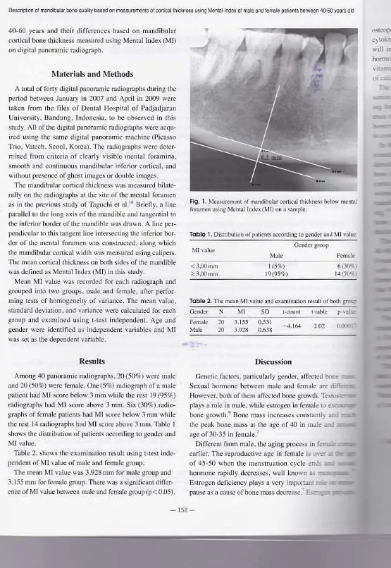
Fig. 1. MC3Suremcn,of mllndibularconicallhidrncssbelow OIenllll
(onunen using MentallndexzyxwvutsrqponmlkjihgfedcbaZYXWVUTSRQPONMLKJIHGFEDCBA(~ 11)onu sample,
Among 40panoramic radiographs. 20 (50%) were male and 20 (50%) were female. One (5%) radiograph of a male pal lent had M[ score below 3 rnrn while the rest 19 (95%) radiographs had.lvITSCOI'Cabove 3 mm. Six(30%) radio-graphs of female patients hnd MI score below 3101llwhile
the rcsll4 radiographs had M(score above 310m. Table I showsthe distribution of parierns according LO gender and
MI value.
Table 2.shows theexamination resultusingt-test
inde-pendent of Ml value of male and female group.
The mean Ml value was 3.928 rnrn for male group and 3.155mm lor female group. There was a significant differ-ence of M I value between male nnd female /,'fOIiP(p< O.()S).
Results
A Lotalof forty digital panoramic radiographs during the period between January in2007 and April in2009 were
taken from the files of Denial Hospital of Padjadjarnn University. Bandung, Indonesia. 10be observed in this study, All of the digital panoramic radiographs were acqu-ired using the same digital panoramic machine (Picasso Trio. Vatcch, Seoul, Korea). The radiographs were deter-mined from criteria of clearly visible mental foramlnn.
smooth and continuous mandibular inferior cortical. and
without presence ofghost images or double images.
The mandibular cortical rhickness was measured bilate-rally on the radiographs at Ihe site of the mental foramen as in the previous study of Taguchi et
aI.'·
Briefly. a line parnllcl 10the long axis of me mandible and tangential 10 theinferior border of [hemandible was drawn. A linepel" pcndicular 10thistangentlineiruersccting the inferior bor-der of the mental foramen WIlS constructed. along which Olemandibular cortical width wus measured using calipers. The mean cortical thickness on both sides of the mandiblewas defined as Memallndex (MI) inthis study,
Mean ~U value was recorded for each radiograph and grouped into two groups. male and female, after
perfor-ming testsof homogeneity of variance. The mean value. standard deviation. and variance were calculated for each SI'OUpnnd examined using l-IeSI independent. Age nnd gender were identified as lndcpcndcnt variables and MJ wasSCIas thedependent vnriublc.
Materials lind Methods
40-60 years and their difference, based on mandibular
cortical bone thickness measured using Mental Index (1\111)
ondigilal panoramic radiograph.
-153-survey of risk, Jakana: Ministry of Health Republic of
Indone-sia:2006,
2. TaguchizyxwvutsrqponmlkjihgfedcbaZYXWVUTSRQPONMLKJIHGFEDCBAf\.SueizyxwvutsrqponmlkjihgfedcbaZYXWVUTSRQPONMLKJIHGFEDCBAY:;San.:.daNt. Ohtsuka M~Nakamoto T, Sumida A. ct ttl. Validation of rleuralpanoramic rudibgruphy
measures for identifying posrmenopausal women W)lh spinal
osteoporosis.AIR AmJRaemgenoI2\104: 183: 17!\~,
3. a.Ongor K. Akarslnn Z, Akdevelioglu f\t, Brienfor,Scrniz tvl.
The precision of m,epanorumic mandibulnr inde:<~
D.enlol'nax-Illofac Radial 2006; 35: <142-6.
4. Schulze R. Krurnmenauer F.Schalldach F. d'Hoedr'B,
Preci-sion and accuracy ofmeasurements in~igit4)
panoramicradio-graphy. Dentornaxillofac RadioI2000;29: 52-6_
5. \Vhuites E.Esscntials.of demalradiography and radiology. 3td
.etl. Edinburgh:'<;;hurchiULivingstone: 2002.1>. J6L-76. Q. Waranabe PC.FarmanA. \Vat~'Ln;lbc/\1G~.lssaJP~ Ractiographic
signals detect jon of systemic disease. Int.IMorphol 2008; 26: 915-26.
? Devlin H. Florner K.Mandibularradiornorphomerric indices
in the diagllosis of reduced skeletal bone mineral density. Os-teopores 1m 20Q2; 13:37:1-8.
8._HildeboJt-tF', Pletcher G. Yokoyama-Crothers .N. Conover ,GL. Vaunier M....V. A omnpurison of the response of storage
phosphor and Iihn radiography tosmall variations in X-niy exposure. Demoniaxillofac Radiol 1997.:26: 147-j I.
9. Sherwood L. Human physiology:fromcells'lp systems. ?Ihcd.
Belmonu Broo.ks/!Cole;201"0..
10. Bozic M, [han Hren N. Osteoporosis and mandibles. Dcnro-m:lxillofae.RadiaI2006; 35: ;78.84.
If. Ganong'VF~ Review
of
medical phYsiQlogy.,22n,dcd. NewYork: McGraw.Hill MOilicaL~2005, .
12. Kumar V,GltianR, Robins'SL. Robbbins tiMic l>itlhQlogy.7th
ed,Philadclphia:'Saunders; 2003.
1'3.Mibunienc -E,AJckn~ VtPeciuliene V. TamulaitieneM,
Mane-ljene R, Relationship between mandibular conical boneheigh1
and bone rnil,end dc,nsityof lumbar spine •.Slonl~l1ologi ja2QOS;
10: 72·5. .
14. Watanabe PC, I$S!;!JP, 'Oliveira T~'L Momeiro
s,..\
zyxwvutsrqponmlkjihgfedcbaZYXWVUTSRQPONMLKJIHGFEDCBAsIyomasa MM. Regalese.
<I01.Morphodigiial-srudy of tbcmandibulartrabecular bone inpanoramic radiographs: lm JMorphol 2007; 25:'875·80.
15. Gulsahi ,..\(YU~UgOIIOB.lnlirr.aliogluP. Gcn~ Y. Assessment
or panoramic radiomorphomerric indices' in Turkish patients
of different age groups. gender and dental StU~lIS'.
Dcntornaxil-IofucRadial 2008; 37: 2$8-92.
16. Taguchi A. Ohrsuka lvi, Tsuda lvi,Nakamoto T, Kodama I. In'ngalO K, ct at. Risk of vertebral osteoporosis in post-mcno-pausnl\vomcn'\vhh alterations of theIntuldiblc.
DeIlIOmfl;<illo-focRadiol2007: 36: 143-8.
17. DUlTl~'V1YangJ.Devlin H,Susin C,Radi<)lnorpholnclric
ind-ices and their relalion lo gendcr. age. and aelual ~ialu-s. Onll
SlIrg Or"110JcdOral Pathal Oral RadioLEndod2tl()5;.99: 479-zyxwvutsrqponmlkjihgfedcbaZYXWVUTSRQPONMLKJIHGFEDCBA
84.
SyMan~Hardantiet al
I. Research and De.velopJnt.:ntCenter of}\tutrienL Rcpon On the
References
osteoporosis by inhibiting the stimulauon cl'fCblon specific
cytokines
in theosteoclast." Decreased
levelof
estrogen will increase the sensitivity·01'
osteoclast to parathyroid hormone. Moreover, estrogen deficiency affects the active vitamin D synthesis in renal tubulesand lead to reduction of calcium nbsorpuon.'!The diagnosis of osteoporosis can be performed by eva],
uation of
the changes in bone-mineral density."BMD
test-ing forall post-meuopausal women can
reduce the incid-ence of the fracture and complications from osteoporosis. however it is still diffiellino perform inclinical practice due tocost
issues, limited facilities. and also limitedexperts.'
In the previous studies. it has been shown that the dec-reased bone mineral density affected the morphometric, densitoruetric, arid architectural properties of mandibular bone in the osteoporotic patlcnts." There. were studies that cortical bone thinning occurred especially in menopausal women on panoramic radiograph" Cortical thinning on mandible
happens-
asa
result fromHaversian
canal widen-ing.!· One technique thai can be used for evaillation'of bone quality is the measurement of mandibular cortical thickness which isMental Index (MI),11,. cortical thicknesses of male and female at the age of 40-60 were significantly different (p
<
0.05) inthis> study. Female groupshowed
thinner cortical thickness comparedwith
male group, The previous study by Dutra et al011dif-fereni
gender, dentitionstams.
and ageof
patientsrevealed
that.Ml was significantly srualler in older fernales, whereas
it
was greater for older males (p<O.OI). Their result was. in accordwith
ourresulr."
.
The threshold of Mlscore to .refer a patient for BMD resting was 3.0 mru. which was based on a previous study
in British women population', Another study on Japanese
population suggests a different threshold value of 2,8 rnm
orless
10refer IIpatient forBlVID Lcsling.'l;This study has
someliminuions
fornot
including factors thai might affect jaw bone quality, such as drugs, regular exercise. smoking. alcoholism. systemicdisease, and
others. Therefore, wecould not compare the cortical thicknesses