The significance of CD105, TGF
b
and CD105/TGF
b
complexes in
coronary artery disease
C.G. Li
a, H. Bethell
b, P.B. Wilson
c, D. Bhatnagar
d, M.G. Walker
e, S. Kumar
a,*
aDepartment of Pathological Sciences,Medical School,The Uni
6ersity,Manchester M13 9PT, UK bDepartment of Cardio
6ascular Medicine,Northwick Park Hospital,Middlesex HA1 3UJ, UK cDepartment of Immunology,St. Mary’s Hospital,Manchester M13 0JH, UK dDepartment of Medicine,Manchester Royal Infirmary,Manchester M13 0JH, UK eDepartment of Vascular Surgery,Manchester Royal Infirmary,Manchester M13 0JH, UK
Received 8 July 1999; received in revised form 25 October 1999; accepted 5 November 1999
Abstract
We have quantified levels of CD105, its ligand TGFband receptor-ligand complexes in sera from healthy individuals (n=31), patients with triple vessel disease documented by coronary angiography (TVD;n=36) and patients with chest pain and a positive exercise electrocardiogram but with normal coronary angiogram (NCA;n=30). Both active TGFb1 and active plus acid-activat-able TGFb1 [(a+l)TGFb1] were significantly depressed in patients with TVD compared with the other two groups (P50.04). CD105 levels in TVD patients were also diminished but elevated in NCA patients. In contrast, patients with TVD had more CD105/TGFb1 complex in their sera than the other two groups, suggesting that this may be the reason why TVD patients had low levels of receptor and ligand. TGFb3 levels were similar in the three groups, but elevated CD105/TGFb3 levels were noted in patients with NCA compared with those with TVD and healthy individuals (P50.02). CD105 was correlated with both active TGFb1 and (a+l)TGFb1 (P=0.02). CD105 also strongly correlated with TGFb3 and CD105/TGFb3 complexes (P=0.001 in both cases). The changes in levels of CD105, TGFb1 and the receptor-ligand complexes in sera of patients with atherosclerosis suggest that these molecules may be important in the pathobiology of the atherosclerotic disease. Further studies on sequential samples from a larger cohort of patients are needed to define a causal relationship between these molecules and the disease progression. © 2000 Elsevier Science Ireland Ltd. All rights reserved.
Keywords:Atherosclerosis; CD105; TGFb; Receptor – ligand complexes
www.elsevier.com/locate/atherosclerosis
1. Introduction
The role of endothelial dysfunction in initiating atherosclerosis is an active area of investigation, be-cause it may reflect the global functional state of vascu-lar disease and thereby help define subjects at increased coronary risk. Circulating markers of endothelial dam-age, such as von Willebrand factor (vWF) [1], vascular cell adhesion molecule-1 (VCAM-1) [2] and plasmino-gen activator inhibitor-type 1 (PAI-1) [3] have been shown to be altered in patients with vascular disease. While these factors may be significant through being
involved in atherosclerosis, there is little definitive evi-dence to support a correlation with the progression of the disease. CD105 (endoglin) is a homodimeric
transmembrane receptor for TGFb1 and TGFb3, which
is particularly abundant in vascular endothelial cells (ECs) [4]. Markedly increased expression of this recep-tor has been demonstrated in activated endothelial cells following irradiation [5] and in tissues undergoing an-giogenesis [6 – 10]. These observations suggest that CD105 may be a novel potential marker for endothelial
damage/repair and activation. Although no signal
se-quence has been identified in CD105 protein, the con-stitutive phosphorylation of the cytoplasmic domain and the formation of the heteromeric complex of
CD105 with the TGFb RI-RII signal complex in
en-dothelial cells implies the involvement of CD105 in
modulating TGFb signalling [11].
* Corresponding author. Tel.: +44-161-2755298; fax: + 44-161-2755289.
E-mail address:[email protected] (S. Kumar).
The role of TGFbin the pathobiology of atheroscle-rosis is being increasingly recognised. Its functional regulation of the vessel wall is by its direct effect on the vascular smooth muscle cells (VSMCs), endothelial cells
and extracellular matrix (ECM). In addition, TGFbhas
a number of other functions including suppression of immune responses [12] and reduction of superoxide anions [13], which are considered to be important in
atherogenesis. TGFb1 and TGFb3, although share 70%
homology, exhibit distinct functions in a range of pathobiological situations, such as wound healing, radi-ation-induced tissue damage and tumour development [14 – 16]. What functional role the two isoforms play in coronary artery disease (CAD) is not known.
Previously two independent studies have reported that CD105 is raised in patients with peripheral
vascu-lar disease [17] and TGFb1 levels are depressed in
patients with atherosclerosis [18]. However, no informa-tion is available in the same cohort of patients about
the possible relationship of soluble CD105, TGFb and
the CD105/TGFbcomplexes. VSMCs participate in the
development and progression of atherosclerosis through their migration, proliferation and secretion of ECM. Each of these aspects of VSMC behaviour can be
modified by TGFb. A recent finding, which
demon-strated the existence of CD105 mRNA and protein in human VSMCs [19], leads to the speculation that in addition to endothelial cells, it may modulate effects of
TGFbon VSMCs. Based on these findings, we
hypoth-esised that the interaction of CD105 with TGFbmight
affect the bioavailability of TGFband hence contribute to the progression of CAD. To examine this hypothesis, in this study we have examined circulating levels of
CD105, TGFb and the CD105/TGFb complexes in
healthy individuals, in patients with established triple vessel coronary artery disease and a group with normal
angiograms but with continued chest pain and positive exercise electrocardiogram.
2. Materials and methods
2.1. Subjects
Two groups of patients were recruited for this study which had ethics committee approval. One group of patients had significant coronary artery disease in all three coronary arteries on coronary angiograms (TVD). Significant coronary artery disease was defined as a reduction of more than 50% in the intraluminal diame-ter of the coronary ardiame-tery. The second group were patients who had continued chest pain suggestive of angina pectoris, a positive exercise electrocardiogram but normal coronary angiograms (NCA). Blood sam-ples were withdrawn through the needle inserted into the femoral artery for angiography before heparin ad-ministration. Control samples were obtained from age matched healthy hospital staff and all were asymp-tomatic for vascular disease. Blood was allowed to clot in plastic tubes for 2 h at room temperature, and the serum was collected after centrifugation. All measure-ments were performed on aliquots of sera stored at
−70°C.
2.2. Detection of acti6e TGFb1
The specific measurement for active TGFb1 by
ELISA was defined by the nature of the antibodies used in the assay. Both the coating antibody (mouse
mono-clonal anti-TGFb antibody, Genzyme, MA) and the
detecting antibody (chicken polyclonal anti-TGFb1
an-tibody, R&D Systems, Abingdon, UK) recognise
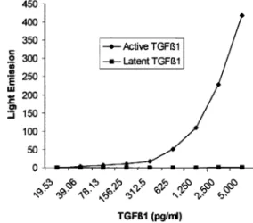
ma-ture active TGFb1 rather than latent forms. The
validity of ELISA for active TGFb1 was further
ver-ified by substituting the active TGFb1 with the latent TGFb1 (both from R&D Systems), indicating that the
cross-reactivity with latent TGFb1 was less than 1%
(Fig. 1). The assay was therefore considered to detect active TGFb1 in serum and the details of procedure are as follows.
A total of 96-well white plates (Dynatech Microfluor,
VA) were coated at 4°C overnight with 100 ml/well
mouse monoclonal antibody against TGFb (Genzyme,
MA) at 1mg/ml in PBS. After blocking with 1% BSA in
0.1 M PBS and 0.1% Tween 20 (PBS-Tween) for 2 h at room temperature, plates were washed three times with
PBS-Tween and 100 ml serum was added to each well.
A standard curve was generated using purified recombi-nant human TGFb1 (R&D Systems). The plate was left at 4°C overnight in a humidified box. Subsequently, the
wells were incubated with 100 ml polyclonal chicken
anti-TGFb1 antibody (R&D Systems), diluted 1/1000
(1 mg/ml) in PBS-Tween, for 3 h at 4°C. Three washes
with PBS-Tween were given between each procedure. After washing, the plates were incubated on a shaker with 100ml per well horseradish peroxidase conjugated rabbit anti-chicken IgG (Jackson ImmunoResearch Laboratories Inc. PA), at 1/2000 dilution (0.2mg/ml) in 1% BSA and PBS-Tween, for 30 min at room
tempera-ture. Finally, the plates were rinsed 3 times, 100 ml
Amerlite signal reagent (Amersham UK) was added to each well and the plate was read immediately in an Amerlite plate reader (Kodak Clinical Diagnostics). All the samples were run in duplicate and the measured values of light emission at 420 nm were converted into
absolute TGFb1 concentrations by reference to the
standard curve.
2.3. Measurement of (a+l)TGFb1
Serum was treated using pH 2.0 buffer to release the mature TGFb1 as described by Grainger et al. [20]. The
TGFb1 determined under this condition represents the
active plus the acid-activatable latent TGFb1 referred to as (a+l)TGFb1. For the activation of the samples, 50ml per well, test serum was added to a 96-well plate
followed by addition of 3ml 150 mM sodium phosphate
buffer at pH 2.0. The plate was incubated for 20 min at room temperature with agitation, then the mixture was
neutralised by addition of 3 ml 150 mM sodium
phos-phate buffer at pH 12.0. Forty-four microlitres of PBS-Tween was added to each well. Thus the sample was diluted 1/2. The acid-activated samples were transferred
to the coated plate to measure TGFb1 using the same
procedures as for the active TGFb1.
2.4. Detection of soluble CD105 in serum
CD105 was measured using an indirect sandwich ELISA wherein purified Mab E9, which specifically reacts with CD105 [21], and biotinylated Mab E9, in conjunction with streptavidin peroxidase, were used as capture and detection reagent [22], respectively. Briefly, 96-well white microtitre plates were coated with Mab E9 (100ml/well) diluted 1/1000 (1mg/ml) in 0.1 M PBS and incubated in a damp box overnight at 4°C. The coated plates were blocked using 1% BSA and PBS-Tween for 2 h at room temperature. Test samples
diluted 1/2 in PBS-Tween were added to the plates in
duplicate. One serum sample with a high level of
CD105 (approximately 100 ng/ml) was titrated to make
a standard curve in each plate. After incubation
overnight, at 4°C, 100 ml/well, biotinylated Mab E9
(1/2000 dilution; 0.2mg/ml) was added to the plates and incubated at 4°C in a humidified box for 3 h, followed by addition of 100 ml/well horseradish peroxidase con-jugated avidin at 1/2000 dilution in PBS-Tween and 1% BSA, which was incubated, with shaking, at room temperature for 30 min. Three washes with PBS-Tween
were carried out between each of the procedures. Fi-nally, 100 ml/well, Amerlite signal reagent (Amersham) was applied to each well and the light emission was immediately measured in an Amerlite plate reader.
2.5. Immunoassay of CD105/TGFb1 complex
The measurement of soluble CD105/TGFb1 complex
was performed using untreated serum. The procedures were the same as described in the assay for active
TGFb1, with the exception that the coating anti-TGFb
antibody was substituted by Mab E9 at 1 mg/ml in
order to capture the complex from the serum [23]. A
serum sample with a high level of CD105/TGFb1
com-plex (100 arbitrary units/ml) was serially diluted from 1/2 to 1/512 to generate a standard curve on each plate.
The assay showed no cross-reaction with CD105/
TGFb3 complex and exhibited a wide range of
detec-tion from 0.05 to 100 units/ml.
2.6. Immunodetection of TGFb3 in serum
The procedure to measure serum levels of TGFb3
was as follows: 96-well white plates were coated with
mouse monoclonal antibody against TGFb at 100 ml/
well (1 mg/ml diluted in PBS), and incubated in a
humidified box overnight at 4°C. The coated plates were subsequently blocked using 1% BSA and PBS-Tween for 2 h at room temperature. Test samples
diluted 1/2 in PBS-Tween were added to the plates in
duplicate. A standard curve was generated on each
plate using purified recombinant human TGFb3
(rhTGFb3; R&D Systems). After incubation at 4°C,
100 ml/well, goat anti-TGFb3 antibody (R&D Systems)
(1 mg/ml) diluted in PBS-Tween, were added to the
plates and incubated for 3 h at 4°C. After washing, the wells were incubated with agitation with 100 ml/well horseradish peroxidase conjugated donkey anti-goat antibody (Jackson ImmunoResearch Laboratories Inc.,
PA) at 0.2mg/ml diluted in 1% BSA in PBS-Tween for
30 min at room temperature. Following washing, Amerlite signal reagent, was added to each well, 100
ml/well, and the light emission immediately measured in an Amerlite plate reader. The measured values of light emission were converted into absolute concentrations
using a TGFb3 standard curve.
2.7. Measurement of CD105/TGFb3 complex
In the assay for CD105-TGFb3 complex, Mab E9
was used as coating antibody to capture the complex from the serum. The ELISA procedure was the same as
described in the assay for TGFb3, with the exception
that mouse monoclonal antibody to TGFb was
re-placed by Mab E9 (100 ml/well) at 1 mg/ml diluted in
Table 1
Some clinical characteristics of controls and patients with TVD and NCAa
Parameters TVD NCA Controls
LDL cholesterol, mmol/l 4.590.8
143923
138921 134922
Systolic blood pressure
Diastolic blood pressure 74915 78911 7899
aData represented as mean9SD or as median and range. Comparisons between groups were made by the Mann–Whitney U test. TVD, triple vessel disease; NCA, normal coronary angiogram; HDL, high density lipoprotein; LDL, low density lipoprotein.
* SignifiesP50.01.
antibody. Thus the molecules determined in this system
contain both CD105 and its ligand TGFb3. The
stan-dard curve was generated using a serum sample con-taining 50 units/ml of the complex on each plate.
2.8. Statistical analysis
Data are presented either as mean (when normally distributed) or median (when skewed) for the various parameters in each group. The logged data were first analysed by the Kruskall – Wallis test. Further compari-sons between groups were conducted using the Mann – Whitney U test. To determine associations between two variables, the Spearman’s correlation test was per-formed. For all the tests, the significance level was set at P50.05.
3. Results
3.1. Clinical data of the subjects
A summary of data related to the clinical tests and some risk factors are presented in Table 1. Compared to normal individuals, both groups of patients with ischemic heart disease (IHD) had higher total and LDL-cholesterol and triglyceride concentration, and
lower HDL-cholesterol levels (P in all cases was 5
0.01). Patients with triple vessel disease had lower levels of HDL-cholesterol compared to patients with normal
coronary angiograms (P50.01).
3.2. Quality assessment of the ELISAs
The quality of the immunoassays was assessed by measuring the sensitivities, intra-and inter-coefficients of variation (CV) and is illustrated in Table 2. The
cross-reactivity between active TGFb1 and latent
TGFb1 was less than 1% as determined by the inter-re-placement assays (Fig. 1). The Mab E9 is specific to
CD105 protein and recognises epitopes encoded by exons 7 and 8 [21].
3.3. Acti6e TGFb1 in serum
The TGFb1 measured in native serum is assumed to
represent the free mature TGFb1 as defined by the
characteristics of the antibodies. Data for active
TGFb1 in serum from the 36 TVD patients, 30 NCA
patients and 31 healthy controls are shown in Fig. 2(a)
and Table 3. Active TGFb1 levels in TVD patients were
significantly depressed compared with NCA and
healthy controls. Seventy-eight% (28/36) TVD patients
had TGFb1 levels below 100 pg/ml. In contrast, only
13% (4/30) NCA and 58% (18/31) controls showed
TGFb1 levels less than 100 pg/ml. No significant
differ-ence in active TGFb1 was seen between NCA and
healthy controls, but its level was significantly higher in these two groups compared with TVD patients (Table 3).
3.4. (a+l)TGFb1 in serum
Data for (a+l)TGFb1 concentrations in sera from
the normal individuals, NCA and TVD patients are
shown in Table 3 and Fig. 2(b). (a+l)TGFb1 levels
were markedly lower in the TVD group compared with
the control and NCA group (P=0.04 and P=0.003,
respectively). The (a+l)TGFb1 levels in NCA were
Table 2
Sensitivities and coefficients of variation (CVs) of the immunoassays
Table 3
Data for CD105, TGFb1, TGFb3 and CD105/TGFbcomplexes for patients with triple vessel disease, normal coronary angiograms and controlsa
Group Active TGFb1 (a+l) TGFb1 CD105 CD105/TGFb1 TGFb3 CD105/TGFb3
52.70 119.00 0.40 1.60 0
(0–15.9)
(27.2–272.0) (68.0–935.0) (0–12.5) (0–3.4) (0.1–15.0) (0–25.0)
Control 275.009100.90 434.969158.0 2.4191.10 1.2690.34 0.6290.24 0.0890.05 0
75.10 118.60 0.91 0.20 0.31
(50.8–4237.3) (0–5.70) (0–6.6) (0–1.1)
aData shown are mean9SEM and median (range). TVD, triple vessel disease; NCA, normal coronary angiograms. Kruskall–Wallis test and Mann–Whitney U test were used for comparisons andPvalues are indicated as: *P=0.04; **P50.02; ***P=0.01; ****P=0.003; *****P= 0.001.
lower than in normal, but the difference was not statis-tically significant (P\0.05).
3.5. Soluble CD105 in serum
The results of soluble CD105 in the three groups are presented in Table 3 and Fig. 2(c). Although values for CD105 in TVD patients were lower compared to both control and NCA group, the difference was only statis-tically significant against the latter group.
3.6. CD105/TGFb1 complex in serum
By using the optimised conditions, the complex levels were measured and data are shown in Table 3 and Fig.
2(d). Markedly increased CD105/TGFb1 complex
lev-els were noted in TVD group (P=0.002) compared
with normal subjects, but no significant difference was found between NCA and TVD or NCA and controls (P\0.05).
3.7. TGFb3 and CD105/TGFb3 complex in serum
Data for TGFb3 and the receptor-ligand complex
levels are presented in Table 3 and Fig. 2(e and f).
Although mean TGFb3 level was nearly threefold
higher in both TVD and NCA compared to controls, this difference was not statistically significant. In con-trast, significant increase in the CD105/TGFb3 complex levels was noted in the NCA group compared with
TVD and normal controls (P=0.02 and P=0.002,
respectively).
3.8. Correlation analysis
Spearman’s correlation test was performed to exam-ine the relationship among the various parameters in TVD and NCA groups. CD105 was weakly but posi-tively correlated with active TGFb1 (r=0.28,P=0.02) and (a+l)TGFb1(r=0.26, P=0.03). Active TGFb1
and (a+l)TGFb1 were strongly correlated with each
other (r=0.72, P=0.001). A positive correlation was
observed between CD105 and TGFb3 (r=0.43, PB
0.001), CD105 and CD105/TGFb3 complex (r=0.67,
PB0.001). In addition, TGFb3 and CD105/TGFb3
were also significantly correlated (r=0.70, PB0.001). None of the other correlations was significant.
4. Discussion
The observation that patients with coronary artery atherosclerotic disease have significantly depressed lev-els of TGFb1 is consistent with the report by Grainger et al. [18], who have suggested that it is a potential cardiovascular protector. The elevated levels of CD105/
TGFb1 complex seen in the present study in patients
with TVD indicate an increased interaction between
CD105 and TGFb1. This could be the reason why
CD105 and TGFb1 levels were decreased in these
pa-tients. The lack of a significant difference in TGFb3 levels in this cohort of patients does not mean that this isoform is not important in atherosclerosis, since it
might have been masked by the formation of CD105/
TGFb3 complexes, which were markedly elevated in
NCA group.
An increasing body of evidence indicates that en-dothelial dysfunction is a key event in the pathogenesis of atherosclerotic lesions. Endothelial injury is said to initiate atherosclerosis through the proliferation and migration of VSMCs, platelet adherence and release of numerous vasoactive growth factors [24]. One such
growth factor is TGFb1, which is involved in the
pathogenesis of atherosclerosis, through its pleiotropic effects. TGFb1 stimulates the synthesis and deposition of extracellular matrix in the vessel wall and suppresses inflammatory processes, which is thought to have
strong implications for atherogenesis [25]. TGFb1 is
potentially a negative growth modulator in a number of cell types. It inhibits the cell proliferation and migration
of VSMCs [26,27]. In addition, TGFb1 may play a role
in repairing ischemic injury by acting as a cardioprotec-tive agent by reducing superoxide anions [13].
Unlike other TGFb receptors, relatively little is
known about the function of CD105. CD105 is capable
of forming heteromeric complexes with the TGFb
re-ceptor I and rere-ceptor II [11]. The observation that patients with peripheral vascular disease but not my-ocardial infarction had elevated serum CD105 levels
[17] suggests that it may be important in only some instances of vascular injury. What could be a reason for this difference? Since the risk profiles (e.g. smoking, lipids, blood pressure, use of blood pressure modifying therapeutic agents) were similar in the two groups, therefore, it can probably be attributed to a difference in anatomy. Further studies on a larger group of pa-tients are warranted, to resolve possible significance of these results and to determine what effect it had on TGFb levels.
In the present study, we have therefore quantified the
soluble CD105, TGFb and the CD105/TGFb
com-plexes in controls and patients with NCA and TVD. In
agreement with a previous study [18], active TGFb1
was found to be markedly depressed in the TVD group compared with both NCA and controls. There was no significant difference between the NCA and control groups. TGFb1 in the circulation exists mainly in latent
forms. Active TGFb1 can be released in vivo from the
latent complexes after activation by plasmin [28] and thrombospondin [29] or conversion in vitro to active molecules by transient acidification using pH 2.0 buffer [20]. Quantification of the active plus acid-activatable
TGFb1 provided information about the proportion of
the active form in sera. The proportion of active
TGFb1 in the TVD group was much lower than in
either NCA or healthy controls, which indicated that
the active TGFb1 might be complexed with its binding
molecules to a greater extent in patients with TVD. Indeed increased levels of CD105/TGFb1 complex were observed in the TVD group. We propose that the
soluble CD105/TGFb1 complex may be responsible for
lowering active TGFb1 in TVD patients.
The assay for soluble CD105 utilising the same anti-body as capture and detection reagent, specifically mea-sures CD105 and cross-reaction with other proteins was undetectable. The presence of high levels of CD105 in NCA suggests that it may be an early event in atherosclerosis, whereas the occurrence of low levels of CD105 in TVD patients could partially be the result of
increased formation of CD105/TGFb1 complexes.
Physical insult, such as irradiation, can up-regulate CD105 expression in endothelial cells [5]. There is enhanced expression also in tissues undergoing
angio-genesis [4]. TGFb1 enhances CD105 expression in
The depression of TGFb1 in TVD patients could have been caused by a number of factors, such as lipoprotein (a) [Lp(a)] and plasminogen activator in-hibitor (PAI-1) [3,26]. As shown in this study, increased levels of CD105 in the NCA group and higher levels of
CD105/TGFb1 complex in TVD suggest that CD105
may make a considerable contribution to lowering TGFb1 levels. This action appears to be operative at an early stage of an atherogenic process.
In conclusion, the data presented here suggest that CD105 may initiate its effects in atherosclerosis at an
early stage through interaction with TGFb. However,
establishment of precise role for these molecules war-rants further investigations using in vitro and in vivo animal models together with examination of sequential blood samples from a large number of patients with atherosclerotic and related cardiovascular diseases.
Acknowledgements
CGL is a Wellcome Trust fellow.
References
[1] Blann AD, McCollum CN. von Willebrand factor, endothelial cell damage and atherosclerosis. Eur J Vasc Surg 1994;8:10 – 5. [2] de Caterina R, Basta G, Lazzerini G, Dell’Omo G, Petrucci R,
Morale M, Carmassi F, Pedrinelli R. Soluble vascular cell adhe-sion molecule-1 as a biohumoral correlate of atherosclerosis. Arterioscler Thromb Vasc Biol 1997;17:2646 – 54.
[3] de Bono D. Significance of raised plasma concentrations of tissue type plasminogen activator and plasminogen activator inhibitor in patients at risk from ischaemic heart disease. Br Heart J 1994;71:504 – 7.
[4] Kumar S, Wang JM, Bernabeu C. CD105 and angiogenesis. J Path 1996;178:363 – 6.
[5] Wang JM, Kumar S, van Agthoven A, Kumar P, Pye D, Hunter RD. Irradiation induces up-regulation of E9 protein in human vascular endothelial cells. Int J Cancer 1995;62:791 – 6. [6] Wang JM, Kumar S, Pye D, Haboubi N, al-Nakib L. Breast
carcinoma: comparative study of tumour vasculature using two endothelial cell markers. J Natl Cancer Inst 1994;86:386 – 8. [7] Krupinski J, Kaluza J, Kumar P, Kumar S, Wang JM. Role of
angiogenesis in patients with cerebral ischaemic stroke. Stroke 1994;25:1794 – 8.
[8] Rulo HF, Westphal JR, van de Kerkhof PC, de Waal RM, van Vlijmen IM, Ruiter DJ. Expression of endoglin in psoriatic involved and uninvolved skin. J Dermatol Sci 1995;10:103 – 9. [9] Seon BK, Matsuno F, Haruta Y, Kondo M, Barcos M.
Long-lasting complete inhibition of human solid tumors in SCID mice by targeting endothelial cells of tumor vasculature with antihu-man endoglin immunotoxin. Clin Cancer Res 1997;3:1031 – 44. [10] Kumar S, Ghellal A, Li C, Byrne G, Haboubi N, Wang JM,
Bundred N. Breast carcinoma: vascular density determined using CD105 antibody correlates with tumour progression. Cancer Res 1999;59:856 – 61.
[11] Yamashita H, Ichijo H, Grimsby S, Moren A, ten-Dijke P, Miyazona K. Endoglin forms a heteromeric complex with the
signalling receptors for transforming growth factor-b. J Biol Chem 1994;269:1995 – 2001.
[12] Massague J, Attisano L, Wrana JL. The TGFbfamily and its composite receptors. Trends Cell Biol 1994;4:172 – 8.
[13] Roberts AB, Sporn MB. Physiological actions and clinical appli-cations of transforming growth factor-b(TGFb). Growth Fac-tors 1993;8:1 – 9.
[14] Li CG, Wilson PB, Barber J, Levine E, Kumar S. TGFb1 levels in pre-treatment plasma identify patients at risk of developing post-radiotherapy fibrosis. Int J Cancer 1999;84:155 – 9. [15] Shah M, Foreman DM, Ferguson MW. Neutralisation of
TGFb1 and TGFb2 or exogenous addition of TGFb3 to cuta-neous rat wounds reduces scarring. J Cell Sci 1995;108:985 – 1002. [16] Kloen P, Gebhardt MC, Perez-Atayde A, Rosenberg AE, Springfield DS, Gold LI, Mankin HJ. Expression of transforming growth factor-b (TGFb) isoforms in osteosarcomas. Cancer 1997;80:2230 – 9.
[17] Blann AD, Wang JM, Wilson PB, Kumar S. Serum levels of the TGFbreceptor are increased in atherosclerosis. Atherosclerosis 1996;120:221 – 6.
[18] Grainger DJ, Kemp PR, Metcalfe JC, Liu AC, Lawn RM, Williams NR, Grace AA, Scholfield PM, Chauhan A. The serum concentration of active transforming growth factor-bis severely depressed in advanced atherosclerosis. Nat Med 1995;1:74 – 9. [19] Adam PJ, Clesham GJ, Weissberg PL. Expression of endoglin
mRNA and protein in human vascular smooth muscle cells. Biochem Bioph Res Co 1998;247:33 – 7.
[20] Grainger DJ, Mosedale DE, Metcalfe JC, Weissberg PL, Kemp PR. Active and acid-activatable TGFbin human sera, platelets and plasma. Clin Chim Acta 1995;235:11 – 31.
[21] Pichuantes S, Vera S, Bourdeau A, Pece N, Kumar S, Wayner EA, Letarte M. Mapping epitopes to distinct regions of the extracellular domain of endoglin using bacterially expressed re-combinant fragments. Tissue Antigens 1997;50:265 – 76. [22] Wang JM, Wilson PB, Kumar S, Pye D, Hunter RD.
Quantita-tion of endothelial cell specific protein E9 employing a single monoclonal antibody in an indirect sandwich ELISA. J Immunol Methods 1994;171:55 – 64.
[23] Li CG, Wilson PB, Bernabeu C, Raab U, Wang JM, Kumar S. Immunodetection and characterisation of soluble CD105-TGFb complexes. J Immunol Methods 1998;218:85 – 93.
[24] Ruschitzka FT, Noll G, Luscher TF. The endothelium in coro-nary artery disease. Cardiology 1997;88:3 – 19.
[25] Waltenberger J, Akyurek ML, Aurivillius M, Wanders A, Larsson E, Fellstrom B, Funa K. Ischaemia-induced transplant arteriosclerosis in the rat: induction of peptide growth factor expression. Arterioscler Thromb Vasc Biol 1996;16:1516 – 23. [26] Kojima S, Harpel PC, Rifkin DB. Lipoprotein (a) inhibits the
generation of transforming growth factor-b: an endogenous in-hibitor of smooth muscle cell migration. J Cell Biol 1991;113:1439 – 43.
[27] Grainger DJ, Kirschenlohr HL, Metcalfe JC, Weissberg PL, Wade DP, Lawn RM. Proliferation of human smooth muscle cells promoted by lipoprotein (a). Science 1993;260:1655 – 8. [28] Sato Y, Tsuboi R, Lyons R, Moses H, Rifkin DB.
Characterisa-tion of the activaCharacterisa-tion of latent TGFbby co-cultures of endothe-lial cells and pericytes or smooth muscle cells: a self-regulating system. J Cell Biol 1990;111:757 – 63.
[29] Schultz-Cherry S, Ribeiro S, Gentry L, Murphy-Ullrich JE. Thrombospondin binds and activates the small and large forms of latent transforming growth factorbin a chemically defined system. J Biol Chem 1994;269:26775 – 82.
[30] Lastres P, Letamendia A, Zhang H, Rius C, Almendro N, Raab U, Lopez L, Langa C, Fabra A, Letarte M, Bernabeu C. Endoglin modulates cellular responses to TGFb1. J Cell Biol 1996;133:1109 – 21.