Clinical and Histopathological Characteristic of Salivary Gland
Carcinoma in Dr. Hasan Sadikin General Hospital in 2009–2012
Fatimah Lidya Andriani,1 Ismet Muchtar Nur,2 Sally Mahdiani3
1Faculty of Medicine Universitas Padjadjaran, 2Department of Anatomical Pathology Faculty of Medicine Universitas Padjadjaran/Dr. Hasan Sadikin General Hospital Bandung, 3Department of Otolaryngology-Head & Neck Surgery Faculty of Medicine Universitas Padjadjaran/Dr. Hasan
Sadikin General Hospital Bandung
Abstract
Background: Salivary gland neoplasm is one of the rare neoplasm. The frequency of this neoplasm is lower than 2% of all type of tumors in human. Malignant salivary gland tumor comprises 6% of all head and neck tumors. Data about salivary gland carcinoma are still limited. The aim of this study was to determine the frequency of malignant salivary gland based on the patients’ age, gender, site of lesion and histopathology type.
Methods: This study was conducted descriptively. There were 97 subjects found from histopathological form that had been examined in Department of Anatomical Pathology, Dr. Hasan Sadikin General Hospital in 2009–2012. Total sampling technique was used and all data about patients’ age, gender, site of lesion and histopathology type were collected and analyzed.
Results: Of 97 cases, age group 50–59 years old had the highest frequency (29%). The prevalence in male was more frequent than female with male:female ratio was 1.4:1. The most common site of carcinoma was found in parotid gland (45%). Mucoepidermoid carcinoma was the most common histopathology type found in this study (28%).
Conclusions: Salivary gland carcinoma is still a rare malignant case in Dr. Hasan Sadikin General Hospital. Carcinoma in parotid gland was the most common site and mucoepidermoid carcinoma was the most common histopathology type. [AMJ.2016;3(1):54–8]
Keywords: Carcinoma, histopathology, salivary gland
Correspondence: Fatimah Lidya Andriani, Faculty of Medicine, Universitas Padjadjaran, Jalan Raya Bandung-Sumedang Km.21, Jatinangor, Sumedang, Indonesia, Phone: +62 8127577725 Email: lidyafla@gmail.com
Introduction
Salivary gland neoplasm is one of the rare neoplasms. Department of Ear Nose Throat (ENT) Wroclaw, Polska reported that 304 salivary glands tumor patient were with 83.9% benign tumor and 16.1% malignant tumor in 2001–2010.1 Its peak incidence was varied according to the previous studies. Most of them reported that salivary gland tumor reached the incident peak in sixth-seven decade of life.2
Five years life expectancy ranged between 5–95% depend on various factors including site of lesion, histopathology type, stage of tumor and the involvement of facial nerve or surrounding structure. Original site of salivary glands cancer is one of significant factors. Cancer that can be excised fully from parotid gland has the better result, followed by submandibular gland, sublingual gland and minor salivary glands.3 Based on the background above and
limited data about salivary gland carcinoma, the study was conducted concerning salivary glands carcinoma based on patients’ age, gender, site of lesion and histopathological type in Department of Anatomical Pathology Dr. Hasan Sadikin General Hospital.
Methods
as salivary gland carcinoma. The incomplete data of histopathological form was excluded from this study. This descriptive study used retrospective methods.
Variables of this study were patients’ age and gender, site of lesion and histopathology type. All cases were classified according to the criteria suggested by 2005 World Health Organization (WHO) histological classification. Data were collected and analyzed using Microsoft Excel 2007. The frequency and percentage of the mentioned variable were calculated. Collected data from medical records were kept confidentially and vanished after the research was done.
Results
During 2009–2012, 97 cases of salivary gland carcinoma were found in Department of Anatomical Pathology, Dr. Hasan Sadikin General Hospital.
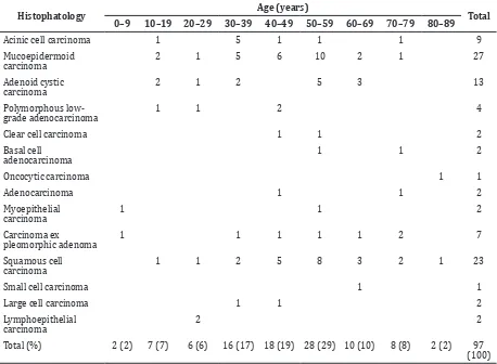
Patients’ age for cases was ranged from 6–86 years old. The cases increased along with
the increasing of age. The peak of salivary gland carcinoma was in the fifth decade of life. The lowest case were found in first and eighth decade of life.
Mucoepidermoid carcinoma was the most common of all carcinoma in salivary gland, followed by squamous cell carcinoma, adenoid cystic carcinoma and acinic cell carcinoma. Mucoepidermoid carcinoma and squamous cell carcinoma were distributed widely in the age group and had the peak in fifth decade of life. The peak of acinic cell carcinoma was in third decade of life (Table 1).
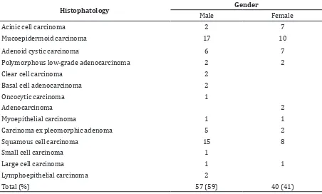
Salivary gland carcinoma was more frequent in male than female with male:female ratio was 1.4:1 and the most common histopathological type of tumor was mucoepidermoid carcinoma in both male and female (Table 2).
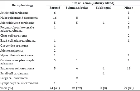
Almost half of the cases were found in parotid gland, followed by minor salivary glands, submandibular gland and sublingual gland. Mucopeidermoid carcinoma was mostly found in parotid gland. Adenoid cystic carcinoma and squamous cell carcinoma
Table 1 Histopathological Type of Salivary Gland Carcinoma based on Age Group
Histophatology Age (years) Total
0–9 10–19 20–29 30–39 40–49 50–59 60–69 70–79 80–89
Acinic cell carcinoma 1 5 1 1 1 9
Clear cell carcinoma 1 1 2
Basal cell adenocarcinoma
1 1 2
Oncocytic carcinoma 1 1
Adenocarcinoma 1 1 2
Myoepithelial
Small cell carcinoma 1 1
Large cell carcinoma 1 1 2
Lymphoepithelial carcinoma
2 2
Table 2 Histopathological Type of Salivary Gland Carcinoma based on Gender
Histophatology Gender
Male Female
Acinic cell carcinoma 2 7
Mucoepidermoid carcinoma 17 10
Adenoid cystic carcinoma 6 7
Polymorphous low-grade adenocarcinoma 2 2
Clear cell carcinoma 2
Basal cell adenocarcinoma 2
Oncocytic carcinoma 1
Adenocarcinoma 2
Myoepithelial carcinoma 1 1
Carcinoma ex pleomorphic adenoma 5 2
Squamous cell carcinoma 15 8
Small cell carcinoma 1
Large cell carcinoma 1 1
Lymphoepithelial carcinoma 2
Total (%) 57 (59) 40 (41)
widely distributed at each site of lesion. Squamous cell carcinoma had the highest case in minor salivary glands. Oncocytic carcinoma and adenocarcinoma only affected parotid gland. Large cell carcinoma was only discovered in submandibular gland (Table 3).
Discussion
In the present review of 97 salivary gland carcinoma, group aged 50–59 years old was the highest case (29%). In 30–39, 40–49 and 60–69 years old group also showed significant number. This result is similar to the reference which reported that frequency of salivary gland carcinoma increase in fourth to seventh decade of life.3,4 The increase in case occured about 6.9% compared to the previous study by Achmad Thohir in Department of Anatomical Pathology Dr. Hasan Sadikin General Hospital in 2003–2006.
Salivary gland carcinoma more often affected male, with male:female ratio was 1.4:1. This result is similar to the study in Iran that reported predominance for men with male:female ratio was 1.4:1.5 In contrast, some references and prior studies reported that women were more often affected than men.4,6,7
According to site of lesion, carcinoma is
more frequent in parotid gland, followed by minor salivary glands, submandibular gland and sublingual gland. This result was similar to the reference that reported that 65–80% salivary gland carcinoma occured in parotid gland, 9–23% in minor salivary glands, 10% in submandibular gland and less than 1% in sublingual gland.3,5 Only 3 cases
were identified in the sublingual gland. This confirmed the rarity of salivary tumors at this site, as mentioned in other studies.5,7–9
Histopathological type that is mostly found was mucoepidermoid carcinoma. In the second place, there was squamous cell carcinoma, followed by adenoid cystic carcinoma. Yu-Long Wang et al.10 reported that mucoepidermoid carcimona was the most common histophatology type (24.6%), followed by adenoid cyctic carcinoma (18%) in Chinesse population. Some reference also reported that mucoepidermoid carcinoma was the most common carcinoma.11 According to WHO, incidence of squamous cell carcinoma was less than 1% of all carcinoma in salivary gland.5 Prior study by Achmad Thohir in Dr. Hasan Sadikin General Hospital reported that adenoid cystic carcinoma was the most common histopathology.
39 years old. This result was similar to the reference that reported that this carcinoma was distributed widely in any group of age.4,12 Mucoepidermoid carcinoma is also distributed widely in age group. Enhancement developed in third to fifth decade of life. This malignant tumor occured in child as well. Some author reported that mucoepidermoid carcinoma is common in child and adult, distributed widely in age group, and had the peak in third to fifth decade of life.12 Adenoid cystic carcinoma also spread in any age group and had the peak in 50–59 years old. This result was similar to the reference that reported that this carcinoma is distributed in all age group and increased in fourth to sixth decade of life.3,4,12 Squamous cell carcinoma was found in almost all age group and increased in 40–79 years old. Previous study reported that squamous cell carcinoma was found from 7–95 years old with peak in seventh decade of life.12
Carcinoma ex pleomorphic and squamous cell carcinoma were most often affected men with male:female ratio was 2:1, as reported previously. Acinic cell carcinoma had higher frequency in women with female:male ratio was 2.8:1. This result was similar to previous study by Ilayaraja2 Mucoepidermoid carcinoma were mostly found in men, while another
study reported that it was more often found in women than men. Polymorphous low-grade adenocarcinoma showed equal case between men and women. However, some authors reported that two to three times is common in women than men.4,12
According to site of lesion, acinic cell carcinoma more often occured in parotid gland and was followed by minor salivary glands, as had been documented that 80% in parotid gland and 17% in minor salivary glands. Mucoepidermoid carcinoma was mostly found in parotid gland, similar to the reference that reported that it was common in major salivary glands, espescially parotid gland.4 The frequency in parotid gland is about 60%.12 Adenoid cystic carcinoma was mostly found in parotid gland and submandibular gland, followed by minor salivary gland and sublingual gland, as mentioned in reference.4 However, other references reported that it was more often affected by minor than mayor salivary gland. Carcinoma ex pleomorphic was also more often found in parotid gland, similar to the reference about 81.7%.12 Polymorphous low-grade adenocarcinoma was more often affected minor salivary gland. This result was similar to the reference that reported 60% occured in minor salivary gland, especially Table 3 Histopathological Type of Salivary Gland Carcinoma based on Site of Lesion
Histophatology Site of Lesion (Salivary Gland)
Parotid Submandibular Sublingual Minor
Acinic cell carcinoma 6 3
Mucoepidermoid carcinoma 16 8 3
Adenoid cystic carcinoma 5 5 1 2
Polymorphous low-grade adenocarcinoma
1 3
Clear cell carcinoma 2
Basal cell adenocarcinoma 1 1
Oncocytic carcinoma 1
Adenocarcinoma 2
Myoepithelial carcinoma 1 1
Carcinoma ex pleomorphic adenoma
5 1 1
Squamous cell carcinoma 5 4 1 13
Small cell carcinoma 1
Large cell carcinoma 2
Lymphoepithelial carcinoma 1 1
palatum.Squamous cell carcinoma is mostly found in minor salivary gland as well, but in contrast with the previous study that reported was more often found in parotid gland and submandibular gland.4,12
Limitations of the study were limited time for data collection and patients’ identity is not completely written in histopathological form.
The conclusion of this study is the frequency of salivary gland carcinoma increased in age group 50–59 years old. Male and parotid gland were the most affected and mucoepidermoid carcinoma was the most frequent lesion, followed by squamous cell carcinoma and adenoid cystic carcinoma.
References
1. Kubacka M, Orendorz-Fraczkowska K, Pazdro-Zastawny K, Morawska-Kochman M, Kręcicki T. Epidemiological evaluation of salivary gland tumors in the Wroclaw ENT Department patients in the years 2001– 2010. Polish Otolaryngol. 2013;67(1):30– 3.
2. Ilayaraja V, Prasad H, Anuthama K, Sruthi R. Acinic cell carcinoma of minor salivary gland showing features of high grade transformation. J Oral Maxillofac Pathol. 2014;18(1):97–101.
3. Faquin WC, Powers CN. Salivary Gland Cytopathology. New York: Springer; 2008. 4. Barnes L, Eveson JW, Reichart P, Sidransky
D, editors. World Health Organization classification of tumours pathology & genetics head and neck tumours. Lyon: IARC Press; 2005.
5. Ansari MH. Salivary gland tumord in an Iranian population : a retrospective study of 130 cases. J Oral Maxillofac Surg. 2007;65(11):2187–94.
6. Kumar V, Abbas AK, Fausto N, Aster JC. Robbin and Cotran pathologic basis of disease. 8th ed.Philadelphia: Elsevier Saunders; 2010. p.756–9.
7. De Oliveira FA, Duarte EC, Taveira CT, Máximo AA, de Aquino EC, Alencar Rde C, et al. Salivary gland tumor: a review of 599 cases in a Brazilian population. Head Neck Pathol. 2009;3(4):271–5.
8. Ito FA, Ito K, Vargas PA, de Almeida OP, Lopes MA. Salivary gland tumors in a Brazilian population: a retrospective study of 496 cases. Int J Oral Maxillofac Surg. 2005;34:533–6.
9. Al-Kahteeb TH, Ababneh KT. Salivary tumors in north Jordanians: a descriptive study. Oral Surg Med Oral Pathol Oral Radiol Endod. 2007;103(5):e53–9.
10. Wang YL, Zhu YX, Chen TZ, Wang Y, Sun GH, Zhang L, et al. Clinicopathologic study of 1176 salivary gland tumors in a Chinese population: experience of one cancer center 1997–2007. Acta otolaryngol. 2012;132(8):879–86.
11. Ovchinsky A, Har-El G. Salivary Gland Enlargement. In: Lucente FE, Har-El G, editors. Essentials of Otolaryngology. 5thed. Philadephia: Lippincot Williams & Wilkins; 2004.