This page
Published by New Age International (P) Ltd., Publishers All rights reserved.
No part of this ebook may be reproduced in any form, by photostat, microfilm, xerography, or any other means, or incorporated into any information retrieval system, electronic or mechanical, without the written permission of the publisher. All inquiries should be emailed to [email protected]
ISBN : 978-81-224-2424-9
PUBLISHINGFORONEWORLD
NEW AGE INTERNATIONAL (P) LIMITED, PUBLISHERS
Knowing others is intelligence;
knowing yourself is true
wisdom. Mastering others is
strength; mastering yourself
is true power.
THIS PAGE IS
FOREWORD
As a foreword, I would like to express that the source of inspiration behind the
crea-tion of this book is Prof. K. Sambamurthy. He is among those pioneers who started the
Biotechnology Department in Andhra University, Visakhapatnam. He also held the most
coveted status of being the Emeritus Fellow, University Grants Commission, New Delhi.
His first book
Pharmaceutical Engineering
was a great success in India and abroad.
The great response from the students and professors encouraged him to initiate another
book on
Pharmaceutical Biotechnology
but unfortunately his sudden untimely demise
stopped the progress of the book half way through.
His dreams are being fulfilled by Prof. Asuthosh Kar who has taken a lot of trouble
and interest in the development and completion of the book.
My heart felt thanks and gratitude to Prof. Asuthosh Kar for making my husband’s
dream a reality.
I hope and believe that this book would be of great help to the students in the
disci-pline of pharmaceutical sciences.
THIS PAGE IS
PREFACE
Biotechnology essentially and predominantly deals with the meticulous application of living organisms or their corresponding products in a variety of large-scale industrial processes. Besides, biotechnology is extremely multidisciplinary in nature ; it has its foundations and domain prominently spread in a wide spectrum of fields, such as : pharmaceutical sciences, microbiology, biology, biochem-istry, molecular biology, genetics, genomics, genetic engineering, chembiochem-istry, and chemical and process engineering. Therefore, it may be genuinely and rightly regarded as a series of ‘enabling technologies’ embracing the practical application of host specific organisms and their respective cellular components to either environmental management or to manufacturing and service industries.
Interestingly, from a historical aspect biotechnology could be regarded as a pragmatic, realistic, and tangible strategy to an ‘art’ more than a ‘science’, which may be enormously exemplified and duly expatiated in the commercial production of wines, beers, cheeses, and the like, whereby the modus
operandi of various techniques involved were well-known and reproducible, but the exact molecular
mechanisms were not known adequately. Nevertheless, at present biotechnology is passing through an amazing growth phase whose ultimate destiny is not too far in sight. With the advent of major advances in the better in-depth knowledge of ‘microbiology’ and ‘biochemistry’, these molecular mechanisms (viz., processes) have been rendered more logically understandable.
Pharmaceutical Biotechnology, based entirely on modern biotechnological techniques, as to date encompass a wider range of altogether newer medicinal compounds, e.g., antibiotics, vaccines, and monclonal antibodies (MABs) that may now be produced commercially using well-defined, optimized, and improved fermentative methodologies. In fact, genetic engineering has brought in a sea change by virtue of the directed construction of microorganisms resulting in a plethora of newer life-saving drugs. The present textbook on ‘Pharmaceutical Biotechnology’ is strictly developed, structured, ex-panded, and expatiated along the guidelines provided by AICTE syllabus for B. Pharmacy–2000. It essentially consists of five main chapters, namely : Immunology and Immunological Preparations ; Genetic Recombination ; Antibiotics ; Microbial Transformations ; and Enzyme Immobilization. In addition to this, there are fiveauxilliary chapters, namely : Advent of Biotechnology ; Biosensor Technology ; Bioinformatics and Data Mining ; Regulatory Issues in Biotechnology ; and Safety in Biotechnology, which have been duly included so as to stimulate the students’ interest and expand their knowledge.
Each chapter has been carefully and adequately supported with a brief introductory note, fol-lowed by theoretical aspects, graphics, neat well-labeled diagrams, explanations, discussions, and pro-fusely supplemented with appropriate examples to make the relevance of each topic more comprehensi-ble to the students of Pharmacy both in India and abroad.
It is earnestly believed that students, learning Pharmaceutical Biotechnology will certainly find this text not only useful but also a good companion for further pursuit of higher knowledge. Besides, research scientists, teachers, food technologists, industrial technical personnels, postgraduate students involved in ‘industrial microbiology’ shall definitely be benefitted from this practical approach to the broader horizons of biotechnology.
The authors solemnly believe that this modern, well documented, lucid and easy presentation of topics contained in the textbook on ‘Pharmaceutical Biotechnology’ will prove to be of immense value to students, teachers, and practising researchers.
K. SAMBAMURTHY
THIS PAGE IS
CONTENTS
Foreword (vii)
Preface (ix)
1. Immunology and Immunological Preparations ... 1
1. Introduction ... 1
2. Principles ... 2
3. Antigens and Haptens ... 3
3.1. Antigens ... 3
3.2. Haptens ... 4
4. Immune Systems ... 5
4.1. Manipulation of Immune System ... 8
4.2. Types of Immunity ... 8
4.2.1. Humoral Immunity ... 8
4.2.1.1. B Lymphocytes ... 9
4.2.1.2. Immunological Peptides (IDPs) ... 10
4.2.1.3. Antigen Presenting Cell (APC) ... 11
4.2.1.4. T Cell Subsets ... 11
4.2.1.5. Class II MHC (Major histocompatibility complex)
proteins ... 25
4.2.2. Cell-Mediated Immunity (CMI) ... 30
4.2.2.1. Immunosuppression ... 32
4.2.2.2. Privileged graft Sites ... 35
4.2.2.3. Graft-Vs-Host Disease (GVHD) ... 37
4.2.3. Innate (or Natural Immunity) ... 40
4.2.4. Acquired Immunity ... 40
4.2.4.1. Actively Acquired Immunity ... 40
4.2.4.2. Passively Acquired Immunity ... 41
4.2.5. Non-Specific Immunity ... 41
5. Antigen Antibodies Reactions and their Applications ... 43
5.1. Antigen Antibodies Reactions ... 44
5.1.1. Antigens ... 44
5.1.2. Antibodies ... 46
5.1.3. Immunoglobulins as Antigens ... 48
5.1.4. Structure of Antibody ... 49
5.1.4.1. Ig A Molecule ... 49
5.1.4.2. Ig G Molecule ... 50
5.1.5. Monoclonal Antibodies (MABs) ... 53
5.1.5.1. MABs in Diagnostics ... 54
5.1.5.2. MABs in Imaging and Therapy ... 54
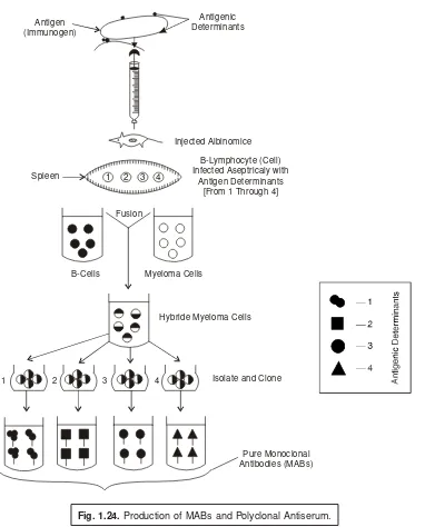
5.1.5.3. Production of Monoclonal Antibodies (MABs) ... 55 5.1.5.4. Application of Monoclonal Antibodies (MABs) ... 58
6. Hypersensitivity Reaction ... 65
6.1. Types of Hypersensitivity Reaction ... 66
6.1.1. Type-I : Anaphylactive Hypersensitivity ... 66
6.1.2. Type-II : Antibody Dependent Cytotoxic Hypersensitivity ... 69
6.1.3. Type-III : Complex Mediated Hypersensitivity ... 71
6.1.4. Type-IV : Cell-mediated or Delayed Type Hypersensitivity ... 75
6.1.5. Type-V : Stimulatory Hypersensitivity ... 76
7. Vaccines : Preparation, Standardization and Storage ... 77
7.1. Definitions ... 77
7.2. Historical ... 78
7.3. Classification of Vaccines ... 78
7.3.1. Synthetic Peptide Vaccines ... 80
7.3.2. Multivaccine System ... 80
7.3.3. Bacterial Vaccines ... 81
7.3.4. DTP-Vaccines ... 81
7.3.5. Typhoid-Paratyphoid A and B Vaccine (TAB-Vaccine) ... 82
7.3.6. Other Bacteria Vaccines ... 82
7.3.7. Typhoid and Tetanus Vaccine ... 83
7.3.8. Anthrax Vaccine ... 83
7.3.9. Q-Fever Vaccine ... 84
7.3.10. Leprosy Vaccines ... 84
7.3.11. Whooping Cough Vaccine (Pertussis Vaccine) ... 85
7.3.12. Diphtheria Vaccines ... 86
7.3.13. Varicella-Zoster Vaccine ... 88
7.3.14. Viral and Rickettsial Vaccines ... 88
7.3.15. Smallpox Vaccine ... 90
7.3.16. Vaccines for Special Protection ... 94
7.3.17. Rabies Vaccines ... 94
7.3.18. Influenza Vaccine (Flu Fever Vaccine) ... 97
7.3.19. Inactivated Influenza Vaccine ... 99
7.3.19.1. Inactivated Influenza Vaccine (Split-Virion) ... 100 7.3.19.2. Inactivated Influenza Vaccine (Surface Antigen) ... 100
7.3.20. Polio Vaccine ... 101
7.3.20.2. Sabin Type Polio Vaccine ... 102
7.3.20.3. Sabin Type Polio Vaccine ... 104
7.3.20.4. Inactivated Poliomyelitis Vaccine ... 105
7.3.20.5. Poliomyelitis Vaccine, Live (Oral) ... 105
7.3.21. Cancer Vaccine ... 106
7.3.22. Birth Control Vaccine for Women ... 106
7.3.23. AIDS-Vaccine ... 107
7.3.24. Pneumococcal Vaccine ... 109
7.3.25. Measles Vaccine, Live ... 109
7.3.25.1. Measles, Mumps and Rubella (MMR) Vaccine ... 110
7.3.25.2. Germanal Measles (Rubella) Vaccine ... 111
7.3.26. Meningococcal Polysaccharide Vaccine ... 111
7.3.27. Future Development Scope of Vaccines ... 112
7.3.27.1. Vaccine against Alzheimeir’s Disease ... 112
7.3.27.2. Vaccine for Meningitis C ... 112
7.3.27.3. Super Vaccine ... 113
7.3.27.4. Immunomodulators ... 114
7.3.27.5. Vaccination with Gas Lighter ... 114
7.3.27.6. Vaccine against Cervical Cancer ... 114
7.3.27.7. Vaccination without Needles ... 114
2. Genetic Recombination ... 119
1. Introduction ... 119
2. Transformation ... 122
2.1. Agrobacterium-Mediated Gene Transfer ... 122
2.1.1. Co-culture with Tissue Explants ... 122
2.1.2. In Planta Transformation ... 124
2.2. Agroinfection ... 125
2.3. Direct Gene Transfers ... 125
2.3.1. Chemical Methods ... 126
2.3.2. Electroporation ... 126
2.3.3. Particle Gun Delivery ... 128
2.3.4. Lipofection ... 130
2.3.5. Microinjection ... 130
2.3.6. Macroinjection ... 131
2.3.7. Pollen Transformation ... 132
2.3.8. DNA Delivery via Growing Pollen Tubes ... 132
2.3.9. Laser-Induced Gene Transfer ... 133
2.3.11. Transformation by Ultrasonication ... 134
3. Conjugation ... 134
3.1. Organismal ... 135
3.2. Cellular ... 135
3.3. Molecular ... 135
4. Transduction ... 139
5. Protoplast Fusion ... 141
5.1. Spontaneous Fusion ... 141
5.2. Induced Fusion ... 142
5.2.1. Sodium Nitrate (NaNO3) Treatment ... 143
5.2.2. Calcium Ions (Ca2+) Treatment at High pH ... 145
5.2.3. Propylene Glycol (PEG) Treatment ... 145
5.2.4. Electrical Impulse (Fusion) ... 145
6. Gene Cloning ... 146
6.1. Cloning Process ... 147
6.1.1. DNA-Cloning ... 148
6.1.2. Cloning Larger DNA Fragments in Specified
Cloning Vectors ... 148
6.1.3. Cloning Eukaryotic DNAs in Bacterial Plasmids ... 149
6.1.4. Cloning Eukaryotic DNAs Phase Genomes ... 151
6.1.5. Cloning cDNAs ... 153
6.1.6. Expression Cloning ... 155
6.1.7. Amplifying DNA : The Polymerase Chain Reaction (PCR) ... 155
7. Development of Hybridoma for Monoclonal Antibodies (MABs) ... 157
7.1. The Principle of Monoclonal Antibody Production ... 158
7.2. Cell Fusion ... 158
8. Drugs Produced by Biotechnology ... 161
8.1. Alteplase ... 162
8.2. Humulin ... 164
8.3. Humatrope : Growth Hormone ... 168
8.4. Hepatitis B [Recombinant HB (Merck)—A Hepatitis B Vaccine] ... 169
3. Antibiotics ... 173
1. Historical Development of Antibiotics ... 173
2. Antimicrobial Spectrum and Methods Used for their Standardization ... 174
2.1. Cylinder-Plate Method ... 179
2.2. Turbidimetric Method ... 179
3. Screening or Soil for Organisms Producing Antibiotics ... 179
3.2. Secondary Screening ... 183
3.2.1. Methodology ... 183
3.2.2. Salient Features of Secondary Screening ... 184
4. Fermentors [or Bioreactors] ... 186
4.1. Salient Features of Bioreactors ... 188
4.2. Classifications ... 189
4.2.1. Solid State Fermentation ... 189
4.2.2. Anaerobic Fermentation ... 191
4.2.3. Aerobic Fermentation ... 192
4.2.3.1. Stirred-tank Type Fermentors (or Stirred Bioreactors) ... 192
4.2.3.2. Air-lift Type Fermentors ... 194
4.2.4. Immobilized Cell Bioreactors ... 194
4.2.4.1. Immurement Cultures ... 195
4.2.4.2. Entrapment Cultures ... 195
4.3. Design and Bioreactors (Fermentor Variants) ... 196
4.3.1. Fermacell (Laboratory) Fermentor ... 196
4.3.2. Bubble-Cap Fermentor ... 198
4.3.3. Loop (Recycle) Bioreactor ... 200
4.3.4. Tower Bioreactor ... 200
4.3.5. Activated Sludge Bioreactor ... 201
4.3.6. Continuous Flow Stirred Tank Bioreactor ... 202
4.3.7. Packed Bed Bioreactor ... 202
4.3.8. Trickling Film Bioreactor ... 203
4.3.9. Mist Bioreactor ... 204
4.3.10. Rotating Drum Bioreactor ... 205
4.3.11. Bubble Column Bioreactor ... 206
4.3.12. Commercial Fermentation Plant ... 207
5. Mutants ... 209
5.1. Isolation of Mutants ... 209
5.1.1. Method of Causing a Mutation ... 210
5.1.2. Somaclonal Variation ... 210
5.1.2.1. Isolation of Somaclonal Variants ... 210
5.2. Factor Influencing Rate of Mutation ... 214
5.2.1. Conditional Mutation ... 214
5.2.2. Radiation Induced Mutations ... 214
5.2.3. Effect of UV Radiation ... 215
5.2.4. Chemically Induced Mutations ... 216
6. Design of Fermentation Processes ... 220
6.1. Quality of Water ... 220
6.2. Quality Control of Raw Materials ... 220
6.3. Nutritional Requirements ... 220
6.4. Sterilization Practices ... 221
6.5. Media Preparation ... 221
6.5.1. Solid Substrate Fermentation ... 222
6.5.2. Submerged Fermentation ... 222
6.5.3. Downstream Processing ... 224
6.5.4. Technology of Mammalian and Plant-cell Culture ... 225
6.5.5. Cell Recycle Technique ... 228
7. Production of Antibiotics (Isolation of Fermentation Products) ... 228
7.1. The Penicillins ... 230
7.1.1. Genes in Penicillin Biosynthesis ... 230
7.1.2. The Penicillin Variants ... 234
7.1.3. Production of Benzylpenicillins [Penicillin G] ... 235
7.1.3.1. Inoculum ... 235
7.1.3.2. Production Media ... 237
7.1.3.3. Biomass Production ... 237
7.1.3.4. Course of Typical Penicillin Fermentation ... 238
7.1.3.5. Penicillin Nucleus : Two Amino Acids ... 240
7.1.3.6. Role of Enzyme Penicillinase ... 241
7.1.3.7. Penicillin Production and Recovery ... 241
7.2. Streptomycin ... 244
7.2.1. Chemical Structure ... 245
7.2.2. Choicest Medium ... 246
7.2.3. Inoculum ... 247
7.2.4. Streptomycin Production ... 247
7.3. The Tetracyclines ... 248
7.3.1. Salient Features of the Tetracyclines ... 248
7.3.2. Nomenclatures ... 249
7.3.3. General Characteristics of the Tetracyclines ... 249
7.3.3.1. Tetracycline ... 251
7.4. Vitamin B12 (Cyanocobalamine; Cobamide) ... 253
7.4.1. Vitamin B12 from Propionobacterium Shermanii ... 256
7.4.2. Vitamin B12 from Pseudomonas Denitrificans ... 256
4. Microbial Transformations ... 261
1. Introduction ... 261
2. Types of Reactions Mediated by Microorganisms ... 262
2.1. Vinegar (Acetic Acid) Production ... 263
2.1.1. Traditional Method ... 263
2.1.2. Aerobic Fermentation Process ... 264
2.1.3. Orleans Process ... 265
2.1.4. Packed-Generator Process ... 265
2.1.5. Trickling Generator ... 269
2.1.6. Submerged Fermentor ... 271
2.2. Gluconic Acid Production ... 273
2.3. Antibiotic Production ... 275
2.4. Single-Cell Proteins (SCPs) from Methanol ... 275
2.5. Lactic Acid Production ... 278
2.6. Kojic Acid ... 281
2.7. Itaconic Acid ... 282
3. Design of Biotransformation Processes ... 283
3.1. Methodologies for Biotransformation ... 289
3.1.1. Growing Cultures ... 289
3.1.2. Resting Cells ... 291
3.1.3. Immobilized Cells ... 291
4. Selection of Organisms ... 291
5. Biotransformation Process and Its Improvements with Special
Reference to Steroids ... 295
5.1. Biotransformation of Steroids ... 296
5.1.1. Types of Transformations/Biotransformations ... 298
5.1.2. Cost-Effective Viable and Important Transformations ... 298
5.1.3. Microbial Cleavage of Sterol-Side Chains (at C-17) ... 303
5.1.4. Mycobacterial Cleavage of Steroid Nucleus (Ring ‘B’) ... 303
5.2. Steroid Bioconversion via Fermentative Procedures ... 304
5. Enzyme Immobilization ... 309
1. Introduction ... 309
1.1. Salient Features ... 309
1.2. Carrier Matrices ... 310
2. Methods of Immobilization ... 310
2.1. Adsorption Method ... 310
2.3. Entrapment ... 317
2.4. Encapsulation ... 319
3. Factors Affecting Enzyme Kinetics ... 323
3.1. Enzyme Activity ... 324
3.2. Michaelis-Menten Constant [Km] ... 326
3.2.1. Kinetics of ES-Complex Formation ... 326
3.2.2. Determination of Km ... 328
3.2.3. Kinetic Characteristic Features ... 329
3.2.4. Parameters Governing Enzymatic Reactions ... 330
3.2.4.1. Maximum Reaction Velocity (Vmax) ... 331
3.2.4.2. pH Activity ... 331
3.2.4.3. Optimum Temperature (Topt) ... 332
3.2.4.4. Stability ... 332
3.2.4.5. Dissociation Constants of Substrate (Ks) and
Product (Kp) ... 333
3.2.4.6. Turnover Number or Rate Constant [Kcat] ... 333
3.2.4.7. Michaelis Constant [Km] ... 333
3.2.4.8. Specificity Constant [Kcat/Km] ... 333
3.2.4.9. Rate Enhancement [Kcat/Knon] ... 333
3.2.4.10. Catalytic Proficiency [(Kcat/Km)/Knon] ... 334
4. Profile of Some Important Enzymes ... 334
4.1. Hyaluronidase ... 335
4.2. Penicillinase ... 336
4.3. Streptokinase ... 336
4.4. Streptodornase ... 338
4.5. Amylases ... 338
4.5.1. α-Amylases [Diastase] ... 339
4.5.2. β-Amylases ... 344
4.6. Proteases [Proteolytic Enzymes] ... 344
4.6.1. Alkaline Proteases ... 345
4.6.2. Neutral Proteases ... 346
4.6.3. Acid Proteases ... 347
4.6.4. Fungal Proteases ... 347
4.6.5. Bacterial Proteases ... 348
5. Immobilization of Bacteria and Plant Cells ... 351
5.1. Immobilization of Bacteria ... 352
6. Advent of Biotechnology ... 359
1. Introduction ... 359
2. Advances in Biotechnology ... 360
2.1. Alcohol Production ... 360
2.2. Algal Biotechnology (Food) ... 363
2.3. Biological Fuel Generation ... 364
2.4. Bioengineered Plant Materials ... 366
2.5. Bioextractive Metallurgy ... 367
2.6. Biopharmaceuticals (Pharmacobiotechnology Based Drugs) ... 368
2.7. Cheese Production ... 369
2.8. Indonesian Temph ... 370
2.9. Immunotoxins ... 370
2.10. Japanese Enzymes ... 371
2.11. Vegetative Products ... 372
2.12. Newer Approaches to Sewage Treatment ... 373
2.12.1. Classical Aerobic Activated Sludge Process ... 373
7. Biosensor Technology ... 377
1. Introduction ... 377
2. Types of Electrodes Used in Biosensors ... 380
2.1. Electrochemical Electrodes [Enzyme Electrodes] ... 380
2.2. Microbial Electrodes ... 381
3. Biosensor Variants ... 383
3.1. Alcohol Biosensor ... 383
3.2. Amorphous Silicon Biosensor ... 384
3.3. Glucose and Carbon Dioxide (CO2) Biosensor ... 384
3.4. Hypoxanthine Biosensor ... 385
3.5. Inosine Biosensor ... 386
3.6. Image Biosensor ... 386
3.7. Integrated Multi-Biosensor ... 387
3.8. Urea Biosensor ... 388
3.9. Thermistor Containing Biosensor ... 389
3.10. Bioaffinity Sensor ... 389
3.11. Opto-Electronic Biosensor ... 390
4. Biochips (or The Biological Computer) ... 390
8. Bioinformatics and Data Mining ... 393
1. Introduction ... 393
3. Web Services ... 397
4. Alignment Tools ... 399
4.1. Local Sequence Alignment Tools ... 399
4.2. Multiple Sequence Alignment Tools ... 402
5. Data Mining ... 403
5.1. Applications of Data Mining ... 404
6. Selected Organization vis-a-vis Interest in Bioinformatics ... 405
7. Wonders of Bioinformatics and Data Mining ... 405
8. Bioinformatics Information Centres in India ... 406
9. Regulatory Issues in Biotechnology ... 408
1. Introduction ... 408
1.1. Biosafety ... 409
1.2. Topic of Concern [Website of ICGEB] ... 411
1.3. Biosafety Guidelines and Regulations ... 412
1.4. Operation/Function of Biosafety Guidelines and Regulations ... 412
1.5. Development of Herbicide Resistant Crops ... 413
2. Intellectual Property Right (IPR) and Protection (IPP) ... 414
2.1. Types of IPR-Protection ... 416
2.1.1. Patents ... 416
2.1.2. Copyrights ... 417
2.1.3. Trade Secrets (Know-how) ... 417
2.1.4. Trade Marks ... 417
2.2. Status and Implication of IPR ... 418
2.3. Protection of Biotechnological Inventions ... 421
10. Safety in Biotechnology ... 423
1. Introduction ... 423
1.1. Biological Precautions ... 423
1.2. Chemical Precautions ... 424
1.3. Personal Precautions ... 425
2. Biosafety ... 426
3. Pathogenic Microorganisms and Fungi ... 427
3.1. Pathogenic Microorganisms ... 427
3.2. Pathogenic Fungi ... 427
Glossary ... 430
1
1
CHAPTER
IMMUNOLOGY AND IMMUNOLOGICAL
PREPARATIONS
1.
INTRODUCTION
The ‘science of immunology’ virtually saw the light of the day through the earnest efforts of Edward Jenner in 1798 who assumed to be true on the basis of reasoning that the value of ‘vaccination’ as a probable means of protection against the cowpox (vaccinia) ailment. In fact, it was Jenner who first and foremost suggested the protective means of ‘vaccination’ with non-virulent cowpox against small-pox infection. It is, however, pertinent to mention here that the science of immunology ultimately got its legitimate recognition as a branch of knowledge requiring systematic study and method only in 1881 when the entire universe witnessed an epoch making spate of progress put forward by two eminent scientists Louis Pasteur and Robert Koch. In reality, Pasteur’s development of a vaccine for anthrax i.e., an acute infectious disease caused by Bacillus anthracis, using attenuated organisms was enor-mously hailed by many scientists across the globe.
Elie Metchnikoff (1833)*, a noted Russian scientist spotted the pivoted role of phagocytes** in causing immunity in the course of an extensive and intensive research due to infection of the popular waterflea, Daphnia, by a specific fungus. Metchinkoff’s observations and findings put forward the well-known phagocytic theory, which essentially postulated that the prevailing ‘inflammatory responses’ in the human body were, in fact, the very outcome of numerous on-going cellularreactions instead of the vasculogenic reactions as suggested earlier by Julius Cohnhein. In short, the preliminary as well as the pioneering work of Metchinkoff not only proved adequately but also established the justified role of the cellular substances present in the blood in accomplishing pathogenic microorganisms to a significant extent.
Interestingly, Metchinkoff’s articulated concept and belief that ‘inflammation produced’ happen to be a ‘protective’ rather than a ‘destructive’ phenomenon. As on date, it has been recognized beyond any reasonable doubt that the phagocytic activity of human WBC designates the primary line of defence against invasion on the body by a host of pathogenic organisms. Virchow and Pfeiffer vehemently opposed Metchinkoff ’sphagocytic theory based on their claim that the entire process caused solely due
* Metchinkoff E., (1893) : Comparative Pathology of Inflammation, Transl FA et. al., Trübner and Co., Lon-don.
to the action of antibodies, which they ultimately laid as the fundamental foundation of the very under-standing of the ensuing immume bacteriolysis.
Salient Features of Metchnikoff’s Doctrine : The various salient features of Metchnikoff’s doctrine are, namely :
(a) importance of activated microphage,
(b) latest concept that certain diseases are controlled by circulating antibodies,
(c) ‘phagocytes’ (specialized cells) active participation in causing protection against other preva-lent diseases,
(d) ‘humoral antibodies’ responsible for the pathogenesis of autoimmuneplus other immunogenic ailments, and
(e) certain diseases are also produced by cell-mediated reactions.
Later on in 1890 Vohn Behring, duly recognized ‘antibodies’ present in serum to diphtheria toxin. Denys and Leclef (1895) observed that phagocytosis invariably get enhanced by immunization to a substantial extent. Bordet in 1899 observed that the lysis of cells by antibody essentially requires the earnest cooperation of various serum factors that are now collectively known as ‘complement’. Landsteiner in 1900, discovered the ABO antigens, a magnificent invention that eventually laid the foundation stone of the ‘science of serology’. Richet and Portier (1902) introduced the terminology ‘anaphylaxis’ i.e., opposite of prophylaxis. In 1903, Almorth Wright based on the clue derived from the
‘humoral theory’ propounded another theory termed as the ‘theory of opsonization’ in relation to
opsonic activity to phagocytosis. In other words, it explains that antibodies as well as phagocytes are equally important and necessary to cause infectious diseases, and supplement each other in the com-plete eradication of pathogenic organisms. His remarkable research outputs conferred on him the Noble Prize in 1908. Almost after a lapse of 22 long years, Zinsser (1925) put forward a well-defined and explicite contrast between immediate and delayed-type hypersensitivity. Lastly, Heidelberger and Kendall (1930-35) carried out an elaborated ‘precipitin* studies’ an antigen-antibody interactions.
2.
PRINCIPLES
The ‘science of immunology’ categorically deals with the specific mechanisms by which the
living tissues invariably react to the so called foreign biological materials, such as : invading pathogenic microorganisms, so that ultimately either immunity or resistance gets developed in vivo to combat the dreadful diseases in the humans and animals. The credibility as well as the integrity of the defence mechanism system of the host, and in turn its ability to undergo critical and specific reaction(s) thereby counteract the possible invasion by microorganisms seems to be of prime and vital importance for the ultimate survival of the individual.
Obviously, the ‘foreign biological materials’ quite often gain entry into a living body through such barriers as : hair, skin or ruptured spaces. Consequently, the innate immune mechanisms of the body trigers its action to offer adequate protection required spontaneously, which is actually carried out by WBC or leucocytes. It has been duly observed that this particular mechanism is absolutely insuffi-cient to afford full protection in most of the instances commonly encountered ; and, therefore, the body does respond through an immune system.
In fact, the entire prevailing ‘immune system’ is essentially made up of cells and macromolecules that usually give rise to a rather complex network of cellular and molecular interactions so designed to negate the adverse effect caused by microorganisms and parasites. In other words, one may explain this intricate immune response as a mechanism explicitely exhibited either by humans or higher organisms, whereby a very critical as well as specific response is invariably drawn out against the probable inva-sion of well-defined pathogenic microorganisms and other foreign substances. Besides, it may also be regarded as a ‘specific physiological response’ which protects human beings against a plethora of dreadful diseases. Interestingly, the ensuing immune response is appreciably signified by memory, specificity and above all the capability to differentiate between ‘self ’ from ‘non-self ’ at the molecular level even.
3.
ANTIGENS AND HAPTENS
The two terminologies viz.,antigens and haptens are intimately associated with immunology; and, hence one may understand and have a clear concept about them as far as possible.
3.1. Antigens
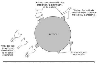
An antigen is either a cell or molecule which will bind with preexiting antibody but will not definitely cause induction of antibody production.
Antigen may also be defined as — ‘a macromolecular entity that essentially elicits an immune response via the formation of specific antibodies in the body of the host’.
In a broader perspective the antigen (or immunogen*) is invariably regarded as the afferent branch of the prevailing immune system, as illustrated in Fig. 1.1 below, and is any cell or molecule which would provoke an immune response** very much in an immunologically viable and competent individual. Generally, immunogens (antigens) must fulfil the following two cardinal characteristic fea-tures, namely :
(a) should be larger than 2000 in molecular weight, e.g., protein, glycoprotein and carbohy-drates, and
(b) must be absolutely foreign to the individual into whom they have been introduced appropri-ately.
Fig. 1.1. Immune Mechanism.
* Sometimes, the term antigen is used synonymously with immunogen, although this usage is incorrect.
Example : The most befitting example of an ‘antigen’ is ones own erythrocytes (WBC). Be-cause, they will not induce antibody formation in oneself but will definitely react with an antibody essentially contained in an improperly matched blood transfusion.
It is, however, pertinent to state here that quite often an antigenis a protein, but it could also be a polysaccharide or nucleic acid or any other substance. Importantly, it may also be possible that a foreign substance (e.g., protein)-not necessarily belonging to a pathogenic microorganism, may act as an antigen so that on being injected into a host, it may induce antibody formation. Besides, they may turn out to be antigenic and thereby cause stimulation of antibody production, incase they are intimately and lightly get bound to certain macromolecules, for instance : proteins, carbohydrates and nucleic acids.
3.2. Haptens
In usual practice, the relatively smaller, less rigid or rather less complex molecules usually are not immunogenetic in their purest form, but may be made so by simply linking them strategically to either larger or more complex structures. Consequently, the smaller molecules are invariably termed as haptens ; whereas, the larger molecules or cells are known as carriers.
Hapten may also be defined—‘as a substance that normally does not act as an antigen or stimu-late an immune response but that can be combined with an antigen and, at a stimu-later time, initiate a specific antibody response on its own’.
Furthermore, small molecules (micromolecular), such as : drug substances, that may serve as
‘haptens’ and can normally be made antigenic by coupling them chemically to a macromolecular
substance e.g., protein, polysaccharide, carbohydrate etc. The hapten is obtained from a non-antigenic compound (micromolecule) e.g., morphine, carteolol etc., which is ultimately conjugated*, covalently to a carrier* macromolecule to render it antigenic.
Morphine should be first converted to the corresponding 3-o-carboxymethyl derivative prior to carbodiimides (CCD) coupling with albumin to provide a functional coupling moiety in the hapten.
Another glaring example is of gastrin (hapten) which is duly coupled to albumin (i.e., protein-carrier) by treatment with carbodiimides (CCD), which couple functional carboxyl, amino, alcohol, phosphate or thiol moieties.
Importantly, the hapten-conjugate thus obtained is normally subjected to emulsification in a highly refined ‘mineral oil’ preparation containing-killed Mycobacterium (Complete Freund’s
*Conjugate : The combined ‘hapten’ and ‘carrier’.
Adjivant*), and subsequently injected intradermally either in healthy rabbits or guinea pigs on several occassions at intervals. Evidently, the serum antibody should have not only high degree of specificity but also a reasonably strong affinity for the prevailing antigens.
It has been observed that a relatively large variety of low molecular-weight chemical substances may cater for as allergenic haptens (partial immunogens) and induce allergy after combining covalently with an appropriate protein carrier. On one hand this serves as a vital and important phenomenon spe-cifically in drug allergy ; however, the most widely found ‘environmentalallergens’. Perhaps the most notable exception in the instance of common allergic contact dermatitis produced by a variety of plants, drugs, clothing additives and other similar substances.
Examples : (a) Urshiols : The allergenic constituents i.e., the oleoresin fraction, derivatives of pentadecycatechol or heptadecylcatechol, that are solely responsible for causing contact dermatitis in North America usually belong to the natural order Anacardiaceae, the genus Toxicodendron (Rhus), and quite often include oak, sumac and poison ivy.
(b) Several plants which essentially belong to the natural order compositae viz.,ragweeds, also responsible for causing contact dermatitis, and the allergens have been duly isolated and characterized as sesquiterpinoid lactones.
4.
IMMUNE SYSTEMS
Immunology, the generation of an immune response or the defence mechanismexclusively depends upon the interaction of the three most vital components of the immune mechanism, namely : (a) immunogen stimulation ; (b) humoral immune system ; and (c) cellular immune system. Since 1901 and 1984 an enormous and substantial research inputs, were made by various scientists across the globe which have enabled the inhabitants of the world to lead a better and safer quality of life through the evolution of ‘immunotechnology’i.e., conglomeration of various immune systems. During the said long period (1901-1984) the wonderful findings of scientists and researchers not only brought them wide recognition through most coveted Noble Prizes but also paved the way towards the introduction of remarkable and most trustworthy remedies for complicated not-so-easy diseases of the present day.
It would be worthwhile to make a brief and comprehensive illustration of some of these meaning-ful contributions in a chronological manner as stated below :
•Emil von Behring (1901) : Awarded with Noble Prize for his interesting discovery that quite a few diseases are caused due to the expression of toxins, which he demonstrated by inoculating healthy animals with diphtheria tetanus toxins to generate antitoxins.
• Jules Bordet (1919) : Bagged the Nobel Prize for proving that erythrocytes may be haemolyzed with particular antibody and complement accordingly. His work further demonstrated quite successfully the strategical involvement of certain biological processes taking place in vivo, namely : bacterial ag-glutination**, neutralization of the precipitin reaction, and complementary mediated immune haemolysis.
* [Jules Thomas Freund, Hungarian-born US immunologist 1890-1960] A mixture of killed microorganisms, usually mycobacteria, in an oil and water emulsion. The material is adiministered to induce antibody forma-tion. Because the oil retards absorption of the mixture, the antibody response is much greater than if the killed microorganisms were administered alone.
•Karl Landsteiner (1930) : Became the Nobel Laureate for his epoch making findings for a deeper and vivid immunological concepts with regard to the discovery of blood group antigens i.e., A-B-O blood groups, that was eventually used for successful transfusions in humans.
• Ehrlich’s Selection Theory : It is directly associated with antibody production, and it was amply revealed that during the intricate phenomenon of acquiring ‘immunity’, the critical substances that are exclusively responsible for the fine specificity of recognition were revealed to be the globular proteins that are strategically located in the γ-fraction of blood serum.Subsequently, Landsteiner ex-perimentally demonstrated the aforesaid theory to be ‘false’, based on blood transfusions. In other words, Ehrlich failed to substantiate his assumption that traces of each individual kind of antibody are carried out in the organism specifically ; whereas, Landsteiner adequately proved that antibodies may only be generated under the influence of foreign substances, i.e.,via an instructive process.
• Linus Pauling’s Instructive Theory (1940s) : Pauling was pioneer in assigning a ‘helical structure’ to the protein molecules ; and, soon after proposed the instructive theory. He even went a step ahead and postulated a ‘template theory’ particularly for the synthesis of antibody molecules that could have a relativeily broad range of diversity in shapes. However, at a later stage a renowned immu-nologist Sir Macfarlane Burnet heavily criticized the ‘template theory’ on logical grounds.
•Natural Selection Theory (1955) : Niels Kaj Jerne put forward the ‘natural selection theory’ for the production of antibody. In fact, his proposed theorization revolves round his logical explanation that — ‘the antigen neither serves as a template nor as an enzyme modifier’. In other words, the antigen is exclusively a highly selection carrier closely associated with a spontaneously circulating antibody to a ‘system of cells’ that is capable of reproducing this antibody. It has been duly observed that globulins* are being synthesized in a large variant of different configuration. At this juncture the introduction of an antigen into the blood stream essentially gives rise to the selective attachment onto the surface of the antigen only such globulin molecules that should exhibit a complementary configuration. In short, Niels Kaj Jarne brought back a new lease of life to the ‘Ehrlich’s Selection Theory’ almost after a long gap of six decades.
•Clonal Selection Theory : Burnet in 1957, put forward clonal selection theory with regard to ‘antibody formation’ that vehemently offered a positive clue that each cell following an usual contact with an antigen yields a clone** of cells that would be exclusively engaged in the production of anti-body of a particular kind. Importantly, Burnet’s theory suggests two responses taking place, namely : primary and secondary, of which the latter one seems to be more powerful by virtue of the fact that ‘antigenic memory ultimately gives rise to colossal clonal expansion in an extraordinarily rapid man-ner in the course of subsequent exposure to antigen. Hence, the said theory has not only been accepted across the globe but also attracted enormous glory, fame and recognition which enabled Burnet to bag the Nobel Prize in 1960 (also shared by Sir Peter Medawar).
It is, however, pertinent to mention here that Niel’s Kaj Jerne also overwhelmingly was honoured with the Nobel Prize in 1984 i.e., almost after a gap of 24 long years for his ever enlightening and wonderful discovery of the following two aspects in the immunological concepts, namely :
* One of a group of simple proteins insoluble in pure water but soluble in neutral solutions of salts of strong acids e.g., serum globulin, fibrinogen, and lactoglobulin.
(a) clonal expansion concept, and
(b) evaluation of idiotype* network in regulatory mechanism of immune responses.
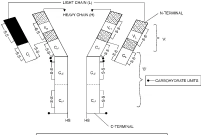
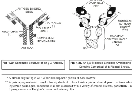
• Rodney R Porter and Gerald M Edelman (1972) : The pioneering discovery made by Porter and Edelman with regard to the elucidation of the structure of antibody molecule,which is extremely vital and important in the elaborated study of antigen-antibody interactions, helped them to be hon-oured with the most coveted and prestigeous Nobel Prize in 1972. Importantly, their findings not only proved but also established the most glaring fact that the four polypeptide chains comprising of each immunoglobulin molecule may be enzytmetically cleaved into three distinct segments, such as : two antibody fragements (Fab) ; and onecrystalline fragment (Fc).
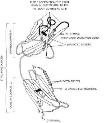
• Wu and Kabat’s Observations : In 1970, these researchers adequately demonstrated the pres-ence of specific ‘hypervariable regions’ strategically located on the ‘antibody molecule’. These criti-cal observations, in fact, virtually paved the way towards the rapid and tremendous progress in the field of immunology ; and, therefore, legitimately caused a geometrical development equally stretched over the two most virulant segments of medicine as well as modern biology, for instance : organ and tissue transplantation, vaccinology, and molecular biology.
• Identification of HLA-Complex : The structure and eventually the specialized role and func-tions of the human leucocyte antigens (HLA) complex were scientifically revealed by Snell, Dausset and Benacerraf in late seventies earned them the Nobel Prize in 1980.
• Somatic Hybridization : The ever sensational production of immunologically homogeneous monoclonal antibodies was accomplished through a wonderful major historical breakthrough by two immunologists : George Kohler and Ceasar Milstein in the year 1975, which bagged them the Nobel Prize in 1982.
•S. Tonegawa’s Immunoglobulin Gene Rearrangement : The evolution of somatic recombi-nation theory logistically suggesting the individual coding in fragments of the ensuing variable and constant regions that are ultimately ressembled to generate an enormous quantum of binding sites ; and, therefore, various binding sites may be adequately obtained due to the different combinations and
permutations of relatively lighter and heavier chains. At this material time Tonegawa’s remarkable
epoch making discovery of the most plausible mechanism of shuffling of several gene segments in the plasma cells (i.e., antibody secreting cells) producing a huge variety of antiody molecules. In short, Tonegawa’s spectacular and superb scientific observations on the immunoglobulin gene rearrangement made an appreciable contribution both in the fields of immunology and molecular biology. Tonegawa was rightfully awarded with the prestigious Nobel Prize in the year 1987.
In a nut-shell, one may interestingly observe and access the tremendous sea-change in the most scientific and logistic progress and development during the period 1901-1987 and even after that in the fields of immunology and molecular biology which, of course, have enormously improved upon the quality of life of humans across the globe irrespective of their caste and creed.
However, the ‘immune systems’ may be viewed from the following two aspects critically, such as :
(a) Manipulation of immune system, and (b) Types of immunity.
4.1. Manipulation of Immune Systems
The ‘immune system’ or the ‘immune defence mechanism’ in an individual person may be sub-jected to a wide spectrum of articulated well-planned manipulation against a host of dreadful and even fatal infectious diseases, caused by highly dangerous and most pathogenic microorganisms, such as : cholera, polio, chicken-pox, small-pox, diphtheria, measles, whooping cough, Hongkong Flu, anthrax, jaundice, hepatitis A, B and C and the like by immunization squarely and effectively. The underlying principle of immunization is solely based on injecting an individual subject with an appropriate dosage with a sterilized and pre-tested/evaluated preparation of a pathogenic microorganism (disease-causing microorganism) that has been rendered harmless absolutely.
It has been duly proved and established beyond any reasonable doubt that immune system pre-dominantly and essentially possess two extremely special characteristic features which not only enable the body in preventing the individual from the dreadful infection but also ensure that he must not suffer from the same infection once again i.e., one normally suffers from certain infectious ailments only once in one’s life-time (small-pox, measles etc.,). Interestingly, the said two special characteristic features are, namely : (a) Memory : and (b) adaptibility.
More explicitely one may understand that the very first encounter of an infections agent, the body starts to learn whether it is either a foreign entity or a nonself entity : and subsequently gets adapted to fight back the caused infection (MEMORY). But, the same individual on being exposed to the infect-ing agent, the body in turn exhibits a substantially increased agent, the body in turn exhibits a substan-tially increased immune response that is actually dependent on the earlier encounter. In the science of immunology these are invariably termed as primary and secondary immune responses. Consequently, as a fundamental characteristic features one may proclaim that the immune system has acquired ad-equate adaptibility.
4.2. Types of Immunity
The various types of immunity that are commonly identified, characterized and studied at length are as stated below :
(i) Humoral Immunity (ii) Cell-mediated Immunity, (iii) Innate (or Natural Immunity), (iv) Acquired Immunity, and
(v) Non-specific Immunity.
These different types of immunities shall now be treated individually in the sections that follows :
4.2.1. Humoral Immunity
Antibodies are immunoglobulin (Ig) molecules (e.g., serum proteins) and they are usually com-prised of several categories designated as IgA, IgD, IgE, IgG, and IgM respectively. However, it is pertinent to state here that each category essentially possesses certain specific characteristic features, such as : size, carbohydrate content, electrophoretic-migration velocity, quantum of antigen-combining sites, im-munological response, and imim-munological objective.
Examples :
(b) IgG : Most versatile important and abundantly available class of antibodies taking part in largest humoral immune reactions. Besides, it happens to cross the placenta thereby pro-viding a newly born baby absolute temporary immunity against whatever immunogens the mother has earlier against IgG.
(c) IgA : Antibodies are invariably found in a plethora of such secretions as : tears, saliva and mucous membranes. These are quite frequently termed as our first-line-of-defense mecha-nism by virtue of the fact that most bacteria, viruses and fungi that eventually gain entry into the body do cross a mucous membrane.
(d) IgE : Antibodies are equally important in our body’s defense against the parasitic worm infections specifically. Prominently and predominantly several allergic manifestations give rise to the release of histamines e.g., allergy due to pollens, house dust, dust mite, human hair, food allergens etc., which in turn afford the apparent discomforts resulting into extrin-sic asthma, hay fever, or hives, or excessive sneezing (during changes of season due to pollens in the air).
(e) Antibodies normally serve as surface receptors strategically located on certain immunologically active cells so as to enable them to bind immunogen.
Importantly, the different types of cells or entities that are held responsible for contributing im-mensely to the humoral immunity are as follows :
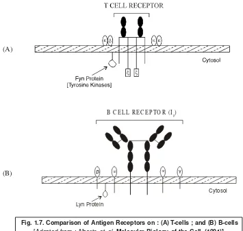
(i) B Lymphocytes (or B Cells), (ii) Immunodominant peptides (IDPs), (iii) Antigen-presenting cell (APC), (iv) T Cell subsets
(v) Class II MHC (major histocompatibility complex) proteins.
It would be worthwhile to have a closer and detailed description and functionalities of each of the cell or entity cited above in the selections that follows :
4.2.1.1. B Lymphocytes [or B cells]
The B lymphocytes or B cells are so named because they were first and foremost found in the Bursa* of Fabricius of birds. It has been observed that in ‘birds’ the multipotent stem cells actually originate in the bone marrow usually migrate to the bursa, and here they virtually get differentiated into specific antibody synthesizing cells. In fact, antibody molecules are normally generated by the plasma cells which are vividly differentiated from B cells. It has been found that B cells are concentrated in various parts of the body, such as : spleen, mucous-associated lymphoid tissue, and regional lymph nodes, where they actually await contact by the foreign epitopes** which promptly initiate the process of conversion into the plasma cells.
Interestingly, the characteristic features of B cells may be enumerated as stated below :
1. B cells possess essentially surface immunoglobulins (sIgs) plus a number of receptors.
2. The sIg form an integral part of B cells ; and, therefore, act as receptor for antigens.
* A padlike sae or cavity found in connective tissue usually in the vicinity of joints.
3. B cells may also have immunoglobulines (Ig) in their cytoplasm.
4. Intially, when B cells are ‘immature’, sIg molecules which are exhibited specially belong to IgM category that do not cross link with other IgMs.
5. IgD molecules appear prominently on the surface showing extremely high levels with B cell marching ahead to its developmental pathway.
6. Activation of B cell initiates loss of IgDs together with other receptors appearing in the membrane that eventually enhance the phenomenon of activation.
7. B cells may undergo activation by the aid of lipopolysaccharide preparations from Gram-negative organisms e.g.,E. coli, Salmonlla. Besides, activation may be followed by the ap-pearance of a plethora of surface receptors for Igs, ocystalline fragment (Fc) component of heavy Ig chain, and also for Epstein-Barr virus*.
8. Antigen critically trigers selection of an appropriate antibody-producing cell and this selec-tion is exclusively based upon the surface receptors.
9. Receptors which are found to be absolutely specific for an antigenic determinant not only provoke but also interact with B cells to initiate strategic proliferation and generate a clone of blast cells**, most of which are capable of giving rise to the same antibody.
10. A portion of the prevailing blast cells get segregated and pass into the plasma cells (i.e., antibody secreting cells), while others do remain behind in lymphoid tissue in the form of memory cells.
11. B cell is characterized by a genetic composition which enables it to produce only one specificityof antibody ; and this is accomplished via random rearrangment of genes which essentially control and monitor both gross and minute antibody structure.
12. B cells are produced continuously in vivo throughout life since the life-span of a mature B cell is only a few days unless contacted by the immunogen for that it remains specific. 4.2.1.2. Immunodominant Peptides (IDPs)
It is, however, an universal truth that ‘antibody production’ invariably takes place through the earnest cooperation and interaction of various types of cells. It has been duly observed that either macrophages*** or other cells having identical lineage encounter predominantly the extracellular foreign immunogen present in the blood or lympth, undergoes phagocytocis, and ultimately meets complete destruction. Importantly, in the course of the ‘phagocytic phenomenon’ the ensuing macrophase criti-cally identifies and recognizes epitope structures present on the immunogen, and notably protects them in the shape of short peptide chains having 10-18 amino acid lengths that are usually referred to as immunodominant peptides (IDPs).
* A member of the herpes virus family, discovered in 1964. It is one of the causes of infections mononucleogis. ** The most popular tool for searching sequence databases is a package called BLAST (basic local alignment
sequence tool).
4.2.1.3.Antigen-Presenting Cell (APC)
The antigen has to be presented correctly and precisely onto the surface of an antigen-present-ing cell (APC) which is normally a macrophage, or similar cell. Most commonly the antigen is pre-sented in association with self Ia molecules duly coded by MHC-genes.
4.2.1.4. T Cell Subsets
T Cells or thymus-derived T lymphocytes do play extremely vital and equally important roles in the domain of immune response mechanism, particularly in the cell-mediated immunity (CMI). In true sense, the different types of immunological functionalities mediated by them usually fall under two distinct categories, namely : (a) effector responses ; and (b) regulatory responses.
Examples of Effector Functions (Responses) : The various types of effector functions are as stated under :
(i) tuberculin reaction (or delayed hypersensitivity response), (ii) destruction of tissue grafts, and
(iii) lymphokines vis-a-vis their ability to perform ‘killer functions’ of other cells due to the secretion of soluble chemical mediators,
Example of Regulatory Function (Response) : The essential kind of regulatory function in-volves their close cooperation with B cells to give rise to the formation of antibodies. Explicitely and precisely but for their excellent mutual cooperation, a good number of antigens would have not suc-ceeded in the induction of antibody formation in animals.
T cells, in general, possess a host of ‘subpopulations’ that essentially contribute remarkable, explicit and above all an excellent array of immune responses, for instance : cytotoxicity, killer proper-ties and significant suppression.
Emergence : The ‘thymus’ is the focal point wherein the actual emergence of pre-T cells invari-ably commence and undergo massive proliferation and differentiation to get converted into immunocomplement T cells. Consequently, the resulting cells undergo migration right into the blood stream and along with B cells help to populate the prevailing secondary lymphoid organs. T cells get matured in thymus, migrate to the peripheral regions of thymus, and ultimately to spleen where they are subjected to further stage of maturation thereby yielding a variety of T cell subsets.
Kinds of T Cell Subjects : There are in all four kinds of T cell subsets that have been duly identified based on their surface markers, namely :
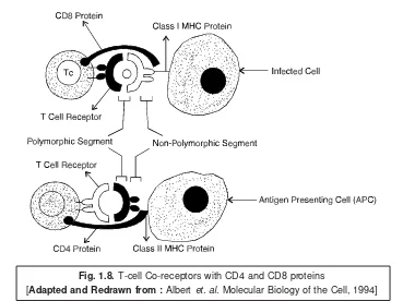
(a) T helper cells (TH cells or CD4 cell) :
An appropriate TH cell is one that essentially possesses a good number of surface receptors arranged in a series and are capable of interacting with the class II major histocompatibility* complex [or class II MHC] of the APC, and also specific for the epitopes present on the IDP. Besides, the T cell receptor series prominently comprises of T cell receptors (TcRs) that are found to be quite specific for :
the prevailing foreign epitopes on the IDP,
CD4 (T4) receptors that essentially interact with class II MHC structures, CD28—which being a costimulatory structure, and
CD3—surface molecules that predominantly cater for a possible communication linkage between TCR, CD4, and the cytoplasm of the TH cell.
Interestingly, the prevailing interaction betweem the APC and TH cells gives rise to the secretion APC yielding a cytokine termed as interleukin-1 (IL-1), which prominently aids to activate the TH cell. Furthermore, the activated TH cell subsequently gives rise to another cytokine known as interleukin-2 (IL-2), that eventually acts as an autocrine* there by raising the TH cell to a relatively higher peak of metabolic activity and also inducing TH cell proliferation. In actual reality, the ensuing activated and duplicated TH cells adequately generate more IL-2 and do help in the production of IL-4, IL-6, IL-13 in addition to quite a few other molecules, including CD4OL, which activate suitable B cells in a concerted manner.
Two subsets of T helper cells (TH) [CF4+] (Th1 and Th2) :
The T helper cells (TH) are of two types, namely : (a) Th1 cells ; and (b) Th 2 cells which would be discussed separately at length as under :
1. Th 1 cells : It is, however, pertinent to state here that since the remarkable discovery of subsets of T helper cells i.e., Th 1 and Th 2, gained wide recognition and acceptance for the first time in 1986, a tremendous and copious volume of work has been duly accomplished and concepts with newer ideas conceived.
Salient features of Th 1 cells : The various salient features of these cells are as follows : (i) They activate macrophages,
(ii) Their clones specifically give rise to interleukin (IL-2), interferon (IFN-γ), and tumour necrosis factor (TNF-β),
(iii) They produce strong delayed-type hypersensitivity reaction (DTH-reaction),
(iv) They invariably generate cytokines for its own proliferation e.g.,autocrine growth factor IL-2 for Th 1, and
(v) They produce cytokines which cross-regulates each other’s development and activity viz., IL-4 and IL-10 suppressing Th 1.
2. Th 2 cells : The salient features of Th 2 cells are as enumerated below :
(i) Their clones give rise to IL-3, IL-4, IL-5, IL-10 and IL-13. However, it was earlier believed that IL-10 to be the product of Th 2, and later on it was demonstrated to be the product of both Th 1 and Th 2.
(ii) They generate weak DTH reaction involving relatively higher level of ‘antibody production’. (iii) They product cytokines for its own proliferation viz.,autocrine growth factor IL-4 for Th 2. (iv) They also produce cytokines, which essentially cross-regulate each other’s development and
activity e.g., IFN-γ suppressing Th 2.
In general, leishmaniasis**, is found to be a ‘model’ for the functional dichotomy of helper T-cells, whereby Th 1 associated with resistance and Th 2 with succeptibility of this ailment.
(b) Cytotoxic T cells [CTLs or Tc] : These represent a subset of T cells that essentially act as ‘killer cells’ and are strategically located in the human peripheral blood. In fact, they kill the cells which are invariably infected either with a virus or any other pathogenic (i.e., disease producing) microorgan-isms. CTLs are also termed as effector T cells, or CD8 cells. Since the Tc cells are recruited to an area infested with viral infection, growing tumour, or foreign organ (i.e., transplanted organ like kidney) ;
and those with TCRs specific for the epitopes of the target shall eventually get bound to the ‘target cell’ which specifically exhibits either the foreign tumour or virus epitopes in perfect harmony with its class I MHC surface structures. Interestingly, this usually affords an activation of the IL-2 receptors upon the recruited as well as attachedTc cells. As a result the ensuing IL-2 generated by the TH cells activates, proliferates, and also differentiates TC cells, thereby enabling their conversion to the corresponding CTL cells ; and, therefore, causing an effective initiation of the ‘effector phase’ of the prevailing immune response mechanism to a considerable extent.
(c) Supperssor-inducer T cells [Ts] : These cells are believed to serve as suppresor of helper T cells (TH) on one hand and in turn cause inhibition of the B cells to generate antibodies significantly. They are essentially found to bear CV8 markers.
(d) Delayed Hypersensitivity T cells [TDH] : It has been recently demonstrated and advocated that the delayed hypersensitivity cells (TDH) essentially form an integral part of a subset of T cells that have been shown to take part actively in delayed hypersensitivity reactions.
The following two Figs. 1.2 and 1.3 evidently illustrate the development of T cells before they are actually exposed to the antigen, and the subsets of T cells that actively participate in the regulatory immune response mechanism respectively.
T HELPER CELL [T ]H
T SUPPRESSOR CELL [T ]S
T CYTOTOXIC CELL [T ]C
T DELAYED HYPER SENSITIVITY CELL [T ]DH THYMUS
PRE-T CELL MULTIPOTENT STEM CELLS
Fig. 1.2. Development of T cells Prior to their Exposure to Antigen.
Explanation of Fig. 1.2 : A situation when the T cells are under the process of ‘differentiation’ and ‘proliferation’ in the thymus, these are invariably termed as thymocytes*. Subsequently, these thermocytes eventually emerge as completely differentiated and fully distinguishable T cells, that essentially undergo clonal expansion on being subjected to interaction with the ensuing antigen, such as : TH ; TS ; TC ; and TDH.
Explanation of Fig. 1.3 : It vividly illustrates the various emerging subsets of T cells that actu-ally take part in the regulatory immune response mechanism thereby giving rise to TS (suppressor T cell), B lymphocyte (B cell), TC (cytotoxic T cell), and macrophage respectively. The ‘arrrows’ indi-cated here designate the activating signals explicitely and squarely.
Fig. 1.4 : Designates the two typical kinds of helper T cells e.g., Th 1 and Th 2, respresenting explicitely the molecules secreted by them, their effector functions together with other similar interac-tions.
Fig. 1.3. Subsets of T cells Participating in Regulatory Immune Response Mechanism.
* A large tissue cell resembling basophil that does not circulate in the blood. It contains hydrolylic enzymes. ** Diseases produced when the body’s normal tolerance of its own antigenic markers on cells disappears.
Effector Functions of Th 1 and Th 2
It has been adequately established and proved beyond any reasonable doubt that Th 1 cells are both essentially and primarily responsible for the cell-mediated immunity (CMI). In other words, ac-quiring a strong level of delayed-type hypersensitivity (DTH) (i.e., killing through phagocytosing the antigen containing cells e.g., viruses loaded cells. Likewise, Th 2 are specifically and solely engaged in the humoral immunity (HI) ; or distinctly possessing weak DTH (i.e., they profusely help both B cells and other cells in generating antibodies adequately. Based on the fact that IFN-γ, usually released by Th 1 cells, causes induction and subsequent conversion of B cells to a good number of IgG isotypes have undoubtedly raised doubt upon the integrity of the aforesaid classification linked to their effector functions. In order to get rid of such an ambiguity the effector functions of Th 1 and Th 2 logically may be classified exclusively based on the observed ‘biological activities’ of the cytokines produced by them.
Examples :
Principal cytokine of Th 1 being IFN-γ : It plays two vital roles : (a) activation of microphages for microbicidal action, and (b) stimulation for the production of IgG antibodies. Principal cytokines of Th 2 being IL-4 and IL-5 :
(a) IL-4 : causes induction of B-cell switching to IgE production, and
(b) IL-5 : causes activation of eosinophils ; and both IgE and eosinophil mediated defense is very vital for eliminating certain, but not all helminths.
Th1 and Th 2 [Subsets of CD4+ helper T-cells] and Diseases
The Th 1 and Th 2 helper cells have been observed to be intimately linked with several dreadful ailments. Evidently, it is on account of the critical indulgence by each separate cytokines that are associ-ated with the management and control of serious pathological immune responses. A few such infec-tious diseases are, namely : (a) leischmaniasis ; (b) allergic disease conditions (which involves mast-cells* and IgE ; and also autoimmune diseases.**
Importantly, one is loaded with so much solid evidences to suggest that modulation of Th1/Th2 equilibriation by the adequate and timely administration of either recombinant cytokines or cytokine antagonists helps to change the flare up and intensity of the disease.
Examples :
(a) IL-12 : When given at the time of infection usually increases significant resistance to a host of several intracellular pathogens, such as : protozoa, viruses, fungi, and microorganisms.
(b) IL-12 : May also be indicated for the effective treatment of cancer, and allergic symptoms, and is also under active investigation in certain antineoplastic vaccine protocols.
* Toxemia caused due to the presence of endotoxins in the blood.
** A disease produced when the body’s normal tolerance of its own antigenic markers on the cells disappears.
into Th 1 pattern, but also augment resistance to ensuing infection and therby suppressing the prevailing Th 2 dependent pathological conditions aprreciably.
(d) IL-10 : Can reasonably suppress lipopolysaccharide-induced endotoxemia* and also in-flammatory bowel disease in experimental animals.
(e) Th 2 cells : On being subjected to ‘selective induction’ may be used for the treatment and cure of tissue autoimmune diseases** e.g.,diabetes mellitus : in which the autoantibodies (AAbs) attack the insulin-producing cells of the pancreases ; rhematoid arthritis : caused by inflammatory changes in the connective tissue of joints ; and multiple sclerosis : produced by AAb destruction of the myclin sheath covering nerves.
Therefore, one may accomplish almost the same rate of success by administering instead of cytokine or cytokine antagonists, disturbing the prevailing balance between Th 1/Th 2 with a particular course of treatment.
Example : It is possible to prevent the incidence of ‘autoimmune diseases’ (as mentioned in ‘e’ above) by the oral administration of antigens which particularly helps in the development of T-cells that essentially produce the transforming growth factor (TGF-β) and Th 2 cytokins.
Future Development : In the light of the aforesaid latest and remarkable achievements to com-bat the dreadful autoimmune diseases, such as : diabetes mellitus, rheumatoid arthritis, and multiple sclerosis, there exists an ample and tremendous scope for a better indepth knowledge vis-a-vis understading of Th 1/Th 2 dichotomy towards the development of ‘prophylactic vaccines’ ; and, there-fore, the therapy for many ailments that are intimately associated with it.
Fig. 1.5. Block Diagram of Thymus Programme T-cells Passing through it to follow T-cell Differentiation in Subset Variants.
Interestingly, in the life-span within the thymus, lasting for nearly three days, the T cells start showing typical and specific surface markers. Besides, these surface markers are nothing but macro-molecules (e.g.,glycoproteins) that have a well-defined clear cut distinction from the glycoproteins that are strategically coded for by the major histocompatibility complex (MHC) genes shared equally by all T-cells. However, a good part of these markers have been duly well defined both in human T cells as well as in mouse T cells.
In actual practice, however, there are several other surface markers that are critically and exclu-sively exhibited on a population of T cells ; and, therefore, these are invariably employed to identify the T cell subsets and their individual prevailing functions as depicted in Table 1 below :
Table 1 : Surface Markers of T-cells and Their Corresponding Subsets.
T-Cells Surface T-Cell Individual Functions
Markers Subsets
T-Cells CD4+ T-helper cell — Regulator cells ;
(TH) — Inducers of TC cells that are CD8- ;
— Stimulate B cell to generate antibodies ;
CD8+ T-cytotoxic — Kill (lyse) antigen-loaded target cells cell (TC) (i.e., effector cells)
CD2+ ; CD8+ ; T-suppressor — Prevent production of antibody by B-cells ; CD3-cell cells (TS) — Exert action on CD4– and TH cells. receptor