National Cataract Surgery Registry
Ministry of Health Malaysia
THE THIRD REPORT OF THE
NATIONAL CATARACT SURGERY REGISTRY
2004
Edited by
Goh Pik Pin Shamala Retnasabapathy
Rajalakshmi Gopal Ronald Arun Das
A publication of the
National Cataract Surgery Registry And
Clinical Research Centre, Ministry of Health
October 2004
© National Cataract Surgery Registry, Malaysia.
Published by the
National Cataract Surgery Registry (NCSR) C/O Clinical Research Centre,
Level 3, Dermatology Block, Kuala Lumpur Hospital, Jalan Pahang,
50586 Kuala Lumpur.
General Line: 603-2698 0310 Fax: 603-2691 1682
Email [email protected]
Website: http://www.crc.gov.my/ncsr
Disclaimer
The data reported here have been supplied by NCSR. The interpretation and reporting of these data are the responsibility of the Editor and in no way should be seen as an official policy or interpretation of the NCSR.
Suggested citation
The suggested citation for this report is as follows:
Goh Pik Pin, Shamala Retnasabapathy, Rajalakshmi Gopal, Ronald Arun Das.(Eds) THE SECOND REPORT OF THE NATIONAL CATARACT SURGERY 2003 Kuala Lumpur, Malaysia 2004
Electronic version
Electronic version of this report can be downloaded at http://www.crc.gov.my/ncsr
PARTICIPATING CENTRES
1. Angkatan Tentera Kem Terendak Hospital
Head, Ophthalmology Department : Lt. Kol. (Dr) Nor Aishah Malik
Dr In Charge : -
Coordinator : Sarjan Jawariah Ali
Participating From : January-November 2002
January-December 2003
2 Alor Setar Hospital
Head, Ophthalmology Department : Dr. Ahmad Mat Saad
Dr In Charge : Dr. Zaharidah Abd Kadir
Coordinator : T/AN Siti Aishah Salim
Participating From : January-December 2002
January-December 2003
3 Duchess Of Kent Hospital, Sandakan
Head, Ophthalmology Department : Dr. Adarsh Bhardwaj
Dr In Charge : -
Coordinator : MA Linjabat Mandasah
Participating From : January-October 2002
January-October 2003
4 Ipoh Hospital
Head, Ophthalmology Department : Dato` Dr. P. Balaravi
Dr In Charge : Dr. Indarjit Singh
Coordinator : MA Bernard
Participating From : January 2002
January-December 2003
5 Kangar Hospital
Head, Ophthalmology Department : Dr. Mohd Nazri Sulaiman
Dr In Charge : -
Coordinator : MA Nasihat Dahaman
Participating From : January-December 2002
January-December 2003
6 Kuala Lumpur Hospital
Head, Ophthalmology Department : Dr. Joseph Alagaratnam
Dr In Charge : Dr. Sharifah Intan
Coordinator : SN Hazizah Mohamed
Participating From : January-December 2002
January-December 2003
7 Kuala Terengganu Hospital
Head, Ophthalmology Department : Dr. Zuraidah Mustari
Dr In Charge : -
Coordinator : SN Juriffah Mohd Amin
Participating From : January-December 2002
8 Umum Sarawak Hospital , Kuching
Head, Ophthalmology Department : Dr. Intan Gudom
Dr In Charge : -
Coordinator : SN Hajah Fatimah Hassan
Participating From : January-December 2002
January-December 2003
9 Melaka Hospital
Head, Ophthalmology Department : Dr. S. Anusiah
Dr In Charge : -
Coordinator : SN Siti Asiah Yusop
Participating From : January-December 2002
January-December 2003
10 Miri Hospital
Head, Ophthalmology Department : Dr. V. Prabhu
Dr In Charge : Dr. Maheran
Coordinator : SN Wong Chu Hiong
Participating From : January-June 2002
2003- Excluded due to absent of
Ophthalmologist
11 SultanahFatimah Hospital,Muar
Head, Ophthalmology Department : Dr. Adnan Abas
Dr In Charge : -
Coordinator : MA Nuruhadi B Ghani
Participating From : January-December 2002
January-December 2003
12 Pulau Pinang Hospital
Head, Ophthalmology Department : Dr. Elizabeth John
Dr In Charge : -
Coordinator : En. Azahari Ahmad (Optometrist)
Participating From : January-December 2002
January-December 2003
13 Queen Elizabeth Hospital, Kota Kinabalu
Head, Ophthalmology Department : Dr. Kong Vui Yin
Dr In Charge : -
Coordinator : SN Euginie
Participating From : January-December 2002
iv 14 Seremban Hospital
Head, Ophthalmology Department : Dr. Bethel Livingstone
Dr In Charge : -
Coordinator : MA Thivagaran
Participating From : January-December 2002
January-December 2003
15 Sibu Hospital
Head, Ophthalmology Department : Dr. Reddy
Dr In Charge : -
Coordinator : MA Morni Morsen
Participating From : January-December 2002
January-December 2003
16 Sultanah Aminah Hospital, Johor Bharu
Head, Ophthalmology Department : Dr. Loh Swee Seng
Dr In Charge : Dr. Siow Yun Ching
Coordinator : MA Tan Eng Chai
Participating From : January-December 2002
January-December 2003
17 Sungei Petani Hospital
Head, Ophthalmology Department : Dr. Foo Sui Wan
Dr In Charge : -
Coordinator : SN Zainab
Participating From : January-December 2002
January-December 2003
18 Taiping Hospital
Head, Ophthalmology Department : Dr. Haji Mohamad Sharif Fahruddin
Dr In Charge : -
Coordinator : SN Halina Sharom
Participating From : January-December 2002
January-December 2003
19 Tawau Hospital
Head, Ophthalmology Department : Dr. Ajit Majunder
Dr In Charge : -
Coordinator : MA Bacho Nordin
Participating From : January-November 2002
20 Teluk Intan Hospital
Head, Ophthalmology Department : D. Vivian Gong Hee Meng
Dr In Charge : -
Coordinator : MA Halim
Participating From : January-December 2002
January-December 2003
21 Tengku Ampuan Afazan Hospital, Kuantan
Head, Ophthalmology Department : Dr. Vasantha Kumar
Dr In Charge : -
Coordinator : MA. Azizi
SN Rozila Bt Ashaari
Participating From : January-December 2002
January-December 2003
22 Tengku Ampuan Rahimah Hospital, Klang
Head, Ophthalmology Department : Dr. Yogan Kanagasabai
Dr In Charge : Dr. Tan Lian Hong
Coordinator : SN Wong Huey Fen
Participating From : January-December 2002
January-December 2003
23 Kota Bharu Hospital
Head, Ophthalmology Department : Dr. Zulkifli Abd Ghani
Dr In Charge : Dr. Sakinah Zakaria
Coordinator : -
Participating From : June-December 2002
January-December 2003
24 Kajang Hospital
Head, Ophthalmology Department : Dr. Chandramalar A/P Santhirethilegan
Dr In Charge : Dr. Norazlina Sabri
Coordinator : -
Participating From : June-December 2002
January-December 2003
25 Putrajaya Hospital
Head, Ophthalmology Department : Dr. Salmah Othman
Dr In Charge : -
Coordinator : SN Sarniah Sidek
Participating From : June-November 2002
vi 26 Batu Pahat Hospital
Head, Ophthalmology Department : Dr. Normah A. Khalid
Dr In Charge : -
Coordinator : MA Zainuddin Ali
Participating From : August-December 2002
January-December 2003
27 Kuala Pilah Hospital
Head, Ophthalmology Department : Dr. Khairul Husnaini Mohd Khalid
Dr In Charge : -
Coordinator : SN Rohaizah Baharin
SN Zalina
Participating From : June-November 2002
January-December 2003
28 Selayang Hospital
Head, Ophthalmology Department : Dr. Mariam Ismail
Dr In Charge : Dr. Thaya A/P K. Sandragasu
Coordinator : Ms. Arini Hanim
Participating From : January-December 2003
29 Bukit Mertajam Hospital
Head,’ Ophthalmology Department : Dr. Sri Kumar
Dr In Charge : Dr. Teoh Hian Jin
Coordinator : SN Masheran Musa
Participating From : January-December 2002
January-December 2003
30 Mentakab Hospital
Head, Ophthalmology Department : Dr. Hanizasurana Bt Hashim
Dr In Charge : -
Coordinator : MA Haji Abdul Akim Sabit Ali
Participating From : January-December 2002
January-December 2003
31 Kuala Lipis Hospital
Medical Officer : Dr. Ahmad Abir B Abd Ghani
Dr In Charge : -
Coordinator : MA Abdul Halim Jamion
32 Klinik Pakar Mata Azman Sdn. Bhd
Head, Ophthalmology Department : Dr. Azman Abd Aziz
Dr In Charge : -
Coordinator : Ms. Lina
Participating From : May-October 2003
33 Universiti Sains Malaysia Hospital
Head, Ophthalmology Department : Dr. Wan Hazabbah
Dr In Charge : Dr. Asokumaran A/L Thanaraj
Coordinator : SN Sarimah Samsudin
Participating From : January-December 2002
January-December 2003
34 Universiti Kebangsaan Malaysia Hospital
Head, Ophthalmology Department : Prof. Dr. Muhaya Haji Mohammad
Dr In Charge : Dr. Jemaima Che Hamzah
Coordinator : -
viii
NATIONAL CATARACT SURGERY REGISTRY ADVISORY COMMITTEE
Dr. Goh Pik Pin Chairperson
Consultant Ophthalmologist,
Ophthalmology Department, Hospital Selayang
Dr. Mariam Ismail Co-Chairperson
Head, Ophthalmology Service, MOH and
Head, Ophthalmology Department, Selayang Hospital
Datuk Dr. Abdul Gani B. Mohammed Din
Director,
Medical Development Division Ministry of Health
Dr. Gomathy Arumugam President,
Ophthalmology Society Malaysian Medical Association
Dato' Dr. P. Balaravi Head,
Ophthalmology Department, Ipoh Hospital
Dr. Bethel Livingstone Head,
Ophthalmology Department, Seremban Hospital
Dr. Joseph Alagaratnam Head,
Ophthalmology Department, Kuala Lumpur Hospital
Dr. Zuraidah Bt. Mustari Head,
Ophthalmology Department, Kuala Terengganu Hospital
Dr. Choong Yee Fong Ophthalmologist,
Ophthalmology Department, Hospital Selayang
Associate Prof. Dr. Muhaya Bt. Mohamad
Head,
Ophthalmology Department, Universiti Kebangsaan Malaysia
Dr. Elias Hussein Head,
Ophthalmology Department, Universiti Sains Malaysia
Dr. Hoh Hong Beng Private,
Pantai Medical Centre, Kuala Lumpur
Dato Dr. Y. C. Lee Private,
Lee Eye Centre, Ipoh, Perak
Dr. Lim Teck Onn Head,
Clinical Research Centre, Kuala Lumpur Hospital
Dr. Jamaiyah Haniff Head
Clinical Registry Unit Kuala Lumpur Hospital
ABOUT NATIONAL CATARACT SURGERY REGISTRY
INTRODUCTION
The National Cataract Surgery Registry is a disease outcome registry. It is a prospective, ongoing systematic collection of data pertaining to patients who have had cataract surgery. Data collected include demography, operative events, post-operative visual outcomes and probable causes for poor outcome. These data are used to calculate cataract surgery rates and to evaluate surgical outcome. Such information is useful for performance audit in each participating ophthalmology department, leading to improvement in cataract surgery service, and to assist Ministry of Health, non-governmental organization, private eye care providers and industry in blindness prevention programme planning and evaluation in the country. Analyzed data is presented in report and is disseminated to contributors and other users of the registry at a timely and regular fashion.
NCSR was established in January 2002. In the first year, there are 30 source data producers (SDP) /participating centres in the registry consisting of 28 ophthalmology departments from the Ministry of Health Hospitals, Hospital Angkatan Tentera Kem Terendak, and Universiti Sains Malaysia Hospital. In the year 2003, there were 32 SDP, with addition of Hospital Kuala Lipis, Hospital Selayang and Klinik Azman , but with temporary exclusion of Hospital Miri, due to the absent of ophthalmologist and thus a small number of cataract surgery performed.
OBJECTIVES
The objectives of National Cataract Surgery Registery are to:
1 Determine the frequency and distribution of cataract surgery in Malaysia. These are useful measures of the health burden arising of cataract and its treatment provision in the country
2 Determine the outcomes, and factors influencing outcomes of cataract surgery. This serves the needs of outcome assessment.
3 Evaluate cataract surgery services. This serves the need of accountability. 4 Stimulate and facilitate research on cataract and its management.
The objectives listed above, while typical of any cataract surgery registry, is clearly rather ambitious and certainly cannot be met right away. Thus the registry is implemented in phases.
Phase 1 of the proposed cataract surgery register shall be limited to Public Hospitals only.
Phase 2 of the proposed cataract surgery register shall expand the coverage of Phase 1 to include university, private hospitals and private ophthalmologists in the country.
SPONSORS OF NCSR
CLINICAL RESEARCH CENTRE
The Clinical Research Centre is the designated collaborating unit to the NCSR. It provides the functional capacity to support the operations of the NCSR.
The CRC is the clinical research arm of the Ministry of Health. Apart from the NCSR, CRC currently also supports the National Renal Registry, National Cancer Registry, National Neonatal Registry, National Mental Health Registry, National HIV/AIDS Treatment Registry and National Transplant Registry.
In recent years, CRC has emerged to become the preferred collaborating partner for medical professional groups to establish disease and treatment registries in the country. This is because CRC possesses sophisticated facility and equipment, state of the art technology, and most importantly the trained human resources such as registry managers, epidemiologists, statisticians, information technology professionals and other supporting staff skilled in registry operations. These resources are consolidated in the Disease and Treatment Registry Unit in the CRC. The unit specializes in assisting medical professionals to establish and operate their registries.
Staff of the Clinical Research Centre (CRC) and Cataract Surgery Registry Unit (CSRU) of Clinical Registry Unit (CRU)
Director Dr. Zaki Morad B Mohamad Zaher
Head Dr. Lim Teck Onn
Head of CRU Dr. Jamaiyah Binti Haniff
Cataract Surgery Registry Manager CSRU
SN Lee Poe Poay
Clinical Registry Assistant CSRU Ms. Sharmila Bt Saari
Clinical Registry Assistant CSRU Mr. Mohamad Fauzan B Jamaluddin Information Security Officer Ms Celine Tsai Pao Chien
Network Administrator Mr. Kevin Ng Hong Heng
Assistant Network Administrator Mr. Adlan Ab. Rahman Database Administrator Ms. Lim Jie Ying
Webmaster/Desktop publisher Mr. Patrick Lum See Kai
Programmer Mr. Sebastian Thoo
CONTENTS
ACKNOWLEDGMENTS ...i
PARTICIPATING CENTRES...ii
NATIONAL CATARACT SURGERY REGISTRY ADVISORY COMMITTEE viii ABOUT NATIONAL CATARACT SURGERY REGISTRY ...ix
INTRODUCTION ...ix
OBJECTIVES ...ix
SPONSORS OF NCSR...ix
CLINICAL RESEARCH CENTRE ...x
CONTENTS ...1
ABBREVIATIONS ...5
GLOSSARY ...5
REGISTRY METHODS...6
1.ORGANISATION AND ADMINISTRATIVE STRUCTURE ...6
2.DATA STANDARDS ...8
3.DATA FLOW PROCESS...8
Overall Data Management Flow...10
Overall Data Flow Process ...11
4.LEGAL ASPECT AND CONFIDENTIALITY...12
REPORT SUMMARY...13
1. PATIENTS’ CHARACTERISTICS...13
2. CATARACT SURGERY PRACTICE ...13
3. CATARACT SURGERY OUTCOMES ...13
3.1 Cataract Surgery Complications -Intra-Operative ...13
3.2 Cataract Surgery Complications -Post-Operative ...13
3.3 Post-Oeprative Follow Up Period...13
3.4 Post-Operative Visual Acuity ...13
3.5 post-Operative Refracted VA Improved By One Or More ...14
Line Snellen Chart ...14
3.6 Factors Contributing To Post-Operative VA Worse ...14
Than 6/12...14
LIST OF TABLE
Table 1.1 : Age Distributions...15
Table 1.2 : Gender Distributions ...16
Table 1.3(a) : Number Of Patients With First Eye And Second Eye Surgery ...16
Table 1.3(b) : Period Of Time Before Second Eye Surgery ...16
Table 1.4 : Number Of Patients With Ocular Co-Morbidity ...17
Table 1.5 : Number Of Patients With Systemic Co-Morbidity ...18
Table 1.6(a) : Pre-Operative Visual Acuity Measurement...19
Table 1.6(b) : Pre-Operative Visual Acuity Measurement By Gender ...20
Table 1.7 : Causes Of Cataract ...20
Table 2.4 : Distribution Of Types Of Cataract Surgery By Centre ...26
Table 2.5 : Distribution Of Combined Surgery By Centre ...27
Table 2.6 : Proportion Of Nature Of Cataract Surgery...28
Table 2.7 : Type Of Anaesthesia ...29
Table 2.8 : Type Of Local Anaesthesia ...31
Table 2.9 : Distribution Of Single And Multiple Local Anaesthesia ...33
Table 2.10 : Type Of Sedation Given To Patient Who Had Local Anaesthesia34 Table 2.11 : Intraocular Lens Implantation ...35
Table 2.12 : Distribution Of Cataract Surgery Without IOL...36
Table 2.13 : Distribution Of IOL- Materials And Types...37
Table 3.1.1 : Distribution Of Intra-Operative Complications By Type Of Cataract Surgery ...38
Table 3.1.2 : Distribution Of Intra-Operative Complications By Combined Surgery...42
Table 3.1.3 : Distribution Of Intra-Operative Complications By Nature Of Cataract Surgery ...43
Table 3.1.4 : Distribution Of Intra-Operative Complications By Type Of Anaesthesia ...44
Table 3.1.5 : Distribution Of Intra-Operative Complications By Type Of Local Anaesthesia ...45
Table 3.1.6 : Distribution Of Intra-Operative Complications By Single Or Multiple Local Anaesthesia ...47
Table 3.1.7 : Distribution Of Intra-Operative Complications By Type Of Sedation ...48
Table 3.1.8 : Distribution Of Intra-Operative Complications By Sedation ...50
Table 3.1.9 : Distribution Of Intra-Operative Complications By Cataract Surgery With IOL...51
Table 3.1.10 : Distribution Of Intra-Operative Complications By Cataract Surgery Without IOL...53
Table 3.1.11 : Distribution Of Intra-Operative Complications By Surgeon Status.. ...55
Table 3.2.1 : Distribution Of Post-Operative Complications ...56
Table 3.2.2 : Distribution Of Post-Operative Complications By IOL Types ...57
Table 3.2.3 : Distribution Of Post-Operative Complication By Material...58
Table 3.2.4 : Post-Operative Complication By Centre ...59
Table 3.3.2 : Median Follow-Up Period In Weeks (Patients With Refracted Vision) ...64 Table 3.4.1 : Distribution Of Post-Operative VA...65
: (a) All Patients, With Primary Cause Of Cataract And Not Combined Surgery...65
: (b) All Patients, With Primary Cause Of Cataract, Not Combined Surgery And Without Ocular Co-Morbidity ...66 Table 3.4.2 : Distribution Of Post-Operative Refracted VA 6/12 Or Better At
The Last Follow Up Among Patients Without Ocular Co-
Morbidities, By Surgery ...69 Table 3.4.3 : Distribution Of Post-Operative Refracted VA 6/12 Or Better In
Relation To Age And Type Of Surgery, Among Patients Without Ocular Co- Morbidities...70 Table 3.4.4 : Distribution Of Post-Operative Refracted VA 6/12 Or Better In Relation To Gender And Type Of Surgery, Among Patients
Without Ocular Co-Morbidities...71 Table 3.4.5 : Distribution Of Post-Operative Refracted VA 6/12 Or Better In Relation To Comorbidity And Type Of Surgery, Among Patients
Without Ocular Co-Morbidities...72 Table 3.4.6 : Distribution Of Post-Operative Refracted VA 6/12 Or Better In Relation To Complication And Type Of Surgery ...73 Table 3.4.7 : Distribution Of Post-Operative Refracted VA 6/12 Or Better In Relation To Nature Of Surgery And Type Of Surgery ...74 Table 3.4.8 : Distribution Of Post-Operative Refracted VA 6/12 Or Better In Relation To Anaesthesia And Type Of Surgery ...75 Table 3.4.9 : Distribution Of Post-Operative Refracted VA 6/12 Or Better In Relation To Combined Surgery And Type Of Surgery...76 Table 3.4.10 : Distribution Of Post-Operative Refracted VA 6/12 Or Better In Relation To IOL And Type Of Surgery...77 Table 3.4.11 : Distribution Of Post-Operative Refracted VA 6/12 Or Better In
Relation To Surgeon Status And Type Of Surgery Without Ocular Co- Morbidity ...78 Table 3.4.12 : Distribution Of Post-Operative Refracted VA 6/12 Or Better In Relation To Centre And Type Of Surgery...79 Table 3.5.1 : Distribution Of Post-Operative Refracted VA Improved By One Or More Line Of Snellen Chart, At The Last Follow Up...81 Table 3.5.2 : Distribution Of Post- Operative Refracted VA Improved By One
Or More Line Of Snellen Chart, With And Without Ocular Co- Morbidity At The Last Follow Up...81 Table 3.5.3 : Distribution Of Post- Operative Refracted VA Improved By One Or More Line Of Snellen Chart With Intra-Op Complication And Without Intra- Op Complication, At The Last Follow Up ...82 Table 3.5.4 : Distribution Of Post -Operative Refracted VA Improved By One Or More Line Of Snellen Chart With Systemic Co-Morbidity And Without Systemic Co-Morbidity, At The Last Follow Up ...82 Table 3.5.5 : Distribution Of Post -Operative Refracted VA Improved By One
LIST OF FIGURE
Figure 1.1 : Age Distributions...15
Figure 1.6 : Pre-Operative Visual Acuity Measurement...19
Figure 2.1 : Number (%) Of Surgery ...21
Figure 2.2 : Number Of Surgery Done By Center ...23
Figure 2.3(a) : Distribution Of Day Care And In Patient By Centre, Year 2003 ..25
Figure 2.3(b) : Distribution Of Day Care And In Patient By Centre (Exclude Children And Those With Combined Surgery), Year 2003 ...25
Figure 2.7 : Type Of Anaesthesia ...30
Figure 3.1.1.1 : Distribution Of Intra-Operative Complication ...39
Figure 3.1.1.2 : Distribution Of Intra-Operative Complication By Posterior Capsule Rupture With Vitreous Loss And Posterior Capsule Rupture Without Vitreous Loss...40
Figure 3.1.1.3 : Distribution Of Intra-Operative Complication By Zonular Dialysis With Vitreous Loss And Zonular Dialysis Without Vitreous Loss...41
Figure 3.1.3 : Distribution Of Intra-Operative Complications By Nature Of Cataract Surgery ...43
Figure 3.1.4 : Distribution Of Intra-Operative Complications By Type Of Anaesthesia ...44
Figure 3.1.6 : Distribution Of Intra-Operative Complications By Single Or Multiple Local Anaesthesia ...47
Figure 3.1.7 : Distribution Of Intra-Operative Complications By Type Of Sedation ...49
Figure 3.1.8 : Distribution Of Intra-Operative Complications By Sedation ...50
Figure 3.1.9 : Distribution Of Intra-Operative Complications By Cataract Surgery With IOL ...52
Figure 3.1.10 : Distribution Of Intra-Operative Complications By Cataract Surgery Without IOL...54
Figure 3.1.11 : Distribution Of Intra-Operative Complications By Surgeon ... Status ...55
Figure 3.4.1.1(b) : Distribution Of Post-Operative VA ...67
Figure 3.4.1.2 : Cumulative Distribution Of Visual Acuity By Pre- And Post- Operative Unaided VA ...68
Figure 3.4.1.3 : Cumulative Distribution Of Visual Acuity By Pre- And Post- Operative Refracted VA ...68
ABBREVIATIONS
CF Counting finger CI Confidence interval CMO Cystoid macular oedema CSRU Cataract surgery registry unit ECCE Extracapsular cataract extraction HM Hand movement
IOL Intraocular lens
ICCE Intracapsular cataract extraction NPL No perception of light
PCO Posterior capsule opacification PCR Posterior capsule rapture PE Phacoemulsification PL Perception of light
SDP Source data producers VA Visual acuity
ZD Zonular dialysis
GLOSSARY
Advisory Committee
A committee, board, council, panel or group thereof that is established by the sponsors of the registry to govern the registry. The Advisory Committee shall direct and control the activities of the designated collaborating unit, which manages the day-to-day operations of the registry.
Advisory Committee member
An individual appointed to serve on an advisory committee. Members may have relevant expertise and/or represent the interest of SDP, users or donor.
Chairperson An advisory committee member who is appointed to preside at committee meetings and ensure that all rules of order and conduct are maintained during each session.
Disease Register The ongoing systematic collection, analysis and interpretation of a specific disease data essential to the planning, implementation and evaluation of clinical and public health practice, closely integrated with dissemination of these data to those who need to know. The final link in the chain is the application of these data to the management, prevention and control of the disease. A registration system includes a functional capacity for data collection, analysis and dissemination linked to clinical and public health programs.
Secretary The individual responsible for an advisory committee’s overall
administrative management. He/she is ordinarily a staff provided by the designated collaborating unit for the purpose.
Source data producer
The individuals or institutions that report the required data to the registry.
REGISTRY METHODS
The following aspects of registry methods are described below. 1. Organization and Administrative structure
2. Data standards 3. Data flow process
4. Legal aspects and confidentiality
1 .ORGANISATION AND ADMINISTRATIVE STRUCTURE
In brief, the organizational structure of NCSR consists of sponsors, advisory committee, cataract surgery registry unit (CSRU), source data producers and target groups/users. The Ophthalmology Service and the Clinical Research Centre, both of the MOH, jointly sponsor the registry. The NCSR is governed by an advisory committee who oversees the operations of registry. The cataract surgery registry unit, which is based at the Clinical Research Centre, MOH, provides the functional capacity to support the operation of NCSR. The source data producers are Departments of Ophthalmology, both public and private, who provide data on patients who have had cataract surgeries. The users or target groups are individuals or institutions to which the regular registry reports are addressed.
The description of the duties and functions of each entity depicted follows.
Sponsor
The registry is jointly sponsored by the Ophthalmology Service and the Clinical Research Centre; both of the MOH.
Sponsors shall
♦ Be responsible to Director General of Health, MOH for the effective, efficient and responsive operations of the registry.
♦ Provide leadership and direction for the registry.
♦ Establish an Advisory Committee, jointly chair the committee and appoint members to the Advisory committee. Membership should represent all interested parties. These must include source data producers, Target groups or users and representative from the Cataract Surgery Registry Unit.
♦ Provide the financial, human and information resources required, if necessary with financial contribution from industry or donor agencies.
Sponsor
Advisory Committee
Source Data Producers
Cataract Surgery Registry Unit
Advisory Committee
An Advisory Committee for cataract surgery register shall be established by sponsors to oversee the operations of registry. Interested parties including source data producers and target groups or users are represented on this committee.
The Committee shall
♦ Provide leadership and direction for cataract surgery registry.
♦ Ensure the continuing relevance of registry.
♦ Determine policy and procedures for the operations of the registry.
♦ Designate a collaborating unit to be the Cataract Surgery Registry Unit.
♦ Oversee the progress of registry.
♦ Facilitate access to data sources.
♦ Galvanize commitment of all stakeholders.
Cataract Surgery Registry Unit (CSRU)
The CSRU in the Clinical Research Centre (CRC) is established to provide functional capacity to support the operation of the NCSR. Here, the collection and analysis of data, and feedback of information collected are performed. CSRU is a sophisticated unit staffed by epidemiologist, statistician, information technology personnel and other supporting staff.
To achieve the objectives of the NCSR, the function of CSRU is to ensure: 1. The complete enumeration of all cataract surgery done at the SDP centres 2. The validity of the data collected
Source Data Producers (SDP)
These are individual Departments of Ophthalmology who collects the required data. It is the most costly and difficult element of the system. As the data collected has to be systematic and uniform, and producers of source data need to be trained and motivated to ensure high data quality.
There are 30 Ophthalmology departments under Ministry of Health (MOH), one under Ministry of Defence and 3 in the local universities. Of these public operated ophthalmology departments, 32 registered as source data producers in the year 2003. This gives a coverage rate of 91% in the government hospitals. If only the MOH hospitals are taken into account, the coverage rate is 97%. Of the 32 SDPs, 30 participated for the full year.
Users or Target groups
These are the individuals or institutions to which the regular registry reports are addressed.
They include
• Public health practitioner
• Health provider
• Industry
• Decision maker
• Researcher
• Press and public
2.DATA STANDARDS
The data collected are patient demography, cause of cataract, first or second eye surgery, prior intra-ocular surgery, pre-existing ocular morbidity and systemic co-morbidity, pre-operative unaided and refracted vision, surgeon’s status, type of admission (day care or non day care surgery), urgency of surgery, type of anesthesia, types of sedation, types of IOL (placement of IOL, material, foldable or non-foldable), and intra-ocular complication, post-operative complications, post-operative best corrected visual acuity by 12 weeks, and possible factors contributing to post-operative refracted VA of worse than 6/12.
3.DATA FLOW PROCESS
Inclusion criteria
All patients, regardless of age, who have undergone cataract surgery, including those who have combined cataract surgery, are included in the registry. Patients who have their lens removal, decided by surgeons while performing the other surgeries, usually during vitreo-retinal surgery were excluded.
Data Collection On Clinical Record Forms
Three clinical record forms are used in NCSR. They are: i. Pre-clerking record
ii. Operative records
iii. Cataract surgery outcomes through 12 weeks post-op record
These forms are used as medical records in the day-to-day patient care at the eye departments, with duplicate copies to be sent to CSRU. By doing so, there is no additional work in data collection.
The pre-clerking records gather information on patient demography, cause of cataract, first or second eye surgery, prior intra-ocular surgery, pre-existing ocular co-morbidity and systemic co-co-morbidity, pre-operative unaided and refracted vision; the operative record forms capture data related to surgical procedure, surgeon’s status, type of admission (day care or non day care surgery), urgency of surgery, type of anesthesia, both local and systemic sedation, types of IOL (placement of IOL, material, foldable or non-foldable), and intra-ocular complication, and the cataract outcome records collect data on post-operative complications and post-operative best corrected visual acuity by 12 weeks, as well as the possible factors contributing to post-operative refracted VA of worse than 6/12. Refer appendix 1 for the clinical record forms.
Data flow
Doctors complete the pre-clerking forms while doing pre-clerking of patients. Upon completion of surgery, the operative records are entered. Post-operative findings and visual outcome findings are filled in the post-operative records by 12 ± 2 weeks post-op. Site coordinators ensure completeness of case ascertainment and completeness of data collection. She/he will send the completed forms, together with the operating list to CSRU in a monthly basis.
Data submission by SDP is tracked by CSRU computer system, which flags any late submission and automatically sends a reminder.
An instruction manual is used as reference and is available at http://www.crc.gov.my/ncsr website . It is also used as a training manual to new doctors and other new staff who join the eye department.
Data Management At CSRU
Visual review, data entry,data update and edit checks
Data received by the CSRU were logged- in and manually reviewed to check for completeness and error. Data without apparent problems were entered into the registry database. Edit checks were performed periodically to identify potential data errors, such as missing data, non-allowed values, out of range numeric values, inconsistent data and error with deduplication. Data queries that are resolved are then updated to the database.
To ensure complete enumeration and validity of data, a series of tasks as shown in the SDP-: EYE DEPARTMENTS
Data collection by doctors, optometrists & paramedics.
Site coordinators monitor and collect completed forms and send to CSRU.
CSRU: CRC
Data analysis & interpretation. Report writing
Users :
OVERALL DATA MANAGEMENT FLOW
( Audited )
( Audited )
( Audited )
No Data Source
Data Receipt
Final Data Editing Pre Entry Manual Review
Data Logging
Data Entry
Edit Checks Run ( Data Query )
Deduplication
Final Data Validation Check Run Final CRF Received
Data Editing
Database required for interim report -data freezing
Statistical Analysis & Reporting
Repeat Flow
End of Period?
Archive
End Data Clarification
Queries
Yes Data Verification
OVERALL DATA FLOW PROCESS
Receive data from SDP
CRF 1 only
*Generate query list to site
End
*Log receipt of CRF 3 in batch
*Enter CRF 1, 2 & 3 data
Statistical Analysis
Descriptive analysis was employed in this report. All data were described in terms of percentages except continuous data, like follow-up period and age, where summary statistics like mean, median, 25th percentile and 75th percentile were calculated.
We ignored the missing data and confined the analysis to available data. Therefore, no imputation was done.
4.LEGAL ASPECT AND CONFIDENTIALITY
Data transfer from source data producers is entirely voluntary. There is no legal provision to compel any individual or institution to report or transfer its data to the CSRU.
The data transferred to CSRU is of course highly sensitive and has to be kept strictly confidential with access only to authorized individual working in the CSRU. Strict data protection procedure will need to be put in place, following standard disease registration practice, and in compliance with applicable regulatory guidelines.
REPORT SUMMARY
The 2003 annual report contains data from 16,815 patients who had cataract surgery performed in January to December 2003 from 32 SDPs/ centres and whose complete set of clinical record forms (CRF) were received by Cataract Surgery Registry Unit by 31st July, 2004. A total 15,821 patients had complete set of three CRFs. As not all the patients who had cataract surgery done had the complete set of CRFs, the number of surgeries did not reflect the true magnitude of cataract surgery performed in each centre. Two-year comparison was possible for 19 centres as they participated fully for the year 2002 and 2003.As returns of CRF continued after the printing of 2002 annual report, the data for 2002 displayed here may not be the same as that in the printed report.
1 . PATIENTS CHARACTERISTICS
2. CATARACT SURGERY PRACTICE
3. CATARACT SURGERY OUTCOMES
3.1 CATARACT SURGERY COMPLICATIONS -INTRA-OPERATIVE
3.2 CATARACT SURGERY COMPLICATIONS -POST-OPERATIVE
3.3 POST-OEPRATIVE FOLLOW UP PERIOD
3.4 POST-OPERATIVE VISUAL ACUITY
3.4 POST-OPERATIVE REFRACTED VA IMPROVED BY ONE OR
MORE LINE SNELLEN CHART
3.5.1
1. PATIENTS’ CHARACTERISTICS
Table 1.1: Age Distributions
Age, Years N=18392
Figure 1.1: Age Distributions
Table 1.2: Gender Distributions
Gender N=18392 % Male 49 Female 51
Table 1.3(a) : Number Of Patients With First Eye And Second Eye Surgery
Type Of Surgery No. %
N 18392 100
First Eye 12911 70 Second Eye 5481 30
Table 1.3(b): Period Of Time Before Second Eye Surgery
Period, Months N=3673
Mean 16.88 Sd 18.84 Minimum 0
Maximum 298.87 Median 10.48
Table 1.4: Number Of Patients With Ocular Co-Morbidity
Patients With Ocular Co-Morbidity No. %
N 18392 100
Patients With Any Ocular Co-Morbidity 6993 38
Patients With Specific Ocular Co-Morbidity Anterior Segment
1. Pterygium Involving The Cornea 349 2 2. Corneal Opacity 183 1
3. Glaucoma 1238 7
4. Chronic Uveitis 80 0 5. Pseudoexfoliation 209 1 Len Related Complication
1. Phacomorphic 118 1
2. Phacolytic 79 0
3. Subluxated/Disclosed 86 0 Posterior Segment
1. Diabetic Retinopathy: Non Proliferative 956 5 2. Diabetic Retinopathy: Proliferative 510 3 3. Diabetic Retinopathy: CSME 163 1 4. Diabetic Retinopathy: Vitreous Haemorrhage 138 1
5. ARMD 308 2
6. Other Macular Disease (Includes Hole Or Scar) 140 1 7. Optic Nerve Disease, Any Type 78 0 8. Retinal Detachment 247 1 9. Cannot Be Assessed 2290 12
Miscellaneous
1. Amblyopia 78 0
2. Significant Previous Eye Trauma 96 1 3. Pre-Existing Non Glaucoma Field Defect 4 0
Table 1.5: Number Of Patients With Systemic Co-Morbidity
Patients With Systemic Co-Morbidity No. %
N 18392 100
Patients With Any Systemic Co-Morbidity 11020 60
Patients With Specific Systemic Co-Morbidity
1.Hypertension 7425 40
2.Diabetes Mellitus 5800 32 3.Ischaemic Heart Disease 1782 10
4.Renal Failure 351 2
5.Cerebrovascular Accident 174 1
6.Coad/Asthma 955 5
7.Hansen's Disease 11 0
8.Allergies 39 0
Table 1.6(a): Pre-Operative Visual Acuity Measurement
Pre-Operative VA Unaided Refracted
N=18222 100% N=2319 100%
Figure 1.6: Pre-Operative Visual Acuity Measurement
Table 1.6(b): Pre-Operative Visual Acuity Measurement By Gender
Pre-Operative VA 3/60 Or Worse Unaided Refracted
No. % No. %
N 11003 100 664 100
Gender
Male 5304 48 320 48
Female 5699 52 344 52
Table 1.7: Causes Of Cataract
Causes Of Cataract No. %
N 18392 100
Primary Cataract
Senile/Age Related 17290 94
Congenital 173 1
Development 209 1
Other 25 0
Secondary Cataract
Trauma 440 2
Drug Induced 84 0
Surgery Induced 56 0
2. CATARACT SURGICAL PRACTICES
Table 2.1: Number (%) Of Surgery Done By Month
Month No. %
Figure 2.1: Number (%) Of Surgery
Table 2.2: Number Of Surgery Done By Centre, 2002-2004
Table 2.3: Distribution Of Day Care Setting By Centre, All Surgery And Those Excluded Children Below 18 Years And Combined Surgery
Day Care Year 2003 Day Care Year 2004 Centre All Surgery Exclude Children
Figure 2.3(a): Distribution Of Day Care And In Patient By Centre, Year 2004 Average day care (all patients)=38%
%
Day care In-patient
AF
Figure 2.3(b): Distribution Of Day Care And In Patient By Centre (Exclude Children And Those With Combined Surgery), Year 2004
Average day care (all patients)=40%
%
Day care In-patient
Table 2.4: Distribution Of Types Of Cataract Surgery By Centre
Table 2.5: Distribution Of Combined Surgery By Centre
Table 2.6: Proportion Of Nature Of Cataract Surgery
Centre Nature Of Cataract Surgery
Table 2.7: Type Of Anaesthesia
Centre Types Of Anaesthesia
Figure 2.7: Type Of Anaesthesia
Average of local anes thesia =93%
Table 2.8: Type Of Local Anaesthesia
Centre Local Anaesthesia
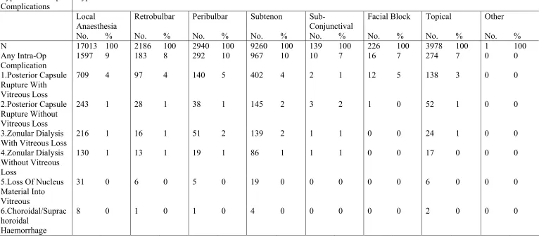
Retrobulbar Peribulbar Subtenon Subconjunctival Facial Block Topical Other N No. % No. % No. % No. % No. % No. % No. % All Centres 17013 2186 13 2940 17 9260 54 139 1 226 1 3978 23 1 0 A 164 37 23 5 3 1 1 149 91 B 1064 617 58 9 1 394 37 2 0 19 2 72 7
C 114 113 99 106 93 1 1
D 729 44 6 3 0 463 64 91 12 1 0 156 21
E 296 1 0 294 99 1 0
F 633 25 4 47 7 480 76 6 1 1 0 94 15
G 192 2 1 120 63 1 1 72 38
H 846 2 0 234 28 616 73 6 1 3 0 788 93 I 1141 2 0 6 1 531 47 7 1 2 0 600 53
J 353 1 0 350 99 1 0 1 0
K 1048 6 1 7 1 1036 99
L 830 53 6 293 35 112 13 39 5 416 50 M 825 16 2 687 83 215 26 4 0 2 0
N 275 248 90 2 1 23 8 2 1 22 8 O 946 412 44 268 28 197 21 1 0 197 21
P 521 17 3 202 39 193 37 111 21
Q 370 1 0 1 0 216 58 157 42
R 200 131 66 68 34 1 1
S 436 1 0 249 57 5 1 219 50
T 422 195 46 63 15 196 46 5 1 26 6 U 683 22 3 120 18 546 80 3 0
V 417 2 0 1 0 362 87 2 0 52 12 1 0
Centre Local Anaesthesia
Retrobulbar Peribulbar Subtenon Subconjunctival Facial Block Topical Other N No. % No. % No. % No. % No. % No. % No. %
X 209 12 6 196 94 1 0
Y 112 112 100
Z 559 3 1 556 99
Aa 140 2 1 95 68 55 39
Ab 1268 175 14 158 12 350 28 1 0 1 0 602 47
Ac 615 8 1 207 34 405 66 1 0
Ad 177 126 71 55 31 46 26 2 1
Af 20 14 70 1 5 5 25 1 5
Ag 602 60 10 131 22 258 43 3 0 21 3 179 30
Table 2.9: Distribution Of Single And Multiple Local Anaesthesia
Centre Local Anaesthesia
Table 2.10: Type Of Sedation Given To Patient Who Had Local Anaesthesia
Centre Types Of Sedation
Table 2.11: Intraocular Lens Implantation 2.11(a) IOL Implantation
No. %
With IOL 17941 98
Without IOL 448 2
All 18389 100
2.11(b) Distribution Of IOL Placement
Table 2.12: Distribution Of Cataract Surgery Without IOL
Centre Cataract Surgery Without IOL IOL Planned But Not
Implanted
No IOL Was Planned
Table 2.13: Distribution Of IOL- Materials And Types
IOL No. %
N 17953 100
Materials
Pmma 9758 54
Silicone 1078 6
Acrylic 7105 40
Other 12 0
N 17950 100
Types
Foldable 8188 46
3. CATARACT SURGERY OUTCOMES
3.1 Cataract Surgery Complications - Intra-Operative
Table 3.1.1: Distribution Of Intra-Operative Complications By Type Of Cataract Surgery
Type Of Intra-Operative Complications
Types Of Cataract Surgery
All Surgeries Lens
5.Loss Of Nucleus Material Into Vitreous
Figure 3.1.1.1 Distribution Of Intra-Operative Complication
%
I n tr a - o p e r a ti v e c o m p l i c a ti o n
02 4 6 8 1 0
A l l s u r g e r i e s
A n y
1
2
3
4
5
6
7
8
Figure 3.1.1.2: Distribution Of Intra-Operative Complication By Posterior Capsule Rupture With Vitreous Loss And Posterior Capsule Rupture Without Vitreous Loss
%
T y p e o f c a ta r a c t s u r g e r y
05 1 0 1 5 2 0
P C R w i th v i tr e o u s l o s s P C R w it h o u t v i tr e o u s l o s s
L A E C C E P E P E t o E C C E IC C E 2 I O L I m p l a n t
LA=Lens Aspiration
Figure 3.1.1.3: Distribution Of Intra-Operative Complication By Zonular Dialysis With Vitreous Loss And Zonular Dialysis Without Vitreous Loss
%
T y p e o f c a ta r a c t s u r g e r y
05 1 0 1 5 2 0
Z D w i th v i tr e o u s l o s s Z D w i th o u t v i tr e o u s l o s s
L A E C C E P E P E t o E C C E IC C E 2 I O L I m p l a n t
LA=Lens Aspiration
Table 3.1.2: Distribution Of Intra-Operative Complications By Combined Surgery
5.Loss Of Nucleus Material Into Vitreous
Table 3.1.3: Distribution Of Intra-Operative Complications By Nature Of Cataract Surgery
Type Of Intra-Operative Complications Nature Of Cataract Surgery
All Patients Emergency Elective
No. % No. % No. %
N 18392 100 106 100 18286 100
Any Intra-Op Complication 1730 9 26 25 1704 9 1.Posterior Capsule Rupture With Vitreous
Loss
760 4 7 7 753 4
2.Posterior Capsule Rupture Without Vitreous Loss
265 1 5 5 260 1
3.Zonular Dialysis With Vitreous Loss 234 1 4 4 230 1 4.Zonular Dialysis Without Vitreous Loss 146 1 2 2 144 1 5.Loss Of Nucleus Material Into Vitreous 34 0 0 0 34 0 6.Choroidal/Suprachoroidal Haemorrhage 10 0 2 2 8 0 7.Significant Trauma To Cornea Or Iris 78 0 0 0 78 0
8.Other 235 1 6 6 229 1
Figure 3.1.3: Distribution Of Intra-Operative Complications By Nature Of Cataract Surgery
%
Intra-operative complication
05 10 15 20 25
Emergency Electiv e
Any
1 2 3 4 5 6 7 8
Table 3.1.4: Distribution Of Intra-Operative Complications By Type Of Anaesthesia
Type Of Intra-Operative Complications Types Of Anaesthesia
All Patients General Local
No. % No. % No. %
N 18392 100 1379 100 17013 100
Any Intra-Op Complication 1730 9 133 10 1597 9 1.Posterior Capsule Rupture With
Vitreous Loss
760 4 51 4 709 4
2.Posterior Capsule Rupture Without Vitreous Loss
Figure 3.1.4: Distribution Of Intra-Operative Complications By Type Of Anaesthesia
%
Table 3.1.5: Distribution Of Intra-Operative Complications By Type Of Local Anaesthesia
Type Of Intra-Op Complications
Types Of Local Anaesthesia
Local Anaesthesia
Retrobulbar Peribulbar Subtenon Sub- Conjunctival
Facial Block Topical Other
Type Of Intra-Op Complications
Types Of Local Anaesthesia
Local Anaesthesia
Retrobulbar Peribulbar Subtenon Sub- Conjunctival
Facial Block Topical Other
No. % No. % No. % No. % No. % No. % No. % No. % 7.Significant
Trauma To Cornea Or Iris
74 0 8 0 9 0 45 0 3 2 0 0 14 0 0 0
8.Other 213 1 21 1 33 1 143 2 0 0 2 1 25 1 0 0
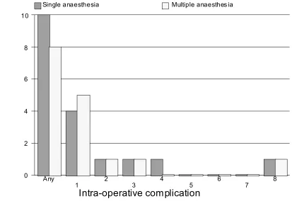
Table 3.1.6: Distribution Of Intra-Operative Complications By Single Or Multiple Local Anaesthesia
Type Of Intra-Operative Complications Local Anaesthesia
Single Multiple
No. % No. %
N 15335 100 1678 100
Any Intra-Op Complication 1460 10 137 8 1.Posterior Capsule Rupture With Vitreous
Loss
633 4 76 5
2.Posterior Capsule Rupture Without Vitreous Loss
221 1 22 1
3.Zonular Dialysis With Vitreous Loss 201 1 15 1 4.Zonular Dialysis Without Vitreous Loss 124 1 6 0 5.Loss Of Nucleus Material Into Vitreous 26 0 5 0
6.Choroidal/Suprachoroidal Haemorrhage 8 0 0 0 7.Significant Trauma To Cornea Or Iris 69 0 5 0
8.Other 202 1 11 1
Figure 3.1.6: Distribution Of Intra-Operative Complications By Single Or Multiple Local Anaesthesia
Single anaesthesia Multiple anaesthes ia
Any
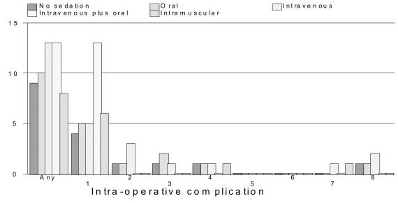
Table 3.1.7: Distribution Of Intra-Operative Complications By Type Of Sedation
Type Of Intra-Operative Complications Types Of Sedation
No Sedation Oral Alone Intravenous Alone
Intravenous Plus Oral
Intramuscular
No. % No. % No. % No. % No. %
N 14031 100 2729 100 144 100 15 100 104 100
Any Intra-Op Complication 1302 9 269 10 18 13 2 13 8 8 1.Posterior Capsule Rupture With Vitreous
Loss
564 4 132 5 7 5 2 13 6 6
2.Posterior Capsule Rupture Without Vitreous Loss
199 1 40 1 4 3 0 0 0 0
3.Zonular Dialysis With Vitreous Loss 174 1 41 2 1 1 0 0 0 0 4.Zonular Dialysis Without Vitreous Loss 111 1 16 1 2 1 0 0 1 1 5.Loss Of Nucleus Material Into Vitreous 26 0 5 0 0 0 0 0 0 0
6.Choroidal/Suprachoroidal Haemorrhage 7 0 1 0 0 0 0 0 0 0 7.Significant Trauma To Cornea Or Iris 66 0 6 0 1 1 0 0 1 1
8.Other 177 1 33 1 3 2 0 0 0 0
Figure 3.1.7: Distribution Of Intra-Operative Complications By Type Of Sedation
%
I n tr a - o p e r a ti v e c o m p l i c a ti o n
05 1 0 1 5
N o s e d a ti o n O r a l I n t ra v e n o u s In tr a v e n o u s p lu s o r a l I n t ra m u s c u l a r
A n y
1 2 3 4 5 6 7 8
Table 3.1.8: Distribution Of Intra-Operative Complications By Sedation
Type Of Intra-Operative Complications Sedation
No Sedation Single Multiple
No. % No. % No. %
N 14031 100 2972 100 10 100
Any Intra-Op Complication 1302 9 293 10 2 20 1.Posterior Capsule Rupture With Vitreous
Loss
564 4 143 5 2 20
2.Posterior Capsule Rupture Without Vitreous Loss
Figure 3.1.8: Distribution Of Intra-Operative Complications By Sedation
No Sedation Single Sedation Mult iple Sedation
Table 3.1.9: Distribution Of Intra-Operative Complications By Cataract Surgery With IOL
Type Of Intra-Operative Complications
Cataract Surgery With IOL
Figure 3.1.9: Distribution Of Intra-Operative Complications By Cataract Surgery With IOL
%
Intra-operative complication
0 20 40 60 80
Posterior Anterior Scleral
Any 1
2
3
4
5
6
7
8
Table 3.1.10: Distribution Of Intra-Operative Complications By Cataract Surgery Without IOL
Type Of Intra-Operative Complications
Cataract Surgery Without IOL
All Patients
3.Zonular Dialysis With Vitreous Loss
44 10 27 16 17 6
4.Zonular Dialysis Without Vitreous Loss
10 2 6 3 4 1
5.Loss Of Nucleus Material Into Vitreous
14 3 11 6 3 1
6.Choroidal/Suprachoroidal Haemorrhage
7 2 4 2 3 1
7.Significant Trauma To Cornea Or Iris
5 1 2 1 3 1
Figure 3.1.10: Distribution Of Intra-Operative Complications By Cataract Surgery Without IOL
%
Intra-operative complication
0 20 40 60 80
IOL Planned No I OL was planned
Any 1
2
3
4
5
6
7
8
Table 3.1.11: Distribution Of Intra-Operative Complications By Surgeon Status
Type Of Intra-Operative Complications
Surgeon Status
All Patients Specialist Gazetting Specialist
5.Loss Of Nucleus Material Into Vitreous
Figure 3.1.11: Distribution Of Intra-Operative Complications By Surgeon Status
%
Spec ialist G azetting s pecialist Medical offic er
3.2 Cataract Surgery Complications - Post-Operative
Table 3.2.1: Distribution Of Post-Operative Complications
Post-Operative Complications No. %
N 18392 100
Patients With Any Post-Op Complication 1670 9 Patients With Specific Post-Op Complication
1.Central Edema Within 4mm Of Visual Axis 77 0 2.Raised Iop Of More Than 30mmhg 22 0
3.Suture Abscess 17 0
4.Severe Iritis With Fibrin 15 0 5.Iris Prolapse/Wound Dehiscence 21 0 6.Vitreous Incarceration Into Wad 4 0 7.Vitreous In Ac Touching Cornea 2 0 8.IOL Decentration/Dislocation 4 0 9.Cystoid Macular Edema 23 0
10.Endophathalmitis 12 0
11.New Retinal Break 1 0 12.Retinal Detachment 10 0 13.Astigmation Of > 3 Diopters 217 1 14.Posterior Capsule Opacification 55 0
Table 3.2.2: Distribution Of Post-Operative Complications By IOL Types
Type Of IOL
Foldable Non-Foldable
Post-Operative Complications N No. % No. %
N 18392 8188 100 9762 100
Patients With Any Post-Op Complication
1670 652 8 979 10
Patients With Specific Post-Op Complication
1.Central Edema Within 4mm Of Visual Axis
77 33 0.4 42 0.4
2.Raised Iop Of More Than 30mmhg 22 1 0 19 0.2 3.Suture Abscess 17 6 0.1 8 0.1 4.Severe Iritis With Fibrin 15 8 0.1 6 0.1 5.Iris Prolapse/Wound Dehiscence 21 2 0 19 0.2 6.Vitreous Incarceration Into Wad 4 2 0 2 0 7.Vitreous In Ac Touching Cornea 2 2 0 0 0 8.IOL Decentration/Dislocation 4 2 0 2 0 9.Cystoid Macular Edema 23 5 0.1 17 0.2
10.Endophathalmitis 12 4 0 8 0.1 11.New Retinal Break 1 1 0 0 0
12.Retinal Detachment 10 2 0 6 0.1 13.Astigmation Of > 3 Diopters 217 18 0.2 196 2 14.Posterior Capsule Opacification 55 17 0.2 37 0.4
Table 3.2.3: Distribution Of Post-Operative Complication By Material Patients With Any Post-Op
Complication
1670 979 10 76 7 576 8 0 0
Patients With Specific Post-Op Complication 1.Central Edema Within 4mm Of Visual Axis
77 42 0.4 8 1 25 0.4 0 0
2.Raised IOP Of More Than 30mmhg
Table 3.2.4: Post-Operative Complication By Centre
Centre
A B C D E F
Post-Operative Complications N No. % No. % No. % No. % No. % No. % N 18392 167 100 1137 100 120 100 816 100 315 100 753 100 Patients With Any Post-Op
Complication
1670 11 7 70 6 6 5 375 46 20 6 69 9
Patients With Specific Post-Op Complication
1.Central Edema Within 4mm Of Visual Axis
77 0 0 12 1 0 0 5 1 0 0 0 0
2.Raised Iop Of More Than 30mmhg 22 1 1 2 0 0 0 1 0 0 0 4 1 3.Suture Abscess 17 0 0 4 0 0 0 1 0 0 0 2 0 4.Severe Iritis With Fibrin 15 0 0 0 0 0 0 4 0 0 0 1 0 5.Iris Prolapse/Wound Dehiscence 21 0 0 1 0 1 1 2 0 1 0 2 0 6.Vitreous Incarceration Into Wad 4 0 0 0 0 0 0 1 0 0 0 0 0 7.Vitreous In Ac Touching Cornea 2 0 0 0 0 0 0 0 0 0 0 0 0 8.IOL Decentration/Dislocation 4 0 0 0 0 0 0 0 0 1 0 0 0 9.Cystoid Macular Edema 23 2 1 0 0 0 0 0 0 1 0 0 0
10.Endophathalmitis 12 1 1 0 0 0 0 0 0 0 0 0 0 11.New Retinal Break 1 0 0 0 0 0 0 1 0 0 0 0 0
12.Retinal Detachment 10 0 0 0 0 0 0 0 0 0 0 1 0 13.Astigmation Of > 3 Diopters 217 1 1 36 3 0 0 8 1 11 3 42 6 14.Posterior Capsule Opacification 55 0 0 11 1 2 2 4 0 3 1 2 0
Centre
G H I J K L
Post-Operative Complications N No. % No. % No. % No. % No. % No. % N 18392 234 100 895 100 1215 100 365 100 112
5
100 889 100
Patients With Any Post-Op Complication
1670 19 8 10 1 34 3 2 1 21 2 6 1
Patients With Specific Post-Op Complication
1.Central Edema Within 4mm Of Visual Axis
77 2 1 0 0 1 0 0 0 2 0 2 0
2.Raised Iop Of More Than 30mmhg 22 1 0 0 0 4 0 1 0 0 0 0 0 3.Suture Abscess 17 0 0 0 0 1 0 0 0 0 0 0 0 4.Severe Iritis With Fibrin 15 0 0 0 0 0 0 0 0 2 0 0 0 5.Iris Prolapse/Wound Dehiscence 21 0 0 0 0 1 0 1 0 0 0 0 0 6.Vitreous Incarceration Into Wad 4 0 0 0 0 0 0 0 0 0 0 0 0 7.Vitreous In Ac Touching Cornea 2 0 0 0 0 0 0 0 0 0 0 0 0 8.IOL Decentration/Dislocation 4 0 0 0 0 0 0 0 0 0 0 0 0 9.Cystoid Macular Edema 23 0 0 1 0 4 0 0 0 2 0 1 0 10.Endophathalmitis 12 2 1 1 0 0 0 0 0 2 0 0 0 11.New Retinal Break 1 0 0 0 0 0 0 0 0 0 0 0 0 12.Retinal Detachment 10 0 0 1 0 1 0 0 0 1 0 0 0 13.Astigmation Of > 3 Diopters 217 0 0 3 0 18 1 0 0 5 0 0 0 14.Posterior Capsule Opacification 55 0 0 1 0 1 0 0 0 2 0 1 0
Centre
M N O P Q R
Post-Operative Complications N No. % No. % No. % No. % No. % No. % N 18392 906 100 300 100 1029 100 526 100 402 100 205 100 Patients With Any Post-Op
Complication
1670 47 5 6 2 83 8 8 2 4 1 14 7
Patients With Specific Post-Op Complication
1.Central Edema Within 4mm Of Visual Axis
77 13 1 0 0 5 0 1 0 0 0 1 0
2.Raised Iop Of More Than 30mmhg 22 1 0 0 0 0 0 0 0 0 0 1 0 3.Suture Abscess 17 0 0 0 0 1 0 1 0 0 0 0 0 4.Severe Iritis With Fibrin 15 4 0 0 0 1 0 0 0 0 0 1 0 5.Iris Prolapse/Wound Dehiscence 21 2 0 0 0 3 0 0 0 1 0 0 0 6.Vitreous Incarceration Into Wad 4 2 0 0 0 0 0 0 0 0 0 0 0 7.Vitreous In Ac Touching Cornea 2 0 0 0 0 0 0 0 0 0 0 0 0 8.IOL Decentration/Dislocation 4 1 0 0 0 0 0 0 0 0 0 1 0 9.Cystoid Macular Edema 23 3 0 0 0 1 0 0 0 0 0 0 0 10.Endophathalmitis 12 1 0 0 0 2 0 0 0 1 0 0 0 11.New Retinal Break 1 0 0 0 0 0 0 0 0 0 0 0 0 12.Retinal Detachment 10 0 0 0 0 1 0 0 0 0 0 0 0 13.Astigmation Of > 3 Diopters 217 14 2 0 0 28 3 4 1 1 0 0 0 14.Posterior Capsule Opacification 55 2 0 0 0 4 0 2 0 1 0 3 1
Centre
S T U V W X Y
Post-Operative Complications N No. % No. % No. % No. % No. % No. % No. % N 18392 458 100 520 100 814 100 444 100 632 100 238 100 120 100 Patients With Any Post-Op
Complication
1670 20 4 213 41 50 6 4 1 30 5 2 1 4 3
Patients With Specific Post-Op Complication
1.Central Edema Within 4mm Of Visual Axis
77 11 2 3 1 6 1 0 0 9 1 0 0 0 0
2.Raised IOP Of More Than 30mmhg 22 1 0 0 0 0 0 0 0 2 0 0 0 0 0 3.Suture Abscess 17 1 0 1 0 0 0 0 0 0 0 0 0 0 0 4.Severe Iritis With Fibrin 15 1 0 0 0 1 0 0 0 0 0 0 0 0 0 5.Iris Prolapse/Wound Dehiscence 21 0 0 0 0 1 0 0 0 0 0 0 0 0 0 6.Vitreous Incarceration Into Wad 4 0 0 0 0 0 0 1 0 0 0 0 0 0 0 7.Vitreous In Ac Touching Cornea 2 0 0 0 0 0 0 0 0 0 0 0 0 0 0 8.IOL Decentration/Dislocation 4 0 0 0 0 0 0 0 0 1 0 0 0 0 0 9.Cystoid Macular Edema 23 0 0 0 0 0 0 0 0 0 0 0 0 1 1 10.Endophathalmitis 12 0 0 0 0 0 0 0 0 0 0 0 0 0 0 11.New Retinal Break 1 0 0 0 0 0 0 0 0 0 0 0 0 0 0 12.Retinal Detachment 10 0 0 0 0 0 0 2 0 2 0 0 0 0 0 13.Astigmation Of > 3 Diopters 217 0 0 4 1 17 2 1 0 2 0 1 0 1 1 14.Posterior Capsule Opacification 55 4 1 0 0 1 0 0 0 6 1 1 0 1 1
Centre
Z Aa Ab Ac Ad Af Ag Ah Post-Operative Complications N No. % No. % No. % No. % No. % No. % No. % No. % N 18392 568 100 145 100 1311 100 678 100 190 100 20 100 630 100 225 100 Patients With Any Post-Op
Complication
1670 24 4 1 1 36 3 8 1 11 6 3 15 453 72 6 3
Patients With Specific Post-Op Complication
1.Central Edema Within 4mm Of Visual Axis
77 1 0 0 0 0 0 0 0 3 2 0 0 0 0 0 0
2.Raised IOP Of More Than 30mmhg
22 0 0 0 0 0 0 1 0 2 1 0 0 0 0 0 0
3.3 Post-Operative Follow-Up Period
Table 3.3.1: Median Follow-Up Period In Weeks (Patients With Only Unaided Vision, Refraction Was Not Performed)
Type Of Surgery N Median 25th Percentile 75th Percentile All Surgeries 941 5.6 1.3 10.9
Lens Aspiration 47 5.4 1.6 9.3
ECCE 379 4.3 1.1 10.4
PE 468 6 1.6 11.8
PE TO ECCE 20 5 2 13.1
ICCE 11 6.3 4.4 11.3
Secondary IOL Implant
16 1.4 1 4.4
Table 3.3.2: Median Follow-Up Period In Weeks (Patients With Refracted Vision)
Type Of Surgery N Median 25th Percentile 75th Percentile All Surgeries 5807 10 7.4 12.1 Lens Aspiration 102 10.1 7.9 13.3
ECCE 2616 10.4 8 12.7
PE 2865 9.1 7.1 11.6
PE TO ECCE 150 10.9 8.7 13.3
ICCE 30 11.5 8.4 13.6
Secondary IOL Implant
3.4 Post-Operative Visual Acuity
Table 3.4.1: Distribution Of Post-Operative VA
(a) All Patients, With Primary Cause Of Cataract And Not Combined Surgery
VA Post Operative Unaided Refracted
N=6228 100% N=5477 100% No. % No. %
6/5 2 0 17 0
6/6 306 5 1601 29
6/9 978 16 2078 38
6/12 1166 19 858 16
6/18 1187 19 392 7
6/24 1054 17 183 3
6/36 704 11 149 3
6/60 434 7 60 1
5/60 46 1 6 0
4/60 31 0 4 0
3/60 56 1 21 0
2/60 39 1 20 0
1/60 63 1 18 0
CF 80 1 27 0
HM 57 1 33 1
PL 15 0 5 0
(b) All Patients, With Primary Cause Of Cataract, Not Combined Surgery And Without Ocular Co-Morbidity
VA Post Operative Unaided Refracted
N=4061 100% N=3605 100% No. % No. %
6/5 1 0 14 0
6/6 238 6 1162 32
6/9 739 18 1465 41
6/12 840 21 585 16
6/18 792 20 207 6
6/24 684 17 85 2
6/36 420 10 53 1
6/60 228 6 9 0
5/60 25 1 2 0
4/60 12 0 1 0
3/60 17 0 5 0
2/60 18 0 6 0
1/60 12 0 1 0
CF 20 0 4 0
HM 9 0 5 0
PL 5 0 1 0
Without Ocular Co-Morbidity
Only Single Cataract Surgery (Combine=None) Primary Only For Cause Of Cataract
(Dr Goh, all the tables from table 3.4.1-3.6.1 included only patients without ocular co-morbidity, single cataract surgery and primary cause of cataract)
Figure 3.4.1.1(b): Distribution Of Post-Operative VA
%
Unaided Refract ed
Figure 3.4.1.2: Cumulative Distribution Of Visual Acuity By Pre- And Post-Operative
Pre-operativ e Post-operativ e
6/5 6/ 6 6/9 6/ 12 6/18 6/24 6/36 6/60 5/60 4/60 3/60 2/ 60 1/60 CF H M PL NPL
Figure 3.4.1.3: Cumulative Distribution Of Visual Acuity By Pre- And Post-Operative Refracted VA
Pre-operativ e Post-operativ e
Table 3.4.2: Distribution Of Post-Operative Refracted VA 6/12 Or Better At The Last Follow Up Among Patients Without Ocular Co-Morbidities, By Surgery
Type Of Surgery Unaided Refracted
N VA 6/12 Or
Figure 3.4.2: Percent Of Patients With Refracted VA 6/12 Or Better At The Last Follow Up, By Surgery
Unaided Refract ed
LA ECC E PE PE t o ECCE ICCE 2 I OL Implant
* La=Lens Aspiration
Table 3.4.3: Distribution Of Post-Operative Refracted VA 6/12 Or Better In Relation To Age And Type Of Surgery, Among Patients Without Ocular Co-Morbidities
Factor Types Of Cataract Surgery
All Surgeries Lens Aspiration ECCE PE PE TO ECCE ICCE Secondary IOL Implant
N No. % N No. % N No. % N No. % N No. % N No. % N No. %
N 3605 3226 89 31 23 74 1479 1257 85 1975 1852 94 97 77 79 4 2 50 19 15 79
Age Group, Year
Table 3.4.4: Distribution Of Post-Operative Refracted VA 6/12 Or Better In Relation To Gender And Type Of Surgery, Among Patients Without Ocular Co-Morbidities
Factor Types Of Cataract Surgery
All Surgeries Lens Aspiration ECCE PE PE TO ECCE ICCE Secondary IOL Implant
N No. % N No. % N No. % N No. % N No. % N No. % N No. % N 3605 3226 89 31 23 74 1479 1257 85 1975 1852 94 97 77 79 4 2 50 19 15 79 Gender
Table 3.4.5: Distribution Of Post-Operative Refracted VA 6/12 Or Better In Relation To Comorbidity And Type Of Surgery, Among Patients Without Ocular Co-Morbidities
Factor Types Of Cataract Surgery
All Surgeries Lens
Aspiration
ECCE PE PE TO ECCE ICCE Secondary IOL Implant
N No. % N No. % N No. % N No. % N No. % N No. % N No. %
All 3605 3226 89 31 23 74 1479 1257 85 1975 1852 94 97 77 79 4 2 50 19 15 79
Ocular Co-Morbidity
Yes 0 0 0 0 0 0 0 0 0 0 0 0 0 0 0 0 0 0 0 0 0
No 3605 3226 89 31 23 74 1479 1257 85 1975 1852 94 97 77 79 4 2 50 19 15 79
Systemic
Co-Morbidities ( Among Patients Without Ocular Co-Morbidity)
Yes 2023 1825 90 7 5 71 805 688 85 1149 1083 94 54 43 80 1 0 0 7 6 86