Original paper
GROWTH RATES OF THE MASSIVE CORAL Porites lutea
EDWARD AND HAIME, ON THE COAST
OF BONTANG, EAST KALIMANTAN, INDONESIA
Supriharyono 1 2*
1
Research Centre for Environment Diponegoro University, Semarang Indonesia 2
Faculty of Fishery and Marine Sciences, Diponegoro University, Semarang Indonesia
Received: 2 May, 2004 ; Accepted: 30 May, 2004
ABSTRACT
Growth rates (linear skeletal extension) and the timing of skeletal band formation were measured in eight specimens of the massive coral Porites lutea at three sites (BK1, BK2, and BK3) and three depths, i.e. 1 m , 3 m, and 5 m in each site. The sites were located in Bontang Kuala Regency, located about 7.5 km from the fertilizing industry, PT Pupuk Kaltim Tbk, Bontang. Growth rates were measured by using two techniques, i.e. X-radiograph and UV-light.
Result of the study indicates that the timing of the high density (HD) and low density (LD) bands is synchronous at the three locations. A one year growth is characterized by three HD bands, one of which is usually very dense. Illumination of the coral slabs by UV-light revealed a distinct fluorescent banding pattern on all coral specimens. The data indicate that the fluorescent bands are usually associated with the high density bands which are accreted during the wet season period. It is characterized by high land run-off containing elevated concentrations of fulvic and humic acid compounds, and this apparently occurred almost through out the year. However fluorescent bands were absent from a number of density couplets, known as “stress bands”. The results suggest that in the present study the linier skeletal extension rates, based on X-ray radiographic techniques, are a more accurate measure of P. lutea growth rates then fluorescence banding.
Comparisons of the skeletal extension rates indicate that the growth rates of P. lutea are not significantly difference (p > 0.05) either between sites or depths. The average of coral growth rates ranged from 0.8-1.2 cm/year. These are significantly correlated (p < 0.01) with the amount of rainfall. While the amount of rainfalls is not correlated with urea production of fertilizing industry, P.T.Pupuk Kaltim Tbk, which some of them are loss as dust (a core for water vapour) during process production.
Key words: Coral growth rate, massive coral’s growth
*Correspondence: Phone 62-24-7460038, Fax, 62-24-7460039, E-mail: pries50@yahoo.com
I
NTRODUCTION
Porites lutea is one of the most common
scleractinian coral species on Indonesian reefs. This species, P. lutea, is known very
exhibits nodular growth form (Chappell, environmental changes over their growth history. Accretion of calcium carbonate by reef building corals depends on a number of environmental factors (i.e. such as sun hour, light transparency, and temperature). These environmental factors affect the accretion of high and low density bands within the skeletal matrix of coral colonies. The high density (HD) and low density (LD) bands are revealed by X-ray radiographic techniques. Generally one year growth consist of two density bands, one HD band and the other LD band. However, some corals may have more than two high density bands during a one year growth period. Similar results were reported for the massive coral Porites lutea from Ko Phuket, Thailand (Charuchinda and Chansang, 1985; Brown et al, 1986), and North and South coast of Central Java, Indonesia (Supriharyono, 1986; 1987; 1998).
Boto and Isdale (1985) have
suggested that organic compounds
produced by freshwater plants in soil, such as fulvic and humic acids, may be incorporated into the coral skeleton. These compounds fluoresce under ultra-violet
light produce an alternate bright/dull banding pattern in corals (Isdale, 1984). Moreover, Isdale (1984) suggested that UV light may be used to analyze coral growth rates. The fluorescent banding
pattern may provide additional
information on the environmental
Coral Collection and Analysis
Eight colonies of Porites lutea were collected from the reef flat in April 2004. Coral specimens were taken from a depth of 1 m (BK11, BK21, and BK31), 3 m (BK1, BK23, and BK33), and 5 m (BK15, BK25, and BK35). After being air dried, each coral head was then cut (with a hack saw machine) parallel to the major growth axis to produce a slab 5-10 mm thick. The coral specimens (slabs) were, then, X-rayed on a medical X-ray machine unit, TOSHIBA, Model DC-12MB-1 using Kodak film at the "Pupuk Kaltim” Hospital, Bontang, East Kalimantan. Exposures were made at 50 kv, 150 mA for 0.03 sec with a source to film distance of 90 cm. X-ray negatives were contact printed on to photographic paper and the positives were used for analysis of annual
bands. Fluorescent banding pattern was analyzed by illuminating all coral slabs with of UV-black light (350 nm). The latter technique was used to compare the banding patterns under the UV-light and X-rays, and to investigate whether land run-off (associate with fluorescent bands) is associated with a high skeletal density bands (i.e. HD bands are formed during the rainy season). In addition, annual growth rates (linear skeletal extension) were measured using the techniques described by several workers (e.g. Dodge and Vaisnys, 1977). One year growth refers to the distant of two HD bands (annual HD bands).
Environmental Assessment
The most of environmental factors may affect on reef corals, among others are
amount of rainfall, sun shine duration, salinity and seawater temperature. These parameters, mainly climatological data, were collected from the nearest climatolog station, located at PT Pupuk Kaltim Tbk, about 7.5 km from the study sites. While other parameters, such as seawater
temperature, water transparency,
suspended solid, and salinity were adopted from the secondary or previous data collection.
R
ESULTS AND
D
ISCUSSION
Environmental Information
The sites are under the influence of a tropical monsoon climate. Generally, the northwest monsoon lasts from about December to February and the southeast monsoon from June to August. The rest of the year consists of two transition periods, from the northwest to the southeast northwest to southeast, brings heavy rainfall. Conversely, the southeast
monsoon is characterized by dry
conditions and it is called the dry monsoon, with the wind blowing from
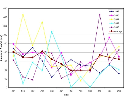
southeast to northwest. Apparently, this phenomenon may not occur in the study however, in average the amount of rainfall is slightly lower during July-September
(Figure 2). This phenomenon of
uncertainly rainfall, may be related to the production of urea by PT Pupuk Kaltim Tbk, Bontang in the study sites. Sasongko
et al (2004) suggests that during process,
Table 1. The mount of rainfall in the study sites, Bontang, 1999-2003
Year Amount of Rainfall (mm)
1999 2000 2001 2002 2003 Average
Jan Feb Mar Apr May Jun Jul Ags Sep Oct Nov Dec
Time
The high amount of rain means that the desalinization may be occurred, and this affects on the decreasing of seawater salinity, with the sequent that the salinity will be lower than the usual. The salinity is recorded about 29.5-32.80‰ in the study sites (PKT-PPLH-UNDIP, 2001), but it is possible that the minimum level will be lower than 29.5‰. As well, the amount of rainfall is also related to the number of land water runoff, with the sequence of sedimentation in coastal areas. Since, the water colour is brown, just looked like “coco” during rainy season. It is reported that the suspended solid went up to more than 50 mg/l, particularly after heavy rain, while in normal condition is seawater salinity, high sedimentation (suspended solids), and water transparency will affect on marine organisms, including reef corals. Although some coral may withstand in lower salinity, for the long exposure they will die (Supriharyono,
1986; Hariyadi, 2004). As well, some
corals may survive on high sedimentation, but the growth rate will be very low (Hubbard and Pocock, 1972; Pastorok and Bilyard,1985; Supriharyono, 1986).
Average seawater temperature
ranges from 25-29ºC in the study sites, it is
normal for reef corals. With exception, seawater temperature goes to about 39-42ºC in the area close to the outlet of cooling system PT. Pupuk Kaltim Tbk. This may be dead point for living marine
organisms, including reef corals
(Neudecker, 1981; Supriharyono, 1996). However, it will not be dangerous for the living coral in the study sites, since the location is far enough from the study sites.
As previously informed, that other that amount of rainfall and sea water temperature, the sun shine duration is also important factor to affect of coral growth.
Banding Pattern
In general the HD band is a
production of CaCO3 duringthe wet season
period (Dodge dan Thomson, 1974;
season period. Therefore, Highsmith
(1979) suggested coral density is
depending on light intensity (hot) and seawater temperature. Moreover, he claims that HD bands may be able to be produced either during high or low temperature. As well, HD bands are possibly accreted when light intensity high or low. While the LD band is generally accreted during medium temperature, 24- 29ºC. Therefore, it may be concluded that high density (HD) bands
are produced as a responce from various environmental factors, while the low density (LD) bands are accreted during the period, when light intensity is very high and range of seawater temperature is low. It is different, with Wellington and Glynn (1983), who worked on coral growth in Eastern Pacific (Panama), they found that various light intensity is more important than seawater temperature in order to affect coral density. Moreover, they concluded that HD bands formation is determined by low level of light intensity.
In the present study, the Porites
lutea produced high density bands either in
the dry season and in the wet season. It season band, while the other two HD bands are considered as stress bands (transition seasons production). This HD band formation apparently happened not only in the study sites. It also occurred in other areas, subject high sedimentation rates, e.g. Bandengan Bay, Jepara, central Java (Supriharyono, 1986), Ko Phuket, Thailand (Charuchinda and Chansang,
HD band
LD band
Fig. 3. Density banding pattern of massive coral
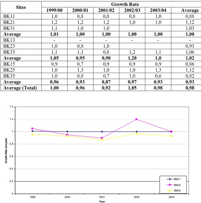
1985; and Brown et al, 1986). It proved that the HD band formation is very determined by water transparency and turbidity. three years growth rate of coral specimens collected from the depths of 1 m, 3 m and 5 m are not significantly different (p > 0.05). As well, the linear skeletal extension growth rates are also not significantly different between sites, BK1, BK2 and BK3 (p> 0.05). Table 2 shows the growth rates of linier extension coral specimens in the study sites. .
Table 2. Growth rates of Porites lutea (cm) in the study sites, Bontang, East Kalimantan
Sites Growth Rate
1999/00 2000/01 2001/02 2002/03 2003/04 Average
BK11 1,0 0,8 0,8 0,8 1,0 0,88
In addition, it is informed that
environmental condition is suitable for
living corals, therefore the coral growth
rates are not significantly different
either sites or depths. Table 3 shows
environmental condition in the study
sites. This Table exhibits that some
“key” environmental parameters, e.g.
salinity,
suspended
solids,
water
transparency, and sea water
tempe-rature, are in optimum condition for
living corals (Supriharyono, 2000). As
well, other environmental factors,
which affect those parameters, e.g.
amount of rainfall (salinity) and sun
shine duration (water temperature) are
the same. It is due very closely to the
distance inter sites, therefore their
effects on reef coral growth are also the
same in the study sites. However, there
is a correlation between amount of
rainfall on coral growth rates.
Table 3. Seawater quality in the study sites
Parameter Unit Ranges1) Optimum level for coral growth rate2)
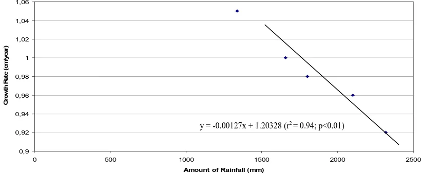
Salinity rainfall (Figure 5). According to analysis of correlation, it is proved that relationship
between those variables, is highly
significant correlated (r2 = 0.94; p<0.01), with regression equation, y = -0.00127x + 1.20328. This proved that amount of rainfall affects on the coral growth rate,
increasing amount of rainfall resulted the decrease of linier skeletal extension rate of corals in the study sites. This is also proved that the amount of rainfall may be dominant factor affects on coral growth rates in the study sites, together with other environmental factors, i.e. sun shine duration, light intensity (Supriharyono, 1986).
0 500 1000 1500 2000 2500
Amount of Rainfall (mm)
Fig 5. Relationship between amount of rainfall and coral growth rates in the study sites
As mentioned previously, that the loss production of urea (as dust) may affect precipitation. Since the production of those
fertilizers are going continuously
throughout the year, the rainfall may always occur in the study sites. While, the amount of rainfall will depend on the amount of (dust) urea, that may be released in the atmosphere. Consequently that the amount of rainfall will be correlated with
the number of urea production.
Unfortunately, the loss urea as dust from PT Pupuk Kaltim Tbk is distributed to all
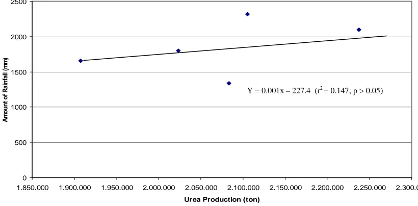
area, surrounding this industry, depending on the wind direction. Therefore the number of urea production is not automatically reflected to number of dust urea in each area, including study sites. This phenomenon, may result that no correlation between the number of urea production and the amount of rainfall in the study sites (r2 = 0.147; p > 0.05). While the amount of rainfall affects significantly on coral growth rates. The data of these variables, moreover, are presented in
Table 4 and Figure 6.
Table 4. Relationship between the number of urea production, amount of rainfall, and the
growth rate of corals in the study sites.
Year Urea Production
(ton/year)1)
Amount of rainfall
(mm/year)2)
Coral growth rates (cm/year)
1999 1,907,551 1,656 1.00
2000 2,237,593 2,107 0.96
2001 2,105,270 2,318 0.92
2002 2,083,587 1,337 1.05
2003 2,023,321 1,801 0.98
Note: 1) PKT. 2003. Performance Pabrik Amoniak dan Urea Tahun 1991-2003.
2) Sasongko et al (2004)
0 500 1000 1500 2000 2500
1.850.000 1.900.000 1.950.000 2.000.000 2.050.000 2.100.000 2.150.000 2.200.000 2.250.000 2.300.000
Urea Production (ton)
Fig. 6. Relationship between urea production of P.T. Pupuk Kaltim Tbk and the amount of
rainfall in the study sites
This figure proves that not all loss urea productions were spread through out the area, it is depending on the wind direction. Some areas may receive a lot of dust urea, while the others may not in the same time. This reflectes that no correlation between the number of urea production and the amount of rainfall in the study sites.
C
ONCLUSION
Conclusion
Based on the results of the study, it may be concluded as follows:
1. Coral growth rates fluctuated with the changes of amount of rainfall in the study sites occurred almost through out the year; the depths and between the sites;
3. Some chemical compounds of
fertilizing industry, P.T. Pupuk Kaltim Tbk, may be loss, particularly urea as dust during process of production. The consequence of this may affect on uncertainty rainfall in the study site;
4. No correlation between total
production of urea and the amount of rainfall (p > 0,05); and
5. Amount of rainfall affects significantly
on coral growth rate (p < 0,01).
Suggestion
Based on the result that may release (loss) of chemical compounds as a dust,
particularly urea, during process
production of fertilizing industry, P.T. Pupuk Kaltim Tbk, on the atmosphere,
some activities may be suggested as follows.
1. Reprocess management to anticipate
the loss of chemical compounds during process of production;
2. Need assessment of the loss the
chemical compounds, mainly urea and ammonia, both in water column, including coral skeleton in reef, and in terrestrial, particularly for human health, in surrounding P.T. Pupuk sampling, oceanographically and climate data around Bontang. zone. Nature. 315: 396-397.
Charuchinda, M. and H. Chansang. 1985. Skeleton extension and banding formation of Porites lutea of natural radiochemical and growth
records in contempory
hermatypic corals from the Atlantic and Caribbean. Earth
Planet. Sci. Lett., 23: 313-322.
Dodge, R.E. and J.R. Vaisnys. 1977. Coral populations and growth patterns : response to sedimentation and
turbidity associated with
dredging. J. Mar. Sci., 35:
715-Hubbard, J.A.E.B. and Y.P. Pocock. 1972. Sediment rejection by recent scleractinian corals a key to paleo-environmental
reconstruction. Geol. Runds. 61: 598-626.
Hudson, J.H., E.A. Shinn, R.B. Halley,
and B. Lidz. 1976.
Sclerochronology: A tool for interpreting past environments.
Geology, 4: 361-364.
Hudson, J.H., E.A. Shinn, and D.M.
Robbin. 1982. Effects of
offshore oil drilling on Philippine reef corals. Bull. Mar. Sci., 32:
Reefs, Great barrier Reef Comn., Brisbane, 2: 277-287.
Neudecker, S. 1981. Growth and survival of scleractinian corals exposed to thermal effluents at Guam. Proc. 4th. Int. Coral Reef Symp.,
PKT-PPLH.2001. Studi pemetaan kondisi biota laut di perairan pesisir dan laut sekitar PT Pupuk Kaltim, Kota Bontang, Propinsi Kali-mantan Timur. Kerjasama antara PT Pupuk Kaltim Tbk dengan
Pusat Penelitian Lingkungan
Hidup, Lembaga Penelitian,
Universitas Diponegoro. Penelitian Lingkungan Hidup, Lembaga Penelitian, Universitas Diponegoro.
Supriharyono. 1987. Growth rate of coral species, Porites lutea, at the west coast of Nusa Kambangan Island, Cilacap, South Central Java,
Indonesia Research Institute,
Diponegoro University Sema-rang.
Supriharyono. 1996. Pengaruh kenaikan suhu air laut terhadap survival rate plankton. Kerjasama antara
Pusat Penelitian Sumberdaya
Alam dan Energi, Lembaga Penelitian UNDIP dengan Pusat
Penelitian Standardisasi dan
Proteksi Keselamatan Radiasi, BATAN, Jakarta.
Supriharyono. 1998. Skeletal banding apttern and growth rates of the massive corals, Porites lutea Edward and Haime on the north coast of Central Java, Indonesia. J. Coast. Dev., 2 : 307-317.
Supriharyono. 2000. Pengelolaan Eko-sistem Terumbu Karang. PT. Penerbit Jambatan, Jakarta.
Weber. J.N., E.W. White, and P.H. Weber. 1975a. Correlation of density banding in reef coral skeletons with environmental parameters : the basis for interpretation of chronolo-logical records preserved in the corolla of corals. Paleobiology, 1: 137-149.
Weber, J.N., P. Deines, E.W. White,
and P.H. Weber. 1975b.
Seasonal high and low density bands in reef coral skeletons.
Nature, 255: 697-698.
Wellington, G.M. and P.W. Glynn. 1983.
Environmental influences on
skeletal banding in Eastern Pacific (Panama) corals. Coral
This figure proves that not all loss urea productions were spread through out the area, it is depending on the wind direction. Some areas may received a lot of dust urea, while the others may not in the same time. This reflected that no correlation between the number of urea
production and the amount of rainfall in the study sites
C
ONCLUSION AND
S
UGGECTION
Conclusion
Based on the results of the study, it may be concluded as follows:
6. Coral growth rates fluctuated with the changes of amount of rainfall in the study sites,
which occurred almost trough out the year;
7. The average of coral growth rates ranged from minimum of 0.8 cm/year to maximum
of 1.2 cm/year. There is no significantly difference (p > 0,05) of the coral specimens, both between the depths and between the sites;
8. Some chemical compounds of fertilizing industry, P.T. Pupuk Kaltim Tbk, may be
loss, particularly urea as dust during process production. The consequence of this may affects on uncertainty rainfall in the study site;
9. No correlation between total production of urea and the amount of rainfall (p > 0,05);
and
10. Amount of rainfall affects significantly on coral growth rate (p < 0,01).
4.2. Suggestion
Based on the result that may releasing (loss) of chemical compounds as a dust, particularly urea, during process production of fertilizing industry, P.T. Pupuk Kaltim Tbk, on the atmosphere, some activity may be suggested :
3. Reprocess management to anticipate the loss of chemical compounds during process
production;
4. Need assessment of the loss the chemical compounds, mainly urea and ammonia, both
LITERATURE CITED
Boto, K. and P. Isdale. 1985. Fluorescent bands in massive corals results from terrestrial fulvic acid inputs to nearshore zone. Nature, London, 315: 396-397.
Brown, B.E., M.D. Le Tissier, L.S. Howard, M. Charuchinda, and J.A. Jacson. 1986. Asynchronous deposition of dense skeletal bands in Porites lutea. Mar. Biol. 93: 83-89.
Chappell, J. 1980. Coral morphology, diversity and reef growth. Nature, London, 286: 249-252.
Charuchinda, M. and H. Chansang. 1985. Skeleton extension and banding formation of
Porites lutea of fringing reefs along the south and west coasts of Phuket Island
(Thailand). Proc. 5th. Int. Coral Reef Cong., tahiti, 6: 83-87.
Dodge, R.E. and J. Thomson. 1974. The natural radiochemical and growth records in contempory hermatypic corals from the Atlantic and Caribbean. Eart and Planet. Sci. Lett., 23: 313-322.
Dodge, R.E. and J.R. Vaisnys. 1977. Coral populations and growth patterns : response to sedimentation and turbidity associated with dredging. J. Mar. Sci., 35: 715-730.
Highsmith, R.C. 1979. Coral growth rates and environmental control of density banding. J. exp. mar. Biol. Ecol., 37: 105-125.
Hubbard, J.A.E.B. and Y.P. Pocock. 1972. Sediment rejection by recent scleractinian corals a key to paleo-environmental reconstruction. Geol. Runds. 61: 598-626.
Hudson, J.H., E.A. Shinn, R.B. Halley, and B. Lidz. 1976. Sclerochronology: A tool for interpreting past environments. Geology, 4: 361-364.
Hudson, J.H., E.A. Shinn, and D.M. Robbin. 1982. Effects of offshore oil drilling on Philippine reef corals. Bull. Mar. Sci., 32: 890-908.
Isdale, P. 1984. Fluorescent bands in massive corals record centuries of coastal rainfall. Nature, London, 310: 578-579.
MacIntyre, I.G. and S.V. Smith. 1974. X-radiographic studies of skeletal development in coral colonies. Proc. 2nd. Int. Symp. Coral Reefs, Great barrier Reef Comn., Brisbane, 2: 277-287.
Pastorok, R.A. and G.R. Bilyard. 1985. Effects of sewage pollution on coral reef communities. Mar. Biol. Prog. Ser., 21: 175-189.
PKT-PPLH.2001. Studi pemetaan kondisi biota laut di perairan pesisir dan laut dekitar PT Pupuk Kaltim, Kota Bontang, Propinsi Kalimantgan Timur. Kerjasama antara PT Pupuk Kaltim Tbk dengan Pusat Penelitian Lingkungan Hidup, Lembaga Penelitian, Universitas Diponegoro.
Sasongko dan Rahmat. 2004. Pemetaan sebaran amodia dan debu ure di udara sekitar PT Pupuk Kaltim, Kota Bontang, Propinsi Kalimantgan Timur. Kerjasama antara PT Pupuk Kaltim Tbk dengan Pusat Penelitian Lingkungan Hidup, Lembaga Penelitian, Universitas Diponegoro.
Supriharyono. 1986. The effects of sedimentation on a fringing reef in north central Java, Indonesia. PhD Thesis, Department of Zoology, University of Newcastle upon Tyne, UK.
Supriharyono. 1987. Growth rate of coral species, Porites lutea, at the west coast of Nusa Kambangan Island, Cilacap, South Central Java, Indonesia. Research Istitute, Diponegoro University, Semarang.
Supriharyono. 1996. Pengaruh kenaikan suhu air laut terhadap survival rate plankton. Kerjasama antara Pusat Penelitian Syumberdaya Alam dan Energi, Lembaga Penelitian UNDIP dengan Pusat Penelitian Standardisasi dan Proteksi Keselamatan Radiasi, BATAN, Jakarta.
Supriharyono. 1998. Skeletal banding apttern and growth rates of the massive corals, Porites lutea Edward and Haime on the north coast of Central Java, Indonesia. Jour. Coastal Development, 2 (1): 307-317.
Supriharyono. 2000. Pengelolaan Ekosistem Terumbu Karang. PT. Penerbit Jambatan, Jakarta.
Weber. J.N., E.W. White, and P.H. Weber. 1975a. Correlation of density banding in reef coral skeletons with environmental parameters : the basis for interpretation of chronolological records preserved in the corolla of corals. Paleobiology, 1: 137-149.
Weber, J.N., P. Deines, E.W. White, and P.H. Weber. 1975b. Seasonal high and low density bands in reef coral skeletons. Nature, London, 255: 697-698.
Wellington, G.M. and P.W. Glynn. 1983. Environmental influences on skeletal banding in Eastern Pacific (Panama) corals. Coral Reefs, 1: 215-222.
I wish to thank to Mrs. Arni Kusumastuti and Mr. Trijoko Laksono who have assisted to collect the coral specimens, as well as to P.T. Pupuk Kaltim Tbk, which has facilitated speed boat for coral sampling, oceanographically and climate data in around Bontang.
ABSTRACT
Mucus is one of a non-specific defense mechanism, since this is the first element of aquatic organisms, which contact physically, chemically, or biologically with the environment. The mucus self defense mechanism investigation was carried out on fresh water fish tilapia (Oreochromis mosambicus). Eight (8) types of lectine were used to examine residual carbohydrate-based protein from mucous component based on histological and histochemical observation method. The review was directed as basic information for detail review about physiology adaptation aspects.
The results showed that mucous in goblet cells from palatal, gills primary lamella, ecophagus and skin reacted with WGA (Wheat Germ Aglutinin) lectine. In another part, mucous from the goblet cells in palatal and esophagus cells reacted with PNA (Peanut Aglutinin). Based on these results, therefore, it can be concluded that mucous from goblet cells in esophagus contains residual of N-asetil glucosamine and/or similar acid β-galactose and α-N-acetyl galactomine. Mucous from goblet cell in palatal contains residual of X-acetyl glucosamine and/or sialat acid and galactose. While mucous in the gills lamella contains carbohydrate residual, namely N-acetyl glucosamine and/or sialat acid.
Key words: Mucus, Tilapia, histochemical analyzes
*) Correspondence: Telp. 024 – 8311525
I
NTRODUCTION
Mucous which coats or covers the outer side of fish and other aquatic invertebrate is known as a non-specific mechanical and chemical defense against environmental
Mucous has several functions; many of these are as a mechanical protection, osmoregulation, and barrier to colonization of parasites, fungus and
bacteria. Mucous contains several
substances, such as immunoglobulin
(Rombourt et al, 1995), lysozyme
(Fletcher and White, 1973), C-reactive protein (CRP) (Ramos and Smith, 1978) and lectine (Suzuki, 1995). The main component of mucous is glicoprotein produced by goblet cells/mucous cell (Pickering, 1974). Glicoprotein in mucous varies depends on the fish species. Histochemical painting by lectine at
superficial skin tissue showed the
existence of glycoprotein mucous in the environment. The investigation was aimed
at examining goblet/mucous cells
distribution in producing mucous and biochemical nature of lectine in Tilapia.
M
ATERIALS AND
M
ETHODS
Tilapia (Oreochromis mosambicus) of 15.2 – 25.0 cm total body lengths and 145 – 250 grams of body weight was used in this experiment.
After anesthesia by 0.01% 2-b-phenoxyethanol, samples from palatal epithelium, gills, esophagus and skin were drawn. These samples were fixed in Bouin liquid for 24 hours at room temperature, and then dehydrated through series of ethanol, infiltrated with paraffin and embedded in hystoparafin. The embedded tissue was cutted in 5-cm thickness and painted with haemotoxylin-eosin. The tissue observed using microscope.
Histochemical painting was done in which each sample was fixed in 4%
paraformaldehide liquid in 0.1 M
phosphate buffer pH 7.2 and stored several
days at temperature at 4o C. These samples
were embedded and cut with thickness
aforementioned. The lectine was
conjugated by FITC (Floresence Immuno
Thiocyanat) that was used for
histochemical painting. In this test, 8 lectine were used. Painting technique orders were done after deparafination
process by xylene continued by
dehydration to series ethanol concentrat from 100% to 70%. Sample was washed 3 times in PBS at pH 7.4, and then incubated by FITC-lectine that was diluted by PBS (1: 1000) in room temperature for 1 hour in dark condition, and then washed 3 times by PBS. After the slides were closed by glass object using 1.4 Diazabicyclo (2-2-2) octane solution (Sigma, St. Louis, MO) liquid that mixed with glycerol (1:1) was then observed by florescent microscope. The sample incubated by PBS without FITC-lectine at room temperature for 1 hour was used as a control.
R
ESULTS AND
D
ISCUSSION
Mucous cell histology
The figures of mucous cell histology from several tissues of Tilapia were shown in
Table 1. The epithelia surface from
palatal cells of Tilapia (Figure 1a) indicates many goblet/mucous cells in different shapes and sizes. Several mucous found in this area has cylindrical shape, but no broken cells in proximal were found.
The mucous cells density was
approximately 47 cells / 0.01 mm2, and the
biggest mucous cell size was 60 m height and 40 m widths. .
No Variable Palatal epithelium Gills Esophagus Skin
1 Type Oval, ellipse Globular, small Oval, ellipse Oval
2 Biggest size ( m) Height 60, width 40 Diameter 10 Height 70, width 50 Small
3 Total (0,01 mm3) 47 12 23 4
In the gills (Figure 1b), mucous cells were relatively low and distributed at apical area. Several cells contain mucous in vesicle shape and swell to oval cells. These mucous cells were relatively small in their shape at the diameter of about 10 m. The number of mucous cells
approximately 12 cells / 001 mm2.
In esophagus (Figure 1c), the epidermis was constructed by cells layer in cuboids shape. In this area, the mucous cells were well developed and varied in their shape, from circle to oval. Mucous cells density about 23 cells / 0.01 mm2, and the biggest cell was 70 m height and 50
m width respectively.
Many mucous cells were found in palatal epitheliums cell and esophagus. According to Drenner et al (1984), tilapia
(Oreochromis esculeritus) was reported as a type of “size-selective suspension feeder’’ fish and the mucous that disposed from digestion duct was related to feeding digestion activity. Furthermore, Sanderson
et al (1996) reported that in Tilapia
(Oreochromis niloticus) the mucous
secretion speed was determined by stimulus response toward particle size of feed.
A thin layer from several epithelia cells built the skin epidermis of tilapia (Figure 1d). The epidermis surface was relatively smooth in its shape covered by several epithelia squamous cells. The number of mucous cells was relatively small, i.e. about 4 cells / 0.01 mm2 which were distributed on the surface of epidermis layers.
b)
( a )
( b )
Fig. 1. Observation histological from several tissues of Tillapia (Oreochromis mosambicus)
a. Goblet cell epithelium from palatal
b. Goblet cell from gills
c. Goblet cell from esophagus
d. Goblet cell from skin
Histochemical of goblet/mucous cells
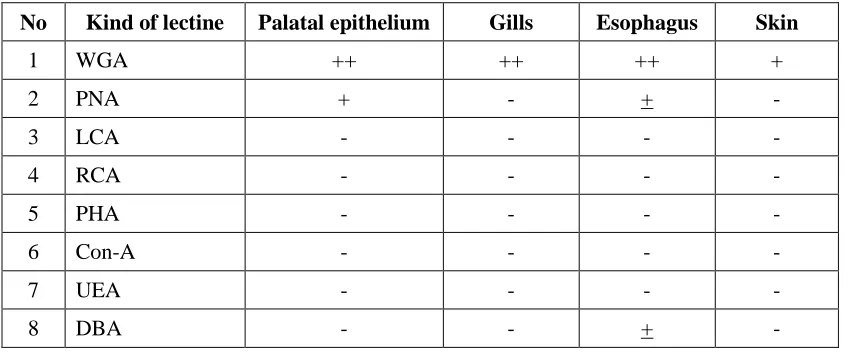
From 8 types of lectine used in the test (Table 2), WGA (Wheat Germ Aglutinine) reacted with epithelium of palatal cells, gills primary lamella, esophagus and skin of Tilapia. Lectine of PNA (Peanut Aglutinine) type reacted with surface of palatal epithelium and esophagus cells. Lectine from DBA (Dilichos Biflofus) type reacted only with esophagus. Painting/ reaction intensity shows that WGA was stronger than PNA and DBA toward mucous cells. The other types of lectine
such as LCA, RCA, OHA, Con-A and UEA showed no reaction with mucous cells.
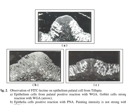
The epithelium cells of palatal, lectine of WGA type was different from PNA such as painting intensity or the number of mucus cells that positively reacted (Figure 2). Lectine of WGA type was stronger in their reaction intensity with mucous cells than PNA. Also lectine of WGA type reacted with mucous cells from the gills, esophagus and skin. Both PNA and DBA lectines weakly reacted with
mucous cells from esophagus.
Table 2. Painting FITC-lectine on several tissues of Tillapia
No Kind of lectine Palatal epithelium Gills Esophagus Skin
1 WGA ++ ++ ++ +
2 PNA + - + -
3 LCA - - - -
4 RCA - - - -
5 PHA - - - -
6 Con-A - - - -
7 UEA - - - -
8 DBA - - + -
Explanation:
+ +: Strongly reacted - : Weakly reacted + : Fairly reacted
According to Gona (1979) the function of mucous layer was very related to type of glycoprotein produced by mucous cells. Lectine of WGA type was found more in mucous cells in rat, monkey, ma, and guinea pig. The lectine
( a )
( b ) ( c )
Fig. 2. Observation of FITC-lectine on epithelium palatal cell from Tillapia
a) Epithelium cells from palatal positive reaction with WGA. Goblet cells strong
reaction with WGA (arrow);
b) Epithelia cells positive reaction with PNA. Painting intensity is not strong with WGA (arrow);
c) Epithelia cells of palatal as control. No reaction with goblet cells (arrow).
Lectine of PNA type found in mucous cells of Tilapia intestine according to Pajak and Danguy (1993) was contained
residual of b-galactose and β
-N-acetylgalactosamine that plays as a viscoelastic barrier that covers mucous from acid and proteolyses environment. Lectine of DBA type was also found in mucous cells of intestine according to Menghi et al (1996), lectine of DBA type was generally found in epithelium and glandule of tilapia’s side, frog and turtle intestine.
C
ONCLUSION
1. The mucous cells in tilapia were found
in palatal cells, gill primary lamellae and esophagus or skin.
2. Lectine of WGA type was found in
mucous cells from palatal cells, gill primary lamella, esophagus and skin. Lectine of PNA and DBA types were found only in mucous cells from tilapia’s esophagus.
Asakawa, M, 1970. Histochemical studies of the mucus on the epidermis of eel, Anguilla japonica. Nippon
Suisan Gakkaishi. 36: 83-87.
Drenner, R.W, Taylor, S.B., Lazzaro, X. and Kettle, D. (1984). Particle-grazing and plankton community impact of an omnivorous cichlid.
Trans. Am. Fish. Soc. 113:
397-402.
Fletcher, T.C. and White, 1973. Lysozyme activity in the plaice
(Pleuro-nectes platessa L.). Experientia
29: 1283-1285.
Gona, O. 1979. Mucus glikoproteins of teleostean fish, a comparative histochemical study. J.
Histo-chem. 11: 709-718.
Menghi, G., Scocco, P. and Ceccarelli, P. 1996. Glycohistochemistry of the tilapia spp stomach. J. Fish Biol. 49: 584-593.
Pajak, B. and Danguy, A. 1993.
Characterization of sugar
moieties and oligosaccharides sequences in the distal intestinal epithelium of the rainbow trout
by means of lectine
histochemistry. J. Fish Biol. 43: 709-722.
Pickering A.D. 1974. The distribution of mucous cells in the epidermis of the brown trout (Salmo trutta L.) and the char (Salvelinus alpinus
L.). J. Fish Biol. 6: 111-118.
Ramos and Smith, A.C. 1978. The-C-reactive protein (CRP) test for the detectionof early disease in fishes. Aquaculture 14: 261-266.
Rombourt, J.H.W.M., N. taverne, M. van de kamp and A.J. Taverne-Thiele., 1995. Differences in
mocus and serum
immu-noglobulin of carp (Cyprinus
carpio L.). Dev. Comp. Immunol.
17: 309-317.
Sanderson, S.L., Stebar, M.C., Ackermann, K.L., Jones, S.H., Batjakas, I.E. and Kaufman, L., 1996. Mucus entrapment of particles by a
suspension feeding tilapia
(Pisces: Cichlidae). J. Exp. Biol. 199: 1743-1756.
Suzuki, Y., 1995. Defense mechanisms in the skin of fish. In: Defense mechanisms in aquatic animals (ed. K. Mori and H. Kamiya), pp.
9-17, Koseisya Koseikaku,