29 UTILIZATION OF ETHYL ACETATE FRACTION KETAPANG LEAF
(Terminalia catappa) AS BIOREDUCTOR IN SYNTHESIS GOLD NANOPARTICLES AND ANALYSIS ANTIBACTERIAL PROPERTIES
Sarwina Hafid1, Muhammad Zakir, Seniwati Dali
Chemistry Departement, Faculty of Mathematics and Natural Sciences, Hasanuddin University, Makassar
90245
Abstrak. Nanopartikel emas disintesis dengan metode bioreduksi menggunakan fraksi etil asetat daun ketapang (Terminalia catappa). Nanopartikel yang terbentuk dikarakterisasi menggunakan spektrofotometer UV-Vis, FourierTransform Infra Red (FTIR) dan X-Ray Diffraction (XRD). Pada analisis spektrofotometer UV-Vis nanopartikel emas dengan konsentrasi larutan prekursor 0,5 mM dan 1 mM masing-masing memiliki serapan antara 533,5-540 nm dan 539-587 nm. Analisis gugus fungsi dalam sintesis nanopartikel emas dianalisis menggunakan Fourier Transform Infra Red
(FTIR). Sistem kristal dari nanopartikel emas adalah kubik dengan ukuran nanopartikel 17,13 nm. Pengujian aktivitas antibakteri dilakukan menggunakan beberapa bakteri uji yaitu Staphylococcus aureus, Escherichia coli, dan Bacillus subtilis. Nanopartikel emas tidak dapat menghambat aktivitas bakteri uji Staphylococcus aureus, Escherichia coli, dan Bacillus subtilis sehingga perbedaan konsentrasi nanopartikel emas tidak mempengaruhi daya hambat bakteri.
Kata kunci: antibakteri, biosintesis, karakterisasi nanopartikel, nanopartikel emas, Terminalia catappa.
Abstract. Gold nanoparticles was synthesized by bioreduction method using ethyl acetate fraction of ketapang leaf (Terminalia catappa). Nanoparticles formed were characterized using spectrophotometer UV-Vis, Fourier Transform Infra Red (FTIR) and X-Ray Diffraction (XRD). In the analysis of spectrophotometer UV-Vis gold nanoparticles with a precursor solution concentration of 0.5 mM and 1 mM each has an absorption between 533.5-540 nm and 539-587 nm. Analysis of functional groups of gold nanoparticles was analyzed by Fourier Transform Infra Red (FTIR). Crystal system of gold nanoparticles is cubic with nanoparticles size of 17.13 nm. The evaluation of test antibacterial activity was performed using several bacteria test such as
Staphylococcus aureus, Escherichia coli, and Bacillus subtilis. Gold nanoparticles can not inhibit the activity of a test bacterium Staphylococcus aureus, Escherichia coli, and Bacillus subtilis so that differences in the concentration of gold nanoparticles did not affect the inhibition of bacteria.
Keyword: antibacterial, biosynthesis, characterization of nanoparticles, gold nanoparticles,
Terminalia catappa.
1
30 INTRODUCTION
Nanotechnology is a branch of science that is growing very rapidly and is an interesting technological innovations in the field of production, size and shape. Nanoparticles are part of nanotechnology which is very popular and increasingly rapid development since the beginning of 2000. This is due to the benefits and applications of nanoparticles are very broad for human life, among others in the field of environmental, biomedical, healthcare, and industrial (Tsuzuki, 2009).
Some research techniques are available for the synthesis of metal nanoparticles, both methods of chemical and physical methods. However, the synthesis method involving a variety of chemicals can cause the presence of a toxic chemical species which can give side effects on biological applications (Mittal et al., 2014). Because of this, various methods have been developed so called experts emerging nanotechnology based green plants as bioreduktor for the synthesis of gold nanoparticles (Singh et al., 2012). Ketapang is a medicinal plant that grows in Indonesia and has been traditionally used to treat cardiovascular disease, skin, liver, respiratory, stomach, gonorrhea and insomnia (Pauly, 2001). Ketapang known to contain medicinal compounds such as flavonoids (Lin et al., 2000), triterpenoids (Gao et al., 2004), tannins (Ankamwar 2010), alkaloids (Mandasari, 2006), steroids (Babayi et al., 2004) and fatty acids (Jaziroh, 2008). Extracts are used in the synthesis of nanoparticles is ethyl acetate fraction of leaf ketapang. Acetate ethyl is a good solvent used for extraction because it can
be easily evaporated, not hygroscopic and has a low toxicity (Ward and Sulistyani, 2012). Ethyl acetate is a solvent that may be interesting compounds alkaloids, flavonoids, tannins and phenols in leaves ketapang (Packirisamy and Krishnamorthi, 2014).
Gold nanoparticles can be applied in the development of antibacterial strategies. This is because the gold nanoparticles are non-toxic and flexibility in surface modification (Cui et al., 2012). Nanoparticles of gold is a metal that is unique among other metals because it is resistant to oxidation and corrosion. Gold tends to be reduced so that in the long term, gold is embedded in the body does not have an adverse effect (Fatimah and Hidajati, 2012) because of the size of the gold nanoparticles are very small be compared with the body's cells, the nanoparticles can exit and enter easily into the cells without interfere with the workings of the cell (Abdullah, 2010).
Based on the description above, conducted research on the synthesis of gold nanoparticles by using ethyl acetate fraction compound leaves of Ketapang
(Terminalia catappa) as bioreduktor and analysis of antibacterial properties of gold nanoparticles were tested against
Staphylococcus aureus, Escherichia coli
and Bacillus subtilis. The third of these bacteria are toxic harmful bacteria and resistant to antibiotics, so it is necessary to control the lives of the bacteria.
MATERIALS and METHODS Materials Research
Materials used gold, ketapang leaf (Terminalia catappa), HCl pa, HNO3 pa,
31 poly acrylic acid (PAA), methanol pa,
ethyl acetate pa, Bovine Serum Albumin (BSA), Nutrient Agar (NA), Medium Muller Hinton Agar (MHA), klorampenikol, cultures of bacteria
Staphylococcus aureus, cultured bacterium Bacillus subtilis, bacterial culture of Escherichia coli, ethanol 95%, paper disc, paper pH universal, aluminum foil, and Whatman filter paper 42.
Research Tools
The tools used are glass tools commonly used in the laboratory, oven SPNI 505 FD, analytical balance, spectrophotometer UV-Vis 2600 Shimadzu, XRD 700 Shimadzu, FTIR IRPrestige-21 Shimadzu, incubator, autoclave, tweezers, magnetic stirrer, funnel , vacuum filters, rotary evaporator Heidolph Hei-Vap Value, spray dryer Armfield, a petri dish (diameter 8.5 cm), 30 mL vial bottle, desiccator, rod stirrer, spatula, spray bottle and scissors.
PROCEDURE
Preparation Extract Ketapang Leaf Plants used in this study is Ketapang (Terminalia catappa). Part of the plant used in this study are the leaves in fresh condition. The leaves were dried at room temperature. Once dried then crushed and sieved. Furthermore weighed. Ketapang leaf dry powder weighed 50 g, was added a mixture of 50 mL MeOH and 50 mL akuabides. Furthermore, sonicated for 1 hour. After the suspension was filtered, and then the methanol evaporated from the filtrate by using a rotary evaporator to obtain a thick extract ketapang leaf (Terminalia catappa).
The Solvent Partition from Extract Ketapang Leaf
Extract thick ketapang leaf in the partition with ethyl acetate (3 x 100 mL). Furthermore, separated between the organic phase and the water phase. The organic phase was concentrated using a rotary evaporator.
Synthesis of Gold Nanoparticles With Ethyl Acetate Fraction Ketapnag Leaf
The synthesis of gold nanoparticles is done by adding a fraction of ethyl acetate ketapang leaves into the solution HAuCl4 (0.5 and 1 mM) and
added PAA 1%. Characterization of the mixed solution in the form of color, pH and UV-Vis spectra. Once characterized, the mixed solution is dried with a spray dryer to obtain a powder of gold nanoparticles. powder gold nanoparticles were characterized by FTIR and XRD.
Coatings Gold Nanoparticles on Paper Disc
Paper disc sterilized in the oven. Paper discs soaked in colloidal gold nanoparticles using a magnetic stirrer, stirring for 5 minutes and then allowed to stand for 1 day. After that, the disc Paper dried in an oven at a temperature of 70 °C for 5 minutes.
Testing the Antibacterial Activity Testing inhibition of gold nanoparticles on the growth of
Staphylococcus aureus, Escherichia coli
32 using sterile tweezers on the surface of
the medium. Subsequently incubated for 24 hours and 48 hours at 37 ° C and then observed and measured its resistance zone with a sliding crossbar.
RESULTS AND DISCUSSION Characterization Color and pH
Characterization color of the solution was conducted to determine the formation of gold nanoparticles
Figure 1. Changes the color in of gold nanoparticles from the first minute until 1 day
Burgundy color that forms indicate the occurrence of reduction of Au3+ into Au0. The color formation caused by the excitation of surface plasmon nanoparticles. The changes that occur during the synthesis of the color indicates the growth of clusters generated even greater, at which time the gold atom has not interact with each other (colorless solution). In a certain amount of gold clusters provide a purple color is followed into the red, while the larger clusters and upon entering the nano-size, gold became red wine. Au atoms will interact with each other and produce metal bonding cluster in very large quantities.
Characterization of the pH measured for increasing time. PH value measurement is done to observe any possible changes in pH during the reaction process. PH measurement starts for a precursor solution, stabilizers, and
bioreduktor with each pH value is 2, 4 and 5. The pH value of the gold nanoparticles is pH 1, even after the seventh day the pH is not changed. This shows the increase in acidity of the gold nanoparticles is characterized by a decrease in pH value.
Characterization Using UV-Vis Spectrophotometer
The Formation of Gold Nanoparticles Based on the UV-Vis spectra (Table 1) wavelength spectrum measurement results, obtained pattern and the wavelength of maximum absorbance which became the basis of the formation of gold nanoparticles. Gold nanoparticles has a wavelength of 500-600 nm. The process of formation of gold nanoparticles can be seen from the shift of the maximum wavelength is 313.00 nm solution HAuCl4 be 540.00 nm and
587.00 nm as shown in Table 1.
Table 1. Results of UV-Vis spectrum analyzer
Samples Wavelength (nm) Absorbance
HAuCl4
Gold nanoparticles 0,5 mM Gold nanoparticles 1 mM
313,00 540,00 587,00
33 The Stability of Gold Nanoparticles
The stability of the colloidal solution of nanoparticles can be seen from the changes in absorption peak. Gold nanoparticles have a tendency to form larger clusters. Therefore, it takes a stabilizer to prevent aggregation process.
(a)
(b)
Figure 2. Spectrum stability of gold nanoparticles (a) AuNp 0.5 mM (b) AuNp 1 mM.
In this study poly acrylic acid is used as a stabilizer. PAA can be used as a stabilizing agent it can be seen from PAA structure that has a -OH group. O atoms in the PAA likes to stick to the gold atom as O atom has a lone pair, while the acrylate groups that have a long chain would be ligands away from the surface of the gold and gold shield so as not to form aggregates.
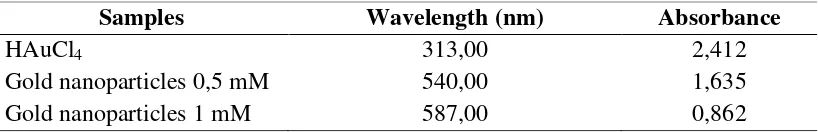
Characterization of Gold Nanoparticles with FTIR
FTIR measurements conducted to identify biomolecules that may play a role in the ethyl acetate fraction of leaf ketapang to reduce ion AuCl4- into Au0.
Phytochemical test on leaves ketapang showed phenolic compounds are compounds that dominant. Phenolic
compounds indicated are tannins. Chen et al., (2000) reported that ketapang leaf contains 21% of tannin.
Figure 3. FTIR spectra of (a) Fraction ethyl acetate ketapang leaves + PAA, (b) gold nanoparticles.
Figure 3 (a) is an FTIR spectrum of ethyl acetate fraction ketapang leaf (Terminalia catappa) with the addition of poly acrylic acid (PAA) shows a prominent absorption band at 1707 cm-1, 1037 cm-1 and 3448 cm-1. At wave number 1707 cm-1 are characteristic of the carbonyl group of carboxylic acids and phenols. vast stretches in the wave number 3448 cm-1 arises because of the free OH in phenol, besides there are local wave number 1037 cm-1 which is a characterization of the CO group.
Figure 3 (b) is an FTIR spectrum of ethyl acetate fraction ketapang reducing gold leaf at wave number 1716 cm-1, 3439 cm-1 and 1035 cm-1. Carbonyl group in 1707 cm-1 shifted into 1716 cm-1. Resonance may be due to binding of gold nanoparticles on the surface. Shifting the CO group (1037 cm-1) has been reduced into a wave number 1035 cm-1. In addition, there is also a shift in the wave number 3448 cm-1 to 3439 cm-cm-1 due to the binding of the hydroxyl group with gold nanoparticles.
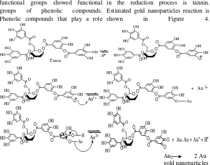
34 functional groups showed functional
groups of phenolic compounds. Phenolic compounds that play a role
in the reduction process is tannin. Estimated gold nanoparticles reaction is
shown in Figure 4. Figure 4. Estimated reaction mechanism of synthesis of gold nanoparticles using ethyl acetate fraction ketapang leaf (Terminalia catappa)
In addition to the involvement of bioactive compounds from ethyl acetate fraction ketapang leaf (Terminalia catappa), the possibility of disproportionation reaction to produce gold nanoparticles. Disproportionation reaction is a redox reaction in which an oxidant and reduktornya the same substance (Shriver and Atkins, 1999). Thus, some of the substances that undergo oxidation and partly reduced.
Colloid formation associated with the emergence of a core in saturated conditions. Once it is formed Au nanoparticles would grow into colloids (Zakir, 2005).
Characterization Gold Nanoparticles by XRD
35 The peaks of the diffraction
pattern of the gold nanoparticles is clearly shown in 2θ value is 38.1750, 34.2975 and 44.3150 respectively with FWHM value of 0.97, 0.745 and 1.11. In addition, Miller index of each peak is (111), (200), (202) and (311). At the orientation (202) and (311) peak looks weak and is much stronger on the orientation (111) and (200). This suggests that the gold nanocrystal dominant on the orientation (111) and (200).
Figure 5. XRD patterns of gold nanoparticles
At Angle 2θ produce several peaks of gold nanoparticles. But which has a high intensity is at an angle 2θ FWHM 0.97 38.1750 with a value that can be used as a reference for the calculation of the size of the gold
nanoparticles. Based on the XRD results obtained gold nanoparticle size is 17.13 nm.
Lembang (2013) managed to synthesize gold nanoparticles of decoction of the leaves extract ketapang with the size of 18.91 nm. It showed that gold nanoparticles were synthesized using a fraction of ethyl acetate resulted in a smaller particle size than the extract of decoction of the ketapang leaves.
Testing the Antibacterial Activity This study uses a bacterial test consisting of Staphylococcus aureus,
Escherichia coli and Bacillus subtilis. Testing inhibition of gold nanoparticles on the growth of bacteria is done by using the agar diffusion method using MHA media as a growth medium.
Based on data from Table 2, the test bacteria Staphylococcus aureus,
Escherichia coli and Bacillus subtilis to the concentration of gold nanoparticles, has no inhibitory zone of incubation time 24 to 48 hours. This shows that the gold nanoparticles are not resistant to test bacteria Staphylococcus aureus,
Escherichia coli and Bacillus subtilis.
Table 2. The average diameter obstacle gold nanoparticles against some bacteria test for 1 x 24 hours and 2 x 24 hours.
No AuNp
Average - average Barriers Diameter (mm)
S. aureus E. Coli B. subtilis.
24 hours
48 hours
24 hours
48 hours
24 hours
48 hours
1 AuNp
(Au3+ 0,5 mM) - - - -
2 AuNp
(Au3+ 1 mM) - - - -
3 Kloramfenikol (+) 11 10 27,3 28,3 24,7 24,9
36
Visually inhibition zone is formed from nanoparticles of gold to test the
bacteria can be seen in Figure 6.
Figure 6. Photo inhibitory zone diameter gold nanoparticles against some bacteria test for 1 x 24 hours (A) and 2 x 24 hours (B).
Label :
1. gold nanoparticles 0,5 mM 3. BSA (-)
2. gold nanoparticles 1 mM 4. Kloramfenikol (+)
CONCLUSION
Based on research that has been done, it can be concluded that:
1. The gold nanoparticles can be synthesized by the method of reduction using ethyl acetate fraction bioreduktor ketapang leaf (Terminalia catappa) with a size of gold nanoparticles is 17.13 nm. 2. The gold nanoparticles can not
inhibit the activity of the bacteria
Staphylococcus aureus, Escherichia coli, and Bacillus subtilis.
3. The difference in the concentration of gold nanoparticles did not affect the activity of the bacteria
Staphylococcus aureus, Escherichia coli, and Bacillus subtilis.
REFERENCES
1. Abdullah, M., 2010, Pengantar Nanosains, Bandung: ITB.
2. Ankamwar, B., 2010, Biosynthesis of Gold Nanoparticles (Green- Gold) Using Leaf Extract of Terminalia Catappa, E. J. Chem., 7(4): 1334-1339.
3. Babayi, H., Kolo, I., Okogun, J.I., Ijah, and U.J.J., 2004, The antimicrobial Activities of Methanolic Extract of Eucalyptus camaldulensis and Terminalia catappa Againt some Pathogenic Microorganisms, An Int. J. Niger. Soc. Exp. Bio., 16(2): 106-111. 4. Chen, P.S., Li, J.H., Liu, T.Y., and
Lin, T.C., 2000, Cancer Letters., 152(2): 115-122.
5. Cui, Y., Zhao, Y., Tian, Y., Zhang, W., Lü, X., and Jiang, X., 2012, The Molecular Mechanism of Action of Bactericidal Gold Nanoparticles on
Escherichia coli, Biomaterials., 33: 2327-2333.
A
37 6. Fatimah, E.N dan Hidajati, N., 2012,
Sintesis Dan Karakterisasi Nanopartikel Emas Sebagai Material Pendukung Aktivitas Tabir Surya Turunan Sinamat, Prosiding Seminar Nasional Kimia Unesa,Surabaya, 25 Pebruari 2012.
7. Gao, J., Tang, X., Dou, H., Fan, Y., Zhao, X., and Xu, Q., 2004, Hepatoprotective Activity of Terminalia catappa L. Leaves and Its Two Triterpenoids, J. Pharm and Pharmacol., 56(1): 1-7.
8. Jaziroh, S., 2008, Isolasi dan Identifikasi Senyawa Aktif dalam Ekstrak n-Heksana Daun ketapang (Terminalia catappa), Jurnal Kimia,
4(2): 61-70.
9. Lembang, M.S., 2013, Sintesis Nanopartikel Emas Dengan Metode Reduksi Menggunakan Bioreduktor Daun Ketapang (Terminalia Catappa), Skripsi (tidak diterbitkan), Program Studi Kimia FMIPA Universitas Hasanuddin.
10. Lin, Y., Kuo, Y., Shiao, M., Chen, C., and Ou, J., 2000, Flavonoid Glycocides from Terminalia catappa L, J. Chin. Chem. Soc., 47(1): 253-256.
11. Mittal, K.A., Bhaumik, J., Kumar, S., and Banerjee, U.C., 2014, Biosynthesis of silver nanoparticles: Elucidation of prospective mechanism and therapeutic potential,
Colloid J. Interf Sci, 415: 39-47. 12. Packirisamy, V., and Krishnamorthi,
V., 2014, Evaluation of Proximate Composition and Phytochemical analysis of Terminalia catappa L. from Nagapattinam Region, Int. J. Sci. Res (IJSR)., 3(12): 877-880.
13. Pauly, G., 2001, Cosmetic, Dermatologycal And Pharmaceutical Use of An Extract of Terminalia catappa, United State Patent Application.
14. Singh, C., Baboota, R.K., Naik, P.k., and Singh, H., 2012, Biocompatible Synthesis of Silver and Gold Nanoparticles using Leaf Extract of Dalbergia sisoo, Res. Article., VBRI Press, India.
15. Tsuzuki, T., 2009, Commercial Scale Production of Inorganic Nanoparticles, Int. J. Nanotecnology., 6(5): 567-578. 16. Wardhani, L.K., dan Sulistyani. N.,
2012. Uji Aktivitas Antibakteri Ekstrak Etil Asetat Daun Binahong (Anredera Scandens (L.) Terhadap
Shigella Flexneri Beserta Profil Kromatografi Lapis Tipis. Jurnal Ilmiah Kefarmasian, 2(1): 1-16. 17. Zakir, M., Sekine, T., Takayama, T.,