GENOMIC DNA EXTRACTION METHOD FROM MATURE LEAF OF LAI
(
Durio kutejensis
Becc.)
Fitri Handayani 1,2*), Rani Agustina Wulandari 1) and Rudi Hari Murti 1)
1) Faculty of Agriculture, University of Gadjah Mada Jl. Flora No. 1 Bulaksumur, Yogyakarta, Indonesia
2) Assessment Institute for Agricultural Technology of East Kalimantan Jl. P.M. Noor Sempaja, Samarinda, East Kalimantan, Indonesia
*) Corresponding author E-mail: [email protected]
Received: September 18, 2015/ Accepted: January 6, 2016
ABSTRACT
Lai (Durio kutejensis Becc.) is an indigeneous germplasm of Kalimantan which has some superiorities compared to its close-relative, durian (Durio zibethinus Murr.). Genetic exploration of lai is important to support its breeding program. According to rapid development in molecular biology, genetic exploration effort of lai will be easier. One of significant step in any molecular biology activities is DNA isolation to produce high quality DNA for further analysis. Leaves of lai, as other perennial crop, contain of high concen-tration of polysaccharides and polyphenol which will be co-extracted with the DNA. These compounds can interfere enzymes activities in subsequent molecular analysis. The aim of this study was to establish an optimal and effective DNA extraction method to obtain high-quality DNA from mature leaf of lai. An established extraction buffer and its modification were used in this study. The result showed that modification 4 could produce high quality DNA, and was considered to be the most effective DNA extraction method for mature leaf of lai.
Keywords: CTAB modification; DNA extraction; lai
INTRODUCTION
Durios family (Durio spp.) is fruit plant with high economic value. The most popular species of this genera is Durio zibethinus Murr. or durian. However, there is another popular durio species in Kalimantan, Indonesia, that is called lai (Durio kutejensis Becc). Lai is an indigeneous germ plasm of Kalimantan, and can be found in most of Kalimantan island i.e. East Kalimantan, South Kalimantan, West Kalimantan, Brunei, Sabah
and Serawak (World Conservation Monitoring Centre, 2013).
There are some differences between lai and durian. Durian has strong and spesific smell, while some lai varieties are odorless and the other have smooth aroma (Antarlina, 2009; Santoso, 2010). Additionally, lai has unique and attractive flesh color from yellow to red (Antarlina, 2009; Santoso, 2010; Hariyati et al., 2013; Hadi et al., 2014), blunt fruit spine, and longer storage period than durian (Antarlina, 2009; Santoso, 2010). So lai has more potential as an export commodity in European market than durian.
Exploration of genetic potency of lai is important to support its breeding program. According to rapid development in molecular biology, genetic exploration effort of lai will be easier. One of significant step in any molecular biology analysis is DNA extraction process to obtain high-quality DNA.
The problem of Durio genus are woody species (Brown, 1997) that generally contain phenolics, polysaccharides and other secondary metabolites that contaminate DNA and interfere with subsequent analysis (Cheng et al., 1997; Angeles et al., 2005; Sari dan Murti, 2015). According to Latief and Amien (2014), the most preferable and frequently used method to extract DNA from polysaccharide and polyphenol-rich leaves is CTAB (Cetyl Trimethyl Ammonium Bromide) method developed by Doyle and Doyle (1990). But it can not be used to extract DNA from mature leaf of sapodilla (Manilkara zapota (L.) van Royen), one of polysaccharide and polyphenol-rich leaves (Sari and Murti 2015). So, some modification in CTAB method is needed.
The modifications that have been carried out were increasing concentration of PVP
Cite this as: Handayani, F., R.A. Wulandari and R.H. Murti. 2016. Genomic DNA extraction method from mature leaf of lai (Durio kutejensis Becc.). AGRIVITA. 38(1): 73-79. doi: 10.17503/agrivita.v38i1.659
Accredited: SK No.81/DIKTI/Kep/2011
(Polyvinyl Pyrrolidone) to suppress polyphenol oxidation (Doyle and Doyle, 1990; Lodhi et al., 1994; Cheng et al., 1997; Syafaruddin and Santoso, 2011; Sari and Murti, 2015), increasing concentration of 2-mercaptoethanol, CTAB and sodium chloride in the extraction buffer to reduce polysaccharide contamination (Doyle and Doyle, 1990; Cheng et al., 1997; Sari and Murti, 2015), modifying repetition and volume of CIAA (Chloroform Isoamyl Alcohol) adding (Sari and Murti, 2015), using a low concentration of spermine to selectively precipitate and purify DNA in final step (Cheng et al., 1997).
To extract DNA from thick and tough leaves, liquid nitrogen is needed to make grinding process easier. Liquid nitrogen has been extensively used for DNA extraction from fresh leaves and or other tissues on coconut (Angeles et al., 2005), sapodilla (Sari and Murti, 2015), temulawak (Utami et al., 2012), apple, austrian pine, barberry, button-wood, cherry, grape, hazelnut, peach, and pear (Cheng et al., 1997). However, liquid nitrogen is not always easily available or convenient to use, so DNA isolation method without liquid nitrogen which still can obtain high quality DNA is needed.
Widiastuti (2010), Sulassih (2011), and Syahruddin (2012) added sterille quartz sand as liquid nitrogen substitution for easier grinding of mangosteen and durian leaves. While Angeles et al. (2005) and Utami et al. (2012) added PVP powder in sample grinding instead of mixed it to the extraction buffer. The aim was to make leaf grinding easier, although without liquid nitrogen.
Some protocols of DNA extraction from woody species require young leaf samples to obtain high quality DNA (Angeles et al., 2005; Mariana et al., 2011; Syafaruddin and Santoso, 2011; Syahruddin, 2012) to avoid accumulation of phenolic compound in mature leaf and and to make sample grinding process easier. However, sometimes leaf sample must be collected from remote areas and shipped for several days, thus using young leaf sample is impossible. For some perennial plants, young leaves were not other perennial crops that contain high phenolic
compound, polysaccharides, and other secondary metabolites. This study used modification in way of PVP and 2-mercaptoethanol adding toisolate DNA of lai. It is important to establish an optimal and effective DNA extraction method to obtain high-quality DNA from mature leaf of lai, as the aim of the study. This is an important early step that will influence subsequent molecular biology analysis in lai.
MATERIALS AND METHODS
Table 2. Treatment in cell lysis and DNA purification step of DNA extraction
Treatment
Cell lysis DNA purification Liquid
nitrogen PVP+Mercaptoetanol Sodium acetate CIAA
Doyle and Doyle Without Buffer included - 2x (@500 µl) Modification 1* With Buffer included - 2x (@500 µl) Modification 2* With Buffer included - 3x (500 µl, 500 µl, 300 µl) Modification 3* Without Buffer included - 3x (500 µl, 500 µl, 300 µl) Modification 4* Without Buffer excluded** - 3x (500 µl, 500 µl, 300 µl) Modification 5* Without Buffer included 3x (@1/10 vol) 3x (500 µl, 500 µl, 300 µl) Modification 6* Without Buffer excluded** 3x (@1/10 vol) 3x (500 µl, 500 µl, 300 µl) Remarks: * = Modification 1-6 using CTAB modification buffer component composed by Sari and Murti (2015); ** =
PVP was ground with leaf sample using mortar and pestle; mercaptoetanol was added to the homogenate before incubation
An equal volume of 3M sodium acetate and CIAA (24:1) as its modification (Table 2) were added to the tube, and the tube was shaken vigorously to form a complete emulsion. The tube was centrifuged at 12,000 rpm for 15 minutes to separate the phases. The aqueous phase (supernatant) was removed with micropipet, and transferred to a new tube. Purification step was repeated as treatment in Table 2.
Sodium acetate 3M (1/10 volume) was added to the supernatant, mixing gently, precipitated with 2/3 volume of cold isopropanol
and incubated in 4˚C for 12-24 hours. The
presipitated nucleic acids were collected and washed twice with ethanol 70%. The pellets were air dried and resuspended in TE buffer. Loading dye (1 µl) was added to 5 µl of DNA sample and electrophoresed on 1% agarose gel to check DNA quality.
DNA Amplification and Visualisation
Each 10 µl reaction volume of DNA amplification contains 5 µl PCR reaction mix, 2.25 µl nuclease free water, 0.25 µl RAPD (Random Amplified Polymorphism DNA) primer, and 2.5 µl
DNA. PCR consists of one cycle of 94˚C for 4
minutes, which was followed by 45 cycles of denaturation (94˚C, 1 minute), annealing (37˚C, 1 minute), and extension (72˚C, 1 minute 30 seconds), completed with one cycle of 72˚C for 7 minutes and 4˚C for 1 minute.
The amplification products were analyzed by electrophoresis using 1.5% agarose gel in TBE buffer 1x for 1.5 hours in 75 Volt. The result was checked by UV transluminator light and documented by digital camera.
RESULTS AND DISCUSSION
DNA isolation is a primary and critical step for molecular analysis of any plant species. The process becomes more difficult when the plant species contains high amount of polysaccharides and secondary metabolytes like polyphenols as contaminants. These contaminants, which are abundance in the foliage of perennial plants, co-extracted with the DNA and interfere with polymerases, ligases, and restriction enzymes (Sarwat et al., 2006; Ogunkanmi et al., 2008). To suppress the interference of the contaminants, some materials like CTAB, NaCl, PVP and 2-mercaptoethanol are used in DNA isolation protocol.
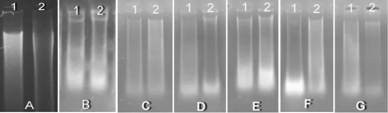
Doyle and Doyle (1990) has used their standard CTAB method successfully on a wide taxonomic sampling of plant families, but it didn’t work for lai. DNA extraction from leaf of lai could not produce high quality DNA when it was extracted by Doyle and Doyle standard buffer, although the purification step with CIAA and washing step with ethanol 70% have been repeated twice (Figure 1A).
Figure 1. Electrophoresis result of various method of DNA extraction: A. standard method of Doyle and Doyle; B. modification-1; C. modification-2; D. modification-3; E. Modification-4; F. Modification-5; G. Modification-6; (1) sample no.1; (2) sample no.2
Quality of the extracted DNA was evaluated agarose gel electrophoresis and RAPD-PCR. Figure 1A shows the electrophoresis result of the DNA extracted by standard CTAB method of Doyle and Doyle. The extraction treatment produced DNA and a lot of residual DNA that indicated high level of polysaccharides (Sari and Murti, 2015). Polysaccharides conta-mination are particularly problematic as they can inhibit the activity of many commonly used molecular biology enzyme such as polymerase, ligase and restriction endonucleases. It is because nucleic acid form tight complexes with polysaccharides, creating a gelatinous pellet, and the embedded DNA inaccessible to the enzyme (Sarwat et al., 2006). So the DNA of lai extracted by standard CTAB method of Doyle and Doyle (1990) can not be amplified in PCR analysis.
The color of the presipitated DNA extracted by standard CTAB method of Doyle and Doyle was brown, instead of white, indicated the presence of high level oxidized phenols (Angeles et al., 2005). Polyphenols are released when the tissues are wounded. In their oxydized form, polyphenol covalently bind to proteins and DNA, giving the DNA a brown color and making it useless for most research applications (Angeles et al., 2005).
To improve the quality of DNA, the standard CTAB method must be modified. In modification treatment of Doyle and Doyle method (modification 1-6), concentration of CTAB, NaCl, PVP and mercaptoethanol has been increased (Table 1). The increasing concentration of PVP was aimed to suppress phenol oxidation (Doyle and Doyle, 1990; Lodhi
Santoso, 2011; Sari and Murti, 2015) by forms complex hydrogen bonds with polyphenolic compound which simplifies their release from DNA strands by centrifugation (Lodhi et al., 1994; Alaey et al., 2005).
While increasing concentration of CTAB, NaCl, and mercaptoethanol was aimed to reduce polysaccharide contamination (Doyle and Doyle, 1990; Cheng et al., 1997; Sari and Murti, 2015). 2-mercaptoethanol also works as an antioxidant agent and forbids oxidation in polyphenol (Lodhi et al., 1994; Alaey et al., 2005; Zidani et al., 2005).
Modification-1 used liquid nitrogen to grind the leaf sample. Its modification produced thicker DNA, but there was still a lot of residual DNA (Figure 1B). The DNA quality produced by modification-1 treatment was not good enough. It also can not be used as template for RAPD-PCR analysis. In modification-2, purification step with CIAA was repeated three times. It resulted brighter final supernatant than the previous one, and so was the precipitated DNA.
In modification-3, liquid nitrogen was omitted while other treatment was identical with modification-2. It produced nearly identic of presipitated DNA color. The weakness of modifi-cation-3 treatment was its grinding process which was very hard because of leaf texture that tough and thick. So, according to Angeles et al. (2005) and Utami et al. (2012), in modification-4 PVP was added in grinding process and was ground simultaneuosly with leaf sample, instead of mix it to the extraction buffer. It made the grinding process easier although did not use liquid nitrogen.
of safety reason. In Safety Data Sheet of 2-mercaptoethanol product, Life Technologies (2014) asserted that 2-mercaptoethanol causes irritation of respiration duct if it was sniffed. Mixing this material in extraction buffer was risky toward evaporation and spreading of its unpleasant odor when the buffer was used to grind the sample. So, minimilize its contact with open air by buffer-excluded using is safer.
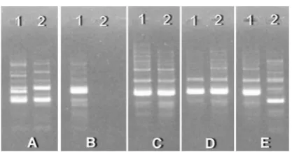
DNA electrophoresis of modification 2, 3 and 4 were not significantly different (Figure 1C, 1D, 1E). In addition to DNA electrophoresis, high quality DNA can be proved by using it as template for PCR analysis. DNA obtained from modification 2, 3, and 4 can be amplified in PCR-RAPD using both OPA 13 and OPB 10 primer, except DNA of sample number 2 from modification 3 (Figure 2 and Figure 3). It was linear with Lodhi et al. (1994), Cheng et al. (1997), Angeles et al. (2005), Syafaruddin and Santoso (2011) and Sari and Murti (2015) that increasing concentration of CTAB, NaCl, PVP, 2-mercaptoethanol and modifying repetition of purification step with CIAA will suppress the interference of the contaminants.
Besides polysaccharides and polyphenols, other contaminant which was often contained in initial DNA extract is protein. Most protein is removed by denaturation and precipitation from the extract using chloroform and/or phenol (Zidani et al., 2005). CIAA is an organic solvent that can dissolve protein, bind, and then precipitate it. CIAA adding followed by centri-fugation will separate protein from aqueous phase containing nucleic acid.
Figure 2. Result of PCR-RAPD analysis using OPA 13; A. modification-2; B. cation-3; C. Modification-4; D. Modifi-cation-5; E. Modification-6; (1) sample no.1; (2) sample no.2
Figure 3. Result of PCR-RAPD analysis using OPB 10; A. modification-2; B. Modifi-cation-3; C. Modification-4; D. Modi-fication-5; E. Modification-6;(1) sample no.1; (2) sample no.2
Modification 5 and 6 added 3M sodium acetate prior to CIAA in every repetition of purification step. Besides using organic solvent, the most common type of protein precipitation is salt induced precipitation. According to Fatchiyah et al. (2012) that salt, like sodium acetate or sodium chloride in high concentration, can be used to separate DNA from protein. Protein solubility depends on several factors. At high concentration of salt, the solubility of protein drops dramatically. When the salt concentration is increased, water molecules are attracted by salt ions, which decreases the number of water molecules available to interact with the charged part of protein. This is termed salting out, and the protein will be precipitated out.
Combination of high salt concentration and CIAA in purification step will precipitate protein perfectly. In modification 5 and 6, the adding of 3M sodium acetate will separate protein from the aqueous phase. So when CIAA added in further step, protein will be precipitated easily, and produce cleaner DNA. But the fact, DNA electrophoresis of modification 5 and 6 (Figure 1F, 1G) were not significantly different with modification 2, 3 and 4 (Figure 1C, 1D, 1E). There was still residual DNA present, although the DNA produced was high enough in quality, and can also be used as template in PCR-RAPD analysis (Figure 2 and Figure 3).
et al. (2012) that liquid nitrogen using was inefficient because it did not produce better DNA bands, while PVP adding for sample grinding produced clear and thick DNA bands.
CONCLUSION
Increasing concentration of CTAB buffer components and repetition in purification step can obtain high quality DNA for subsequent molecular analysis. Extraction method of modifi-cation-4 was considered to be the most effective for DNA extraction of mature leaf of lai. It did not need liquid nitrogen for grinding process, substitute by modification in way of PVP adding.
ACKNOWLEDGEMENT
We would like to thank to Agricultural Research and Development Agency of Ministry of Agriculture for funding this research, Rita Elfianis for technical assistance, and Rujiansyah for providing the plant materials.
REFERENCES
Alaey, M., R. Naderi, A. Vezvaei, A. Khaligi and A. Salami. 2005. Comparing study between four different methods of genomic DNA extraction from Cyclamen persicum Mill. International Journal of Agriculture and Biologi. 7(6): 882-884.
Angeles, J.G.C., A.C. Laurena and E.M. Tecson -Mendoza. 2005. Extraction of genomic DNA from the lipid-, polysaccharide-, and polyphenol-rich coconut (Cocos nucifera L.). Plant Molecular Biology Reporter. 23(3): 297-298. doi: 10.1007/BF02772760 Antarlina, S.S. 2009. Physically and chemically
identification of local fruit of Kalimantan (In Indonesian). Buletin Plasma Nutfah. 15(2): 80-90.
Brown, M.J. 1997. Durio - A bibliographic review. New Delhi: International Plant Genetic Resources Institute (IPGRI). p. 188. Cheng, F.S., S.K. Brown and N.F. Weeden. 1997.
A DNA extraction protocol from various tissues in woody species. Hortscience. 32(5): 921-922.
Doyle, J.J. and J.L. Doyle. 1990. Isolation of plant DNA from fresh tissue. Focus. 12(1): 13-15.
Fatchiyah, S. Widyarti, E.L. Arumningtyas and S. Permana. 2012. Practical workbook of molecular biologi analysis (In Indonesian). Malang: Fakultas MIPA, Universitas Brawijaya. p. 49.
Hadi, S.K., S. Lestari, S. Ashari. 2014. Variability and similarity prediction of 18 durian progenies from crossing between Durio zibethinus and Durio kutejensis (In Indonesian). Jurnal Produksi Tanaman. 2(1): 79-85.
Hariyati, T., J. Kusnadi and E.L. Arumingtyas. 2013. Genetic diversity of hybrid durian resulted from cross breeding between Durio kutejensis and Durio zibethinus based on random amplified polymorphic DNAs (RAPDs). American Journal of Molecular Biology. 3: 153-157. doi: 10.4236/ajmb.2013.33020
Ibrahim, R.I.H. 2011. A modified CTAB protocol for DNA extraction from young flower petals of some medicinal plant species. Geneconserve. 10(40): 165-182.
Latief, W. and S. Amien. 2014. Early study of using RAPD marker to determine the truth of patchouli cultivars (In indonesian). Bionatura-Jurnal Ilmu-ilmu Hayati dan Fisik. 16(2): 109-113.
Life Technologies. 2014. Safety Data Sheet (In Indonesian). Available at tools.lifetech nologies.com/content/sfs/msds/2014/21 985023_MTR-APLT_ID.pdf. Accessed on June 18, 2015.
Lodhi, M.A., G.N. Ye, N.F. Weeden and B.I. Reisch. 1994. A simple and efficient method for DNA extraction from grapevine cultivars and Vitis species. Plant Molecular Biology Reporter. 12(1): 6-13.
Mariana, B.D., A. Sugiyatno and A. Supriyanto. 2011. Genetic diversity of local accessions of Dimocarpus longan revealed by ISSR markers. Buletin Plasma Nutfah. 17(1): 25-29.
Ogunkanmi, A.L., B. Oboh, B. Onifade, A.A. Ogunjobi, I.A. Taiwo and O.T. Ogundipe. 2008. An improved method of extracting genomic DNA from preserved tissues of Capsicum annuum for PCR amplification. EurAsian Journal of BioSciences. 2(14): 115-119.
Sari, V.K. and R.H. Murti. 2015. An effective method for DNA extraction of mature leaf of sapodilla (Manilkara zapota (L.) van Royen). Agrivita. 37(1): 18-23. doi: 10.17503/Agrivita-2015-37-1-p018-023 Sarwat, M., M.S. Negi, M. Lakshmikumaran,
A.K. Tyagi, S. Das and P.S. Srivastava. 2006. A standardized protocol for genomic DNA isolation from Terminalia arjuna for genetic diversity analysis. Electronic Journal of Biotechnology. 9(1): 86-91. doi: 10.2225/vol9-issue1-fulltext-3 Sulassih. 2011. Relationship analysis between
mangosteen (Garcinia mangostana L.) and its relatives based on morphological and molecular (ISSR) markers (In Indonesian). Master Thesis. Institut Pertanian Bogor. Bogor.
Syafaruddin and T.J. Santoso. 2011. Optimation of DNA isolation and purification techniques on Reutalis trisperma (Blanco) Airy Shaw (In Indonesian). Jurnal Penelitian Pertanian Tanaman Industri. 17(1): 11-17. Syahruddin, K. 2012. Variability analysis of several
genotypes of durian (Durio zibethinus Murr.) using morphological and molecular (ISSR) markers (In Indonesian). Master Thesis. Institut Pertanian Bogor. Bogor.
Utami, A., R. Meryalita, N.A. Prihatin, L. Ambarsari, P.A. Kurniatin and W. Nurcholis. 2012. Variation methods of DNA isolation from leaf of temulawak (Curcuma xanthorrhiza Roxb.) (In Indonesian). Proceedings from National Chemistry UNESA (February, 25 2012). Surabaya: Universitas Negeri Surabaya.
Widiastuti, A. 2010. Genetic diversity analysis of mangosteen (Garcinia mangostana L.) irradiated by gamma-ray based on morphological, anatomical and ISSR markers (In Indonesian). Master Thesis. Institut Pertanian Bogor. Bogor.
World Conservation Monitoring Centre. 2013. Durio kutejensis. The IUCN Red List of Threatened Species. http://www.iucnred list.org/details/34568/0. Accessed on April 4, 2014. doi: 10.2305/IUCN.UK. 1998.RLTS.T34568A9876029.en Zidani, S., A. Ferchichi and M. Chaieb. 2005.