* corresponding author: [email protected]
Histocompatibility evaluation of combination
of gypsum with carbonated hydroxyapatite
as bone substitutes in subcutaneous tissue
Dyah Listyarifah1*, Rina Susilowati2, Ika Dewi Ana1
1Laboratory of Biomedic, Faculty of Dentistry, 2Department of Histology and Cell Biology, Faculty of Medicine, Gadjah Mada University, Yogyakarta, Indonesia
ABSTRACT
Combination of gypsum with carbonated hydroxyapatite (CHA) has shown to improve bone healing process. Soft tissue biocompatibility test is required prior to clinical application of bone substitutes since the materials contact with the soft tissues upon application and can cause severe inflammatory response. The aim of this study was to evaluate histocompatibility of gypsum compared to combination of gypsum-CHA in subcutaneous tissue. Disks of gypsum and gypsum-CHA were implanted on paravertebral subcutaneous tissue of 25 male Wistar rats. Histological section were stained with Hematoxylin Eosin then evaluated and scored with a histological grading scale for soft-tissue implants. A two-way ANOVA was used to analyze the histomorphometrical results with 95% confidence interval (p<0.05). The results showed that acute inflammatory cells were found in both groups at 6 hour and on day 5 and 7 after implantation at similar level. Chronic inflammatory cells and capsule were observed on day 7, 14, and 21. Moreover, there was no significant difference (p>0.05) of histomorphometrics score between both implants in each implantation periods. It could be concluded that histocompatibility level of gypsum and combination of gypsum-CHA to soft tissue was the same until day 21 iof mplantation.
Key words: gypsum - carbonated hydroxyapatite – biocompatibility – histocompatibility - subcutaneous implant
ABSTRAK
Kombinasi gipsum dan karbonat hidroksiapatit (CHA) mampu meningkatkan proses penyembuhan tulang. Uji biokompatibilitas jaringan lunak diperlukan sebelum bahan substitusi tulang digunakan di klinik oleh karena bahan tersebut akan kontak dengan jaringan lunak dan dapat menimbulkan respon inflamasi yang lebih serius. Tujuan penelitian ini adalah mengkaji histokompatibilitas gipsum dibandingkan dengan kombinasi gipsum-CHA di jaringan subkutan. Cakram yang mengandung gipsum atau kombinasi gipsum-CHA diimplantasikan pada jaringan subkutan paravertebral dari 25 tikus Wistar jantan. Potongan preparat histologi diwarnai dengan pewarna Hematoksilin Eosin untuk selanjutnya dievaluasi dan diskor berdasarkan skala tingkat histologi untuk implan pada jaringan lunak. Data histomorfometrik dianalisis dengan ANOVA dua jalan dengan tingkat kepercayaan 95% (p<0,05). Hasil penelitian menunjukkan sel inflamasi akut ditemukan pada kedua kelompok pada jam ke 6, dan hari ke 5 dan 7 setelah implantasi dengan derajad yang sama. Sel inflamasi kronis teramati pada hari ke 7, 14, dan 21. Tidak terdapat perbedaan skor histomorfometrik secara bermakna antara gipsum dan kombinasi gipsum-CHA pada masing-masing periode implantasi. Dapat disimpulkan bahwa gipsum dan kombinasi gypsum-CHA mempunyai tingkat histokompatibilitas yang sama pada jaringan lunak sampai pada hari ke 21 setelah implantasi.
INTRODUCTION
Bone damage is one of health problems that can cause chronic disability.1 Extensive bone damage can not heal spontaneously hence bone substitute materials are needed.2 Bone substitutes can be obtained from patient’s own tissue (autograft), other person (allograft), animals (xenograft), or from synthetic materials. Autograft, allograft, and xenograft have several drawbacks such as donor morbidity, tissue rejection and disease transmission. Recently, synthetic materials such as gypsum and CHA have been developed to solve the problem. Both materials have advantages and disadvantages. Gypsum can fill irregular and small defect and evokes minimal inflammatory response, nevertheless it is resorbed in short time before sufficient new bone growth occurs. Carbonated hydroxyapatite is a newly developed synthetic material that has excellent osteoconductivity and good resorbtion rate.3,4 Combining those two materials may produce better materials for inducing bone regeneration. Gypsum can fill irregular defect and set in situ while the CHA gives a slower biodegradation rate.
Biocompatibility test is required prior clinical application of materials in order to evaluate body rejection to the materials. Numerous studies showed a favorable bone response to the materials, nevertheless there is not much of publication concerning soft tissue response to the materials. Since bone substitutes contact not only with the bone, but also with soft tissues, information about biocompatibility of bone substitutes in soft tissue is also essential. Soft tissue gives more severe inflammatory response than bone.5 In this study, soft tissue biocompatibility test was performed to evaluate materials histocompatibility of combination of gypsum with CHA in subcutaneous tissues of the rats.
MATERIALS AND METHODS
This was an experimental study. The experiments were conducted at Laboratory of Histology and Cell Biology of Faculty of Medicine, Laboratoly of Biomedic of Faculty of Dentistry, and Integrated Research and Testing Laboratory, Gadjah Mada University, Yogyakarta. The protocol of this study has been approved by the Medical and Health
Research Ethics Committee, Faculty of Medicine, Gadjah Mada University, Yogyakarta.
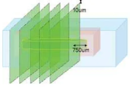
Twenty five male Wistar rats were divided into 5 groups with 5 rats on each group based on duration of implantation i.e. day 0, 5, 7, 14 and 21. Gypsum implants preparation was prepared by mixing 2 mg of calcium sulphate hemihydrates with 1 µL of aquadest. Gypsum-CHA implant preparation was prepared by mixing both materials with a ratio of 7:3. Two mg of gypsum-CHA mixture was then added with 1 µL of aquadest. Both materials were inserted into teflon cast to achieve a disc-shaped mold with a diameter of 6 mm and 0.8 mm thickness. These two types of implant materials were then sterilized with a UV sterilizer before implanted into subcutaneous tissue of the right and left paravertebral areas. Five rats from each group were sacrificed on day 0, 5, 7, 14, and 21 after implantation. Implant and tissue around the implant were dissected out and processed into paraffin sections stained with hematoxylin eosin. Five serial sections (FIGURE 1) from each block were observed by two independent observers. Each slide was evaluated in 4 fields in 700x magnification and its histomorphometry was scored according to previous criteria.12 The average scores were analyzed by two way ANOVA with 95% confidence of interval.
FIGURE 1. Histological serial section of paraffin block
RESULTS
Visual evaluation
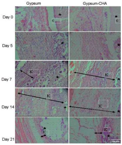
FIGURE 2. The histological soft tissue post implantation of gypsum and combination gypsum-CHA
implantation. Postoperative wound on day 0 was still wet and blood was visible. On day 5, the wound started to dry and covered by crust. No blood or exudate and only minimal edema can be observed. On day 7, the wound was dry. Most crust were already peeled and only left a thin crust. On day 14, rat’s fur in the operation site began to grow and the incision scar has closed. On day 21, the scar was not visible since thick fur had grown covering the area of the scar.
Microscopic evaluation
On day 0, tissue reactions profile on both type of implantation i.e. gypsum or combination gypsum-CHA looked similar. The shape of implanted materials was still intact. The implants were porous without any newly developed connective tissue around or within the implants. Erythrocytes and the scattered acute inflammatory cells such as polymorphonuclear leukocytes could be observed in surrounding tissues and in the pores of both implanted materials.
On day 5, the surface of both implants (gypsum or combination gypsum-CHA) was started to degrade and no erythrocytes can be observed in the surface. Soft tissue began to contact with implant’s surface. Compared to day 0, more polymorphonuclear leukocytes were observed. However, there was no surrounding capsule could be observed.
On day 7, the amount of the degraded parts of the implant increased. Few tissues containing fibroblasts and inflammatory cells has grown inside the implants. Occasionally, foreign giant cells were seen. Only a few scattered polymorphonuclear leukocytes were found. A thin connective tissue capsule was formed. Capsule surrounding gypsum was thicker than in gypsum-CHA.Both implants were more degraded leaving more pores filled by connective tissue. Fibrous capsules surrounding gypsum and gypsum CHA were thinner than an day 7.
On day 14, Both implants were more degraded leaving more pores filled by connective tissue. Blood vessels and chronic inflammatory cells such as lymphocytes and foreign giant cells scattered in the connective tissue of the capsule and the implant’s pores. More foreign giant cells were found in gypsum than in gypsum-CHA implanted tissue.
On day 21, Fibrous capsule surrounding gypsum on day 21 was thinner than on day 14 and also thinner compared to fibrous capsule surrounding gypsum-CHA on day 21. The degradation process continued and only some remaining materials can be found in the tissue. The space left by the degraded materials was filled by more abundant connective tissue containing blood vessels and inflammatory
cells. More inflammatory cells were found in the pores of implanted materials than in the capsule. However, there were less inflammatory cells observed compared to day 14’s tissue samples.
Histomorphometric evaluation
a. Capsule thickness
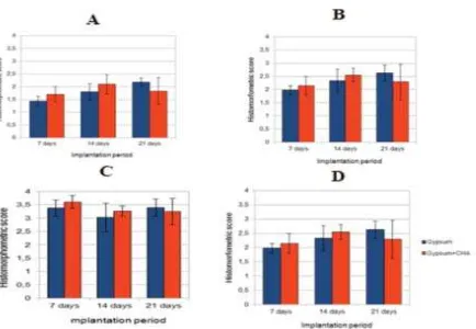
Histomorphometric evaluation was not determined in tissue samples obtained on day 0 and 5 since there was no clear capsule around the implants could be observed. The mean score of capsule thickness of gypsum-CHA group in each period of implantation (FIGURE 3A) was always higher than the gypsum except on day 21 but this was not statistically significant (p>0.05). That higher score indicated that better histocompatibility can be observed on gypsum-CHA group.
The mean score of capsule thickness that surrounds gypsum group increased along the implantation period. On the other hand, the mean score of capsule thickness in gypsum-CHA tissue samples increased on day 14, but it decreased on day 21. The significantly difference of mean scores of capsule thickness between implantation periods was observed in this study (p<0.05) especially between day 7 with day 144and between day 7 with day 21.
b. Capsule quality
FIGURE 3. Histomorphometric bar carts. A. Capsule tickness; B. Capsule quality; C. Interface quality; D. Total score
c. Interface quality
Small numbers of foreign body giant cells were found along the outer surface of both groups of implants on day 7. The number increased on day 14 but then decreased on day 21. The mean scores of interface quality in tissues implanted with gypsum group always increased along with the implantation periods, while in tissues implanted with gypsum-CHA the scores decreased on day 21. However, there was no statistical differences between the scores in gypsum and gypsum-CHA groups (p>0.05) and between implantation periods (p>0.05).
d. Total score
The pattern of total score of gypsum and gypsum-CHA groups in each implantation period and between implantation periods were similar with capsule thickness and capsule quality (FIGURE 3D). The total score of gypsum-CHA group was always higher than gypsum group except on day 21, but this was not significantly different (p>0.05). The mean total score of gypsum group always increased with the time periods but in gypsum-CHA
group, it decreased on day 21, however there was no statistical difference (p>0.05).
DISCUSSION
than in gypsum on day 7 and 21, since the amount of gypsum in gypsum-CHA was less than in gypsum implant.
The level of acute inflammation triggered by gypsum and gypsum-CHA was similar. It was shown by the amount of polymorphonuclear leukocytes infiltrates that almost the same in both implants materials. Acute inflammatory response could be observed in the early implantation period (day 0, 5, and 7) by the presence of monocytes and polymorphonuclear leukocytes, whereas chronic inflammatory response was observed in subsequent periods (day 14 and 21) by the presence of macrophages, foreign body giant cells, and lymphocytes. When implanted into soft tissues with the same implantation procedure, gypsum and combination of gypsum-CHA stimulated inflamma-tory response, either acute or chronic. The presence of an implant can provide a continuous inflammatory stimulus.
The presence of polymorphonuclear leukocytes which started from 6 hours (0 days) after treatment showed that an acute response was already in progress. Polymorphonuclear leukocytes, will work early to eliminate cellular debris. Along with increasing time, other inflammatory cells such as monocytes, will flock the implantation area simultaneously. The number of inflammatory cells that come into the area of inflammation depends on the injury circumstances and the implant materials ability to stimulate inflammation. The type and amount of inflammatory cells in the implantation site will be different at each period of time.6 On day 14 and 21, inflammatory cells were dominated by macrophages, foreign body giant cells, and lymphocytes. Monocytes that come into the inflammation area in acute period will turn into macrophages. Macrophages will phagocyte debris and foreign materials. Some macrophages will fuse with foreign giant cell to increase its phagocytosis ability. The presence of macrophages has impact on implant material resorption rate.7,8
The interface quality, evaluated by the presence of foreign body type giant cells on implanted gypsum and gypsum-CHA decreased on day 14 and increased again on day 21. On day 7, the number of monocytes that came to the implant area and turned into macrophages or foreign body type giant cells was still a few on the acute phase. On day 14, more
monocytes would come and turned into macrophages and foreign body giant cells. The number of macrophage increased and some macrophages fused to form a foreign giant cell in attempt to clean the implant materials and cellular debris. On day 21, half of macrophages and foreign body giant cells underwent apoptosis or followed the lymphatic flow due to lack of remaining gypsum.5 While in the gypsum-CHA implants, the number of macrophages and foreign body giant cells on day 21 was relatively constant. This was because the particles resulted from CHA degradation on day 21 still triggered macrophages and foreign body cells activation. Activated macrophages, foreign body giant cells, monocytes, and other inflammatory cells will produce growth factors and other inflammatory mediators. These growth factors will activate fibroblast to proliferate and produce extracellular matrix surrounding implant materials.9
Capsule thickness surrounding gypsum subsequently decreased on day 7, 14, and 21 since less activated macrophages released growth factors within implantation periods. On the other hand, capsule thickness surrounding gypsum-CHA on day 21 was thicker than on day 14, since increase in activated macrophages released more growth factor due to stimulation by particles from CHA degradation. The mean score of capsule thickness of gypsum and gypsum-CHA ranged from 1.10 to 2.35. These ranges of scores were almost the same with score range of capsule thickness in cement implantation á-tri calcium phosphate and precipitated hydroxyapatite that reported to have a good compatibility and caused minimal inflammatory response with that score range.5
Gypsum and combination of gypsum-CHA showed the ability to be adhered by surrounding fibroblast cells since there was no gap between material and surrounding connectives tissue. In addition, both of these implant materials were able to stimulate tissue growth into materials pores. Those indicated that both gypsum and gypsum-CHA were not toxic to the soft tissue.9
inflammatory response became less with the decrease in content of gypsum in the soft tissue. Total score of gypsum-CHA increased on day 14 and decreased on day 21 because CHA in this combination began to degrade on day 21. Degradation of the CHA stimulated the inflammatory response in the surrounding soft tissue.
No statistical difference between gypsum and combination of gypsum-CHA implants in capsule quality, interface quality, capsule thickness, and total score indicated that gypsum and combination of gypsum-CHA had the same histocompatibility level in soft tissue until day 21 of implantation.
CONCLUSION
Gypsum and combinations of gypsum-CHA had the same level of histocompatibility in soft tissue until day 21 of implantation. Other compatibility testing of the materials such as cytocompatibility, infectability and hemocompatibility will be conducted with longer implantation period, different implantation procedure and a more suitable histological technique. Measurement of residual materials will also performed to evaluate the speed of degradation of the implant materials.
ACKNOWLEDGMENT
We would like to thank Head of Laboratory of Histology and Cell Biology of Faculty of Medicine, Head of Laboratory of Biomedic of Faculty of Dentistry, and Director of Integrated Research and Testing Laboratory, Gadjah Mada University, Yogyakarta for all research facilities so that the study can successfully be performed.
REFERENCES
1. World Health Organization. Global burden of disease: global programme on evidence for health policy discussion
paper 50. Geneva : World Health Organization, 2000. 2. Phelps JB, Hubbard GB, Wang X, Agrawal CM.
Microstructural heterogeneity and the fracture toughness of bone. J Biomed Mat Res 2000; 51:735-41.
3. Jamali A, Hilpert A, Debes J, Afshar P, Rahban S, Holmes R. Hydroxyapatite/calcium carbonate versus plester of Paris: a histomorphometric and radiographic study in a rabbit tibial defect model. Calcif Tissue Int 2002;71:172-78.
4. Orsini G, Ricci J, Scarano A, Pecora G, Petrone G, Iezzi G. Bone-defect healing with calcium-sulfate particles and cement: an experimental study in rabbit. J Biomed Mater Res Part B 2004; 199–208.
5. Ooms EM, Egglezo EA, Wolke JGC, Jansen JA. Soft-tissue response to injectable calcium phosphate cements. Biomaterials 2003; 24:749–57.
6. Shina H, Ruhe PQ, Mikos AG, Jansen JA. In vivo bone and soft tissue response to injectable, biodegradable oligo(poly(ethylene glycol) fumarate) hydrogels. J Biomaterials. 2003; 24:3201–11.
7. Muelen JVM and Koerten HK. Inflammatory response and degradation of three types of calcium phosphate ceramics in a non osseus environment. J Biomed Mater Res 1994; 28:1455-63.
8. Zhao Q, Topham N, Anderson JM, Hiltner A, Lodoen G, Payet CR. Foreign-body giant cells and polyurethane biostability: in vivo correlation of cell-adhesion and surface cracking. J Biomed Mater Res 1991; 25:177–83. 9. Anderson JM and Schoen FJ. In vivo assessment of tissue
compatibility. Biomedical Science, 2nd ed. San Diego:
Elsevier Academic Press, 2004.
10. Thomas MV, Puleo DA. Calcium sulfate: properties and clinical applications. J Biomed Mater Res 2009;88B: 597-610.
11. Wick G, Backovic A, Rabensteiner E, Plank N, Schwentner C, Sgonc R. The Immunology of fibrosis: innate and adaptive responses. Trends Immunol 2009; 31:110-19.
12. Jansen JA, Dhert WJA, Waerden JPCM, Recum AF. Semi-quantitative and qualitative histologic analysis method for the evaluation of implant biocompatibility. J Invest Surg 1994; 7:123-34.
13. Babensee JE, Anderson JM, Larry VM, Mikos AG. Host response to tissue engineered device. Adv Drug Deliv Rev 1998; 33:111-39.