BJuma et al.
97
Three new compounds from Erythrina lysistemon and their antimicrobial,
radical scavenging activities and their brine shrimp lethality
.
Benard F. Juma† and Runner R. T. Majinda†
Department of Chemistry, University of Botswana, Private Bag UB 00704, Gaborone, Botswana.
Abstract
Three new compounds, 4',7-dihydroxy-2
'',2
''-dimethylpyrano [5
'',6
'':5,6]-isoflavone (1), (7E)
(8,2')- 3,7,9,5',9'-pentahydroxy-4,4'-dimethoxyneolign-7-ene (2) and (9E,11Z)
14-hydroxyoctadecan-9,11-dienoic acid (3), along with other known flavonoids, benzenoids,
alkaloids and phenylpropanoids were isolated from the twigs, leaves, stem bark, stem wood and
flowers of Erythrina lysistemon. Their structures were established on the basis of spectroscopic
evidence. Some of these compounds have shown high lethality against brine shrimps (Artemia
salina), moderate radical scavenging ability in the DPPH assay, moderate antifungal activity
against Candida mycoderma, moderate activity against the Gram-positive (Bacillus subtilis and
Staphylococcus aureus) and weak activity against Gram-negative bacteria (Escherichia coli).
Key words: Erythrina lysistemon, flavonoids, neolignane, phenolics, radical scavenging,
lethality, antimicrobial.
Introduction
Previous studies have shown that Erythrina lysistemon (Leguminosae) elaborates erythrinaline
alkaloids, some of which are distributed in several parts of this plant [1,2]. The other major
group of compounds is the flavonoids, especially prenylated ones, and these compounds are
prevalent in the stem and root bark [3,4]. The extracts from this plant have been used in
traditional medicine and have also shown antiviral, anticancer and cytotoxic activities [4,5].
O
The present paper reports on the non-erythrina alkaloid contents of the Erythrina lysistemon and
their lethality against brine shrimp, radical scavenging ability and antimicrobial activities. Three
†
BJuma et al.
98
new compounds, 4',7-dihydroxy-2
'',2
''-dimethylpyrano [5
'',6
'':5,6]-isoflavone (
1), (7E)
(8,2')-3,7,9,5',9'-pentahydroxy-4,4'-dimethoxyneolign-7-ene (
2), the first neolignan in the genus
Erythrina, and (9E,11Z) 14-hydroxyoctadecan-9,11-dienoic acid (
3), have been isolated from
this plant. These have been obtained along with other known compounds including the
flavonoids daidzein (
4), genistein (5), wighteone (6), 4',5,7-trihydroxy-6-(2
''-hydroxy-3
''-methylbut-3
''-enyl)isoflavone (
7), alpumisoflavone (8), derrone (9), 6,8-di-prenylgenistein (10),
erysenegalensein E (
11), lysistisoflavone [isoerysenegalensein E] (12),
2',5,7-trihydroxy-4'-methoxy-5-prenyl isoflavanone (
13), apigenin (14), liquitirigenin (15), medicarpin (16)
phaseollidin (
17), cristacarpin (18), sandwicensin (19), 2,4,4'-trihydroxychalcone (20). The other
non-flavonoid compounds isolated were caffeic acid (
21), coumaric acid 4-glucoside (22),
octadecanyl (E) ferulate (
23), 3-methoxy-
α
,
β
-dihydrocoumaric acid (
24), coumaric acid (25), the
isoquinoline alkaloid precursor norprotosinomenine (
26), the indole alkaloid hypophorine (27),
4-hydroxy-3-methoxybenzaldehyde (
28), 4-hydroxy-3-methoxybenzoic acid (29) and inositol
(
30). Some of these compounds have shown high lethality against brine shrimps, strong radical
scavenging ability against DPPH and antifungal activity against the yeast Candida mycoderma.
Weak activity has also been exhibited against the Gram-negative bacteria (Escherichia coli) and
moderate activity against Gram-positive bacteria (Bacillus subtilis and Staphylococcus aureus).
Discussion
The twig (ethyl acetate) and stem wood (chloroform) extracts of E. lysistemon were worked up
as shown in the experimental to give compounds
1 & 8 and 2 & 3, respectively.
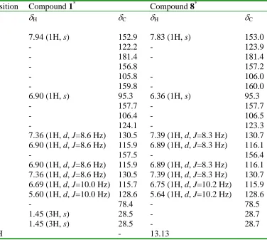
Compound
1 exhibited a peak at m/z 336.1 in its EIMS spectrum and displayed 20
non-equivalent carbon signals in the
13C-NMR spectrum and a molecular formula C20H16O5
was
suggested. The
1H- and
13C-NMR spectra showed signals at
δ
H7.94 (1H, s;
δ
C152.9) interpretive
of H-2 proton and C-2 carbon resonance of isoflavone [6,7,8]. The peak at 270 nm in the UV
(MeOH) spectrum was assigned to the benzoyl moiety (A-ring) of the isoflavone. The
1H-NMR
spectrum of this compound further displayed an AA'BB' spin system 7.36 (2H, dd, J = 8.5, 2.2
Hz) and 6.90 (2H, dd, J = 8.5, 2.2 Hz)], which was associated with a 4'-hydroxyl substituted ring
B of an isoflavonoid. The presence of a dimethylpyrano ring moiety was shown in the same
spectrum by the signals [
δ
H atδ
H 5.60 (1H, d, J = 10.1 Hz, H-3''), 6.69 (1H, d, J = 10.1 Hz,
H-4
'') and 1.45 (6H, s, Me). The
13C-NMR spectrum displayed signals at
δ
C128.6 (C-3
''), 115.7
(C-4
''), 78.4 (C-2
'') and 28.5 (C-2
''Me) representing this group. This was supported by the
EIMS spectrum which exhibited a fragment ion peak at m/z 321 [M-CH3]
+, showing a loss of
methyl group and typical of 2,2-dimethylchromenes [9]. H-4
'', resonating at 6.69, correlated
with the carbon signals at 159.8 (C-7), 156.8 (C-5) and 105.8 (C-6) in the HMBC spectrum,
indicating that this moiety was attached at C-6. The
1H- and
13C NMR spectra for this compound
compared well with those of co-isolated compound
8 [5]. The absence of the chelated C-5
hydroxyl hydrogen signal indicated that the prenyl group was cyclized to the 5-COH group
rather than the 7-COH group contrary to
8. Further, from the UV spectrum a bathochromic shift
of +5 was observed for band II, indicating a free 7-OH [6]. Total assignment for this compound
was accomplished by a close examination of the HMBC and HMQC spectra. This data enable
compound
1 to be identified as 4,7-dihydroxy-2
'', 2
''-dimethyl-dipyrano [5
'', 6
'':5,6]-isoflavone.
BJuma et al.
99
fact that the OH group at C-7' was easily lost to give this fragment ion. Its
13C-NMR spectrum
exhibited 20 carbon signals, which were distinguished as two methoxy, two oxymethylene and
nine methine (one oxymethine). The following signals were observed,
δ
H4.21 (
δ
C62.8) and
δ
H3.87, 3.77 (
δ
C63.8) [oxymethylenes]
δ
H5.52 (
δ
C88.3) [oxymethine] and
δ
H3.51 (
δ
C54.1)
[methane]. The two sets of signals appearing at
δ
H6.77 (
δ
C115.1), 6.84 (
δ
C118.7) and 6.95 (
δ
C109.5) displaying and AMX spin system and
δ
H6.96 (
δ
C115.5) and 6.98 (
δ
C111.1) [singlets],
demonstrated the existence of two aromatic rings in
2. The presence of an olefinic moiety in the
molecule was indicated by the signals appearing at
δ
H6.24 ([1H, ddd, J=15.7, 11.7, 5.9 Hz]
δ
C126.3) and
δ
H6.54 ([1H, br d, J=15.7 Hz]
δ
C131.0) the size of the coupling constant indicated a
trans-configuration. The proton signal at
δ
H6.24 correlated (HMBC) with the carbon at
δ
C62.8
(oxymethylene) indicating direct connection to the olefinic moiety. The other olefinic proton
resonating at
δ
H 6.54 correlated with the carbon signals atδ
C 111.1, 129.3 and 131.5. Theseobservations revealed a C-2, C-4 and C-5 substituted phenylpropanoid system. The methine
signal at
δ
3.51 correlated with the second oxymethylene carbon (
δ
63.8), the oxymethine carbon
(
δ
88.3) and an aromatic carbon (
δ
131.5), while oxymethine proton signal at
δ
5.52 correlated
with the carbon signals at
δ
54.1, 133.5 and 118.7 in the HMBC spectrum. These observations
revealed the existence of a second phenylpropanoid system whose C
3side chain was
hydroxylated at positions C-1' and 3' and its C-2' attached to the aromatic ring of the first ArC
3system. Intense fragment ion peaks at m/z 151 and 180 representing cleavage that gives
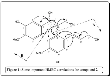
fragments A and B (Figure 3) respectively further strengthens the proposal. Other important
HMBC correlations are shown in figure 1. From these observations,
2 was assigned the structure
(7'E) (8,2')-3,5',7,9,9'-pentahydroxy-4,4'-dimethoxyneolign-7'-ene.
Figure 1: Some important HMBC correlations for compound 2
'
B
A
BJuma et al.
100
the HRESI-MS gave a sodiated ion ([M+Na]
+) exact mass at m/z 319.2244 (calc. 296.4449)
which assigned for molecular formula C
18H
32O
3. The presence of two double bonds in the
molecule was indicated by the signals observed at
δ
H6.51 (
δ
C126.2),
δ
H5.67 (
δ
C133.2),
δ
H5.44
(
δ
C136.2) and
δ
H5.98 (
δ
C128.2). The signal at
δ
C179.6 was assigned to a carboxylic acid group.
Further the
1H and
13C-NMR spectra displayed signals at
δ
H4.18 and
δ
C73.4 (oxymethine). The
proton signal
δ
H4.18 (
δ
C73.4), showed HMBC correlations with carbons signals at
δ
C25.5, 37.6
and 126.2, while the olefinic proton at
δ
H6.51 correlated with the carbons at
δ
C37.6, 136.1 and
133.2. Close examination of the data showed that the oxymethine group was separated by one
methylene group (
δ
H1.56,
δ
C37.6) from the diene moiety. The data available enabled the
identification of
3 as (9E,11Z) 14-hydroxyoctadecan-9,11, -dienoic acid.
Radical scavenging activity of Erythrina lysistemon against DPPH
The radical scavenging activities of the crude extracts and isolated compounds were assessed
using 2,2-Diphenyl-1-picrylhydrazyl (DPPH). To establish the level of activity, of a given
sample, a plot was made of absorbance verses concentration (in
µ
g/ml). The concentration of the
sample at which the absorbance at 517 nm decreases to half its initial value was taken as the IC
50value of the sample in question. Ascorbic acid was used as the standard and its activity was
examined in the same manner.
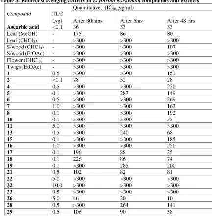
BJuma et al. 102 The pterocarpans showed relatively higher radical scavenging ability compared to the isoflavones. Two of these, phaseollidin (17) and cristacarpin (18) were fast acting showing activity at IC50 values of 196 and 226 µg/ml, respectively, after only 30 minutes. After 6 and 48 hours, compounds 17, 18 and 19 exhibited activities at IC50 values 88 & 25, 86 & 74 and 285 & 200 µg/ml, respectively. Comparing 17 and 19, the methylated pterocarpan is four times less active after 6 hours and after 48 hours its activity is shown to be weaker by a factor of eight. These observations indicated that a free C-9 hydroxyl in pterocarpans is important both in enhancing activity and also the rate of radical scavenging. Also interesting to note in comparing compounds 18 and 19 is the enhancement of activity by the introduction of a hydroxyl at position C-6a.
The neolignan 2 and benzylisoquinoline alkaloid 26 the exhibited the highest activities at IC50 values of 78, 32 & 28 µg/ml and 46, 20 &10, respectively, after 30 minutes, 6 and 48 hours, which were higher than those of the standard ascorbic acid. Apart from possessing free phenolic hydroxyl groups, these compounds possess benzyl electronegative atoms (O and N) groupings, which as in the case of compound 18 could be the reason for their enhanced activities (Table 3).
BJuma et al. 103 Table 3: Radical scavenging activity of Erythrina lysistemon compounds and extracts
Quantitative, (IC50, µg/ml)
Compound TLC
(µg) After 30mins After 6hrs After 48 Hrs
Ascorbic acid <0.1 36 33 33
Leaf (MeOH) - 175 86 80
Leaf (CHCl3) - >300 >300 >300 S/wood (CHCl3) - >300 >300 107 S/wood (EtOAc) - >300 >300 >300 Flower (CHCl3) - >300 >300 >300 Twigs (EtOAc) - >300 >300 >300
1 0.5 >300 >300 151
2 <0.1 78 32 28
4 0.5 >300 >300 230
5 0.1 >300 287 149
6 0.5 >300 >300 269
7 1.0 >300 >300 163
8 0.1 >300 >300 192
10 0.1 >300 >300 55
11 5.0 >300 >300 >300
13 0.5 >300 240 68
15 0.1 >300 >300 185
16 1.0 >300 >300 250
17 0.1 196 88 25
18 0.1 226 86 74
19 0.1 >300 285 200
21 0.5 102 82 81
22 5.0 >300 >300 >300
22 10.0 >300 >300 >300
23 0.5 >300 >300 >300
26 5.0 46 20 10
28 0.5 >300 264 141
29 0.5 106 90 58
Antibacterial properties of Erythrina lysistemon compounds
Antimicrobial analysis was done as outlined in the experimental section for compounds in appreciable quantities. In the test against the Gram-negative bacteria Escherichia coli, the isoflavonoids showed significantly high activities compared to other classes of compounds. The highest activity against this organism was shown by compounds 6, 7, 8 and 11 at loadings of 10 µg per spot. These activities were however much lower compared to those observed for the standard which was active at 0.001 µg. Compounds 3, 4, 7, 12, 17, 21 and 22 showed quite appreciable activity against the Gram-positive bacteria Bacillus subtilis at 5. Hydroxylation on the isoprenyl group seems to increase activity against this organism as shown in compounds 6 & 7 and 11, 12 & 10 (Table 4). Against the Gram-positive bacteria
Staphylococcus aureus, compound 4 showed an activity much closer to that of the standard at
1 µg per spot. Flavone 14 showed a higher activity (0.5 µg) than the known antifungal miconazole (1.0 µg) against the yeast Candida mycoderma.
BJuma et al. 104 Both the crude extracts and the pure compounds isolated from E. lysistemon were subjected to lethality test using brine shrimps (Artemia salina) as outlined in the experimental section. The pterocarpans exhibited extremely high activities against the shrimps. Compounds 17 and
19 had LD50 values of 6.9 ppm and 4.7 ppm (Table 4), which were even higher that those exhibited by standards potassium dichromate (25.39 ppm) and the bufadienolide scallaren A (190.85 ppm). The isoflavones also showed relatively high toxicity. Amongst this group of compounds, the highest activities were observed for compounds 1, 5, 8, 10 and 12 whose activities were exhibited at LD50 values lower than 100 ppm. The benzoic acid derivative 29 however was quite active exhibiting an LD50 value of 17.78 ppm. All the crude extracts also showed appreciable activity with the methanol extract of the leaf exhibiting the highest activity (LD50 23.9 ppm). It is from this that the pterocarpan 17 that showed high activity together with isoflavones were isolated and as such must significantly contribute to this observation.
Table 4: Antimicrobial and cytotoxicity activities of compounds isolated from Erythrina
lysistemon.
Standards: - Lethality - Scillaren A = 190.85 µg/ml, Potassium Dichromate = 25.39 µg/ml
Experimental section.
BJuma et al. 105 0.0400 mm mesh). Gel filtration: - Sephadex LH-20 on glass columns. Prep-TLC plates (0.5 mm thick): - silica gel (Merck) 60 HF254+366 on 20 x 20 cm glass plates. 1H-NMR, (300 or 600 MHz), 13C-NMR (75 or 150 MHz), DEPT, COSY, HMBC, HMQC: - Bruker Avance DXP 300 spectrometer using standard pulse sequences and referenced to residual solvent signal. EIMS: - Finnigan MAT SSQ 7000 single stage quadruple analyzer at 70 eV. The HRESI were obtained from a Bruker Apex III Fourier transform ion cylotron resonance (FT-ICR) mass spectrometer (Bruker Daltonics, Billerica, USA) equipped with an infinity cell, a 7.0 Tesla superconducting magnet (Bruker, karlshule, Germany). UV: - Shimadzu UV-2101PC spectrometer.
Plant Material: The various parts of E. lysistemon were collected between July 2001and
May 2002 in Gaborone (University of Botswana grounds), Botswana and its voucher specimen (No. EL 0701) is preserved in the Department of Biological Sciences, Faculty of Science, University of Botswana.
Extraction and Isolation: Twigs of E. lysistemon were air-dried and ground before being
extracted by soaking in EtOAc for 24 hours three times. The resulting extracts were combined and the solvent removed in vacuo using rotary evaporator to give 12 g of dark green organic material. This extract was subjected to column chromatography (silica gel) eluting with n-Hex/CHCl3 followed by CHCl3/MeOH mixtures with increasing polarities to give 30 eluents ca 50 ml, which were combined into six combined fractions A to G, based on TLC profile. The viscous fraction B (eluted with 4% MeOH/CHCl3, 1.5 g) was chromatographed on silica gel column (50 x 3) eluting with the following gradient: CHCl3, MeOH/CHCl3 (1:99, 1:49, 1:19, 1:9, 1:4) and MeOH. A total of 50 fractions of ca 50 ml were collected and combined on basis of TLC composition. Residue resulting from the fraction eluted with 1:99 MeOH/CHCl3 (80 mg) was subjected to prep-TLC (1:99 MeOH/CHCl3) followed by purification using Sephadex LH-20 column (40 x 2) to give compounds 1 (20 mg) and 8 (7 mg). Compounds 10 (7 mg), 11 (5 mg) and 12 (4 mg) were obtained from the 1:49 MeOH/CHCl3 eluted fraction (40 mg) through multiple developments on prep-TLC (1:99 MeOH/CHCl3) and cleaning using Sephadex LH-20. Compound 7 (11 mg) was obtained from the fraction eluted with 1:9 MeOH/CHCl3.
4,7-Dihydroxy-2 '' ,2 '' -dimethylpyrano[5 '' ,6 '' :5,6]-isoflavanone (1): pale yellow paste,
EIMS m/z (rel int.) 336 [M]+ (5), 321 [M-CH3]+ (11), 302 (3), 286 (24), 270 (24), 254 (100), 243 (4), 216 (3), 197 (4), 153 (15), 137 (60). UV (MeOH) λmax (nm) (log ε): 271 (3.36); + NaOMe: 280; + AlCl3: 281; + AlCl3/HCl: 280; + NaOAc: 276; + NaOAc/H3OBO3: 276. 1 H-NMR (300 MHz, CDCl3) and 13C-NMR (75 MHz, CDCl3) see Table 1.
BJuma et al. 106 The methanol extract was also subjected to flash chromatography eluting with CHCl3/MeOH with increasing amounts of MeOH in CHCl3 to obtain combined fractions A to F. Compounds 26 (14 mg) and 30 (40 mg) were obtained from fractions E and F (8% MeOH/CHCl3 eluent), respectively, through fractional crystallization (solvent CHCl3/EtOAc/MeOH).
Previously dried and ground stem wood material (1.8 kg) was extracted successively with EtOAc and MeOH as described above to obtain 10.5 g and 40.9 g, respectively, of light brown organic material. The EtOAc extract was subjected to step-wise gradient elution chromatography on a column (50 x 5) using n-Hex/CHCl3 and then CHCl3/MeOH while increasing the amounts of CHCl3 and MeOH resulting in fractions A to G. Fractions A was subjected to repeated gel filtration using Sephadex LH 20 on a column (30 x 4) to obtain compound 3 (50 mg). Fraction C (eluted with CHCl3) was subjected to gradient elution chromatography using n-Hex/CHCl3 and then CHCl3/MeOH (column 30 x 2). Compound 5 (7 mg) was obtained from fractions D by fractional crystallization. The methanol extract was on the other hand treated to flash chromatography (silica gel column; 70 x 6) using CHCl3 then CHCl3/MeOH while increasing MeOH. Through a combination of Column chromatography (silica gel), gel filtration (Sephadex LH-20), preparative-TLC and fractional crystallization compound 19 (16 mg) was obtained from B (eluted with 2% MeOH/CHCl3); 2 (21 mg), 24 (7 mg) and 28 (11 mg), were obtained from fraction C (eluted with 5% MeOH/CHCl3); 15 (5 mg), 16 (8 mg), 20 (17 mg) were obtained from fraction D (eluted with 8% MeOH/CHCl3); 18 (21 mg) was obtained from fraction E (eluted with 8% MeOH/CHCl3).
(7'E) (8,2')-3,5',7,9,9'-Pentahydroxy-4,4'-dimethoxyneolign-7'-ene (2): brownish solid,
mp 68-71 °C. EIMS m/z (rel. int.) 358 [M-H2O]+ (100), 340 [M-2H2O]+ (83), 328 (48), 325 (24), 310 (10), 279 (10), 272 (5), 267 (5), 235 (4), 210 (5), 180 (26), 151 (29), 137 (88). 1 H-NMR (300 MHz, MeOD) and 13C-NMR (75 MHz, MeOD) see Table 2.
(9E,11Z) 14-Hydroxyoctadecan-9,11-dienoic acid (3): oily substance, HRESI-MS m/z (rel
int.) [M+Na]+ 319.2244 (296.4449); EIMS m/z (rel int.) 296.2 [M]+ (15), 280 (100), 262 (5), 224 (9), 206 (14), 171 (6), 151 (10), 135 (5), 113 (10), 99 (20), 81 (10); 1H-NMR (300 MHz, CDCl3) 6.51 (1H, dd, J=15.2, 11.0 Hz, H-12), 5.99 (1H, t, J=10.9, Hz, H-9), 5.67 (1H, dd,
J=15.5, 7.0 Hz, H-11), 5.44 (1H, ddd, J=15.4, 10.7, 7.7 Hz, H-10), 4.18 (1H, dd, J=12.9, 6.3
Hz, H-14), 2.35 (3H, t, J=7.4 Hz, H-2), 2.18 (1H, d, J=7.1 Hz, H-8), 1.61 (4H, m, H-3), 1.56 (m, H-13), 1.33 (15H, m, H-4, 5, 6, 7, 15, 16, 17), 0.90 (3H, t, J=6.6 Hz, H-18). 13C-NMR (75
MHz, CDCl3) 179.6 (C=O), 136.1 11), 133.2 10), 128.2 9), 126.2 12), 73.3
(C-14), 37.6 (C-13), 34.3 (C-2), 32.2 (C-16), 29.7 (C-7), 29.3 (C-5), 29.2 (C-6), 27.9 (C-8), 25.5 (C-15), 25.0 (C-3), 22.9 (C-17), 14.4 (C-18: CH3).
BJuma et al. 107 The butanol extract (50 g) of the flowers of this plant was adsorbed on 50 g silica gel and subjected to flash chromatography and eluted with CHCl3 and CHCl3/MeOH mixtures with increasing polarities. The fraction eluted with 5% MeOH/CHCl3 (2.1 g), was subjected to column chromatography and the fraction resulting from 3% MeOH/CHCl3 was subjected to prep-TLC to obtain compounds 14 (15 mg) and 25 (11 mg). Further elution of the column gave fraction D (1.78g, 7% MeOH/CHCl3) from where it was possible to obtain compound 21 (300 mg) through fractional crystallization (solvent n-Hex/CHCl3/EtOAc). Through fractional crystallization it was possible to obtain compounds 22 (860 mg) and 27 (34 mg) from fraction I (2.2 g) and J (300 mg), respectively. These fractions had previously been eluted from the main column using 20% MeOH/CHCl3.
The methanol extract of the pods (42 g) was adsorbed on 42 g silica gel and subjected to column chromatography on a column packed with silica gel (300 g) under CHCl3. Separation was achieved by step wise gradient elution using CHCl3 then CHCl3/MeOH with increasing amounts of MeOH in CHCl3. Compound 4 (24 mg) was obtained from fraction (10% MeOH/CHCl3 eluent, 260 mg) by use of prep-TLC followed by purification using Sephadex LH-20.
Radical scavenging assay using DPPH
2,2-Diphenyl-1-picrylhydrazyl (DPPH), molecular formula C18H12N5O6, was obtained from Fluka Chimie AG, Bucks. The method used by Kumarasamy et al. (2002) [11]and Naik et al. (2003) [12] was adopted. Radical scavenging activity was carried out for both crude extracts and pure compounds isolated from E. lysistemon. This was done at two levels, first the rapid TLC screening (Qualitative assay) followed by quantitative assay where the reaction of DPPH with the test compounds, indicated by reduction in absorbance was monitored by used of UV at 517nm. In evaluating DPPH inhibition amount, varying amounts (ranging from 0.1 to 50 or 100 µg) of the samples were spotted on TLC after which the plate was sprayed with 0.2% of DPPH solution in methanol. Activity was shown by yellow spots on purple background. On the other hand, for quantitative evaluation of radical scavenging ability, 1.5 ml of 500 µM DPPH in methanol was added to equal volumes of test compounds at concentrations ranging from 0 to 300 µg/ml, mixed well and kept in the dark for 30 minutes after which absorbance was measured. Further measurements were done after 6 and 48 hours. Blank experiment was also carried out to determine the absorbance of DPPH at 0 concentration of sample. A plot was then made of absorbance verses concentration. The concentration of sample in µg/ml at which the absorbance at 517 nm decreases to half its initial value is used as the IC50 value of a given compound. Gallic and ascorbic acids were used as the standards and their activities were examine in the same manner.
Lethality Tests.
BJuma et al. 108 The LC50 were determined graphically from the percent lethality against the log conc. and LC50 derived from the best-fit line obtained by linear regression analysis [13].
Antimicrobial test using Agar Overlay Technique. Microorganisms and culture media.
Strains of the gram-negative bacteria Escherichia coli and gram-positive bacteria Bacillus
subtilis and Staphylococcus aureus together with the yeast Candida mycoderma, were
obtained from anti-microbial culture banks at the Microbiology Department, University of Botswana. The bacteria were broth cultured on Nutrient Agar (NA) while the yeast was maintained on Sabourand Dextrose Agar slants and petri dishes. All these cultures were introduced using a heat sterilized wire loop into 250 ml Erlenmeyer flasks containing 100 ml Nutrient Broth. These were shaken at room temperature on a rotary shaker at 200 rpm for 24 hours to achieve homogenous distribution of the organisms. All media were autoclaved at 120°C for 20 minutes.
Samples tested and inoculums for assay.
The known antibacterial chloramphenical was used as a standard antibacterial agent while miconazole was used as the standard anti-fungal agent. The antibacterium was tested between 0.0001 and 10 µg, while the antifungal was tested between 0.01 and 50 µg. The isolated pure compounds were tested at amounts between 0.1 and 50 µg for both antifungal and antibacterial activity. These amounts were spotted on to glass backed TLC plates coated (25 mm thickness) with silica gel G 60 F254 at varying amounts for each sample. The plates were then dried with a hair dryer for complete removal of solvents.
Solid media overlay used for bacteria was Malt Nutrient Agar while Sabourand Dextrose Agar (Oxoid) was used in the case of fungi. The molten agar was maintained at 40°C to keep it from solidifying. 10 ml of nutrient broth containing the microorganisms was seeded in 100 ml of malt nutrient agar. This gave a concentration of approximately 107 cells/ml as established by the use of a UV/VIS spectrophotometer, which gave an optical density value at 540nm (OD540) equivalent to 1 [13]. The final concentration in the solid medium was expected to be approximately 105 cells/ml. The seeding was done immediately before carrying out the overlay.
Bioautography.
The spotted plates were placed on a hot plate maintained at 35°C while the inoculums was rapidly spread over the TLC plates using a sterile Pasteur pipette. After solidification of the medium, the TLC plates were incubated overnight at 37°C for the bacteria and 25°C for the fungi in polythene boxes lined with moistened chromatography paper. After this the bioautograms were sprayed with an aqueous solution (2.5 mg/ml) of thiazolyl blue (3-[4,5-dimethylthiazol-2-yl]-2,5-diphenyltetrazolium bromide; MTT) (Fluka) and then incubated for 4 hours at 37°C for the bacteria and 25°C for the fungi. For activity, clear inhibition zones were observed against a purple background [14].
Acknowledgements: BFJ thanks DAAD-NAPRECA for a scholarship. RRTM thanks IFS
BJuma et al. 109 References
1. Juma, B. F. and Majinda, R. R T. (2004); Erythrinaline Alkaloids from the Flowers
and Pods of Erythrina lysistemon and their DPPH radical scavenging properties
Phytochem. 65: 1397-1404.
2. Amer, M. E.; Shamma, M. and Freyer, A. J. (1991); The tetracyclic erythrina
alkaloids. J. Nat. Prod. 54: 329-363.
3. El Masry, S; Amer, M. E.; Abdel Kader, M. S. and Zaatout, H. H. (2000); Prenylated
Flavonoids of Erythrina lysistemon Growing in Egypt: J. Pharm. Pharmacol. Suppl.
52: 259-265.
4. McKee, T. C.; Bokesch, H. R.; Mc Cormick, L. J.; Rashid, A; Spiel Vogel, D.;
Gustafson, K. R.; Alavanja, M. M. Cardelina II, J. H and Boyd, M. R. (1997); Isolation and Characterisation of New Anti-HIV and Cytotoxic Leads from Plants, Marine and Microbial Organisms. J. Nat. Prod. 60: 431-438.
5. El-Masry, S; Amer, M. E.; Abdel-Kader, M. S. and Zaatout, H. H. (2002); Prenylated
flavonoids of Erythrina lysistemon growing in Egypt: Phytochem. 60: 783-787. 6. Markham, K. R. (1982); Techniques of flavonoid identification; Academic press:
London New York Paris Santiago San fransisco San Paulo Sydney Tokyo Toronto; pp 36-51.
7. Agrawal P. K. (1989); Carbon-13 NMR of flavonoids. Elsevier, Amsterdam.
8. Wandji, J.; Fomum, Z. T.; Tillequin, F.; Seguin, Elisabeth and Koch, M. (1993);
Two Isoflavonoids from Erythrina senegalensis. Phytochem. 35: 245 – 248.
9. Tanaka, H.; Tanaka, T.; Hosoya, A.; Kitade, Y. and Etoh H. (1997); Three
Isoflavonoids from Erythrina orientalis. Phytochem. 48: 355-357.
10.Iinuma, M.; Okawa, Y.; Tanaka, T.; Ho, F.; Kobayashi, Y. and Miyauchi, K. (1994);
Anti-oral microbial activity of isoflavonoids in root bark of Ormosia monosperma.
Phytochem. 37: 889-892.
11.Kumarasamy, Y.; Fergusson M. E.; Nahar L and Sarker, S. D. (2002); Bioactivity of
Moschamidole from Centaurea moschata; Pharm. Bio. 40: 307-310.
12.Naik, G. H.; Priyadarsini, K. I.; Satav, J. G. Banavalikar, M. M.; Sohoni, D. P.;
Biyani, M. K. and Mohan, H. (2003); Comparative antioxidant activity of individual herbal components used in Ayurvedic medicine; Phytochem. 63: 97-104.
13.McLaughlin, J. L.; Chang', C. J. and Smith, D. L. (1991); "Bench -top"Bioasseys for
the discovery of natural products: an update; in: Studies in natural products chemistry;
Attaur-rahman edition. 9 pp 383
14.Rahalison, L.; Hamburger, M., Hostettman, K.; Monod, M. and Frenk, E. (1991); A