Indones J Anim Vet Sci. 20(4): 297-307
Molecular Identification Technique of Trypanosoma evansi by Multiplex
Polymerase Chain Reaction
Sawitri DH1, Wardhana AH1, Wibowo H2, Sadikin M2, Ekawasti F2
1Indonesian Research Center for Veterinary Sciences, RE. Martadinata St. 30 Bogor 16114 2Faculty of Medicine, Universitas Indonesia, Jakarta
E-mail: [email protected]
(received 29-10-2015; revised 14-12-2015; accepted 30-12-2015)
ABSTRAK
Sawitri DH, Wardhana AH, Wibowo H, Sadikin M, Ekawasti F. 2015. Pengembangan teknik identifikasi molekuler Trypanosoma evansi dengan Polimerase Chain Reaction Multipleks. Indones J Anim Vet Sci. 20(4): 297-307. DOI: http://dx.doi.org/10.14334/jitv.v20i4.1248
Trypanosoma evansi adalah parasit hemoflagella yang menginfeksi ternak dan dikenal sebagai penyebab Surra. Beberapa spesies trypanosoma lainnya pada hewan mamalia adalah T. equiperdum, T. b. rhodesiense, T. b. gambiense, T. vivax, T. congolense, T.theileri. Beberapa spesies tersebut cukup sulit untuk dibedakan secara morfologi dengan T. evansi melalui teknik konvensional (preparat ulas darah). Teknik molekuler dengan polymerase chain reaction (PCR) dilaporkan memiliki kemampuan yang akurat dalam mengidentifikasi, mengkarakterisasi dan mendiagnosis Trypanosoma. Namun demikian PCR tunggal yang digunakan adalah relatif mahal karena setidaknya diperlukan dua atau lebih pasang primer untuk menentukan spesies T. evansi. Tujuan penelitian ini adalah untuk mengembangkan teknik identifikasi spesies T. evansi dengan PCR multipleks/mPCR (tiga jenis pasang primer dalam satu reaksi) sehingga dibutuhkan waktu yang realtif cepat dan murah. Sebanyak 31 isolat T.evansi
yang diperoleh dari Bblitvet Culture Collection (BCC) dan Departemen Parasitologi BBLitvet digunakan pada penelitian ini. Isolat-isolat tersebut mewakili isolat yang berasal dari daerah endemis dan Wabah Surra yang diisolasi tahun 1988-2014. Ekstraksi DNA dilakukan pada setiap sampel, termasuk isolat Bang 87 yang telah dimurnikan sebagai kontrol positif. Primer yang digunakan adalah spesifik untuk T. evansi, yaitu ITS-1, Ro Tat 1,2 VSG dan ESAG 6/7. Sebelum melakukan mPCR, masing-masing primer dioptimasi dengan menggunakan PCR tunggal. Hasil penelitian menunjukkan bahwa ketiga primer tersebut dapat dikombinasikan dalam satu reaksi dengan teknik mPCR dan mengamplifikasi masing-masing fragmen DNA target dengan sempurna, sehingga 31 isolat teridentifikasi sebagai T. evansi. Teknik ini dapat diaplikasikan di lapang dengan biaya yang lebih murah dan waktu yang lebih cepat.
Kata Kunci: Trypanosoma evansi, Identifikasi, Multipleks PCR
ABSTRACT
Sawitri DH, Wardhana AH, Wibowo H, Sadikin M, Ekawasti F. 2015. Molecular Identification Technique of Trypanosoma evansi by Multiplex Polymerase Chain Reaction. Indones J Anim Vet Sci. 20(4): 297-307.DOI: http://dx.doi.org/10.14334/jitv.v20i4.1248
Trypanosoma evansi is a Hemoflagella parasite that infects cattle and is known as the agents of Surra. Several other trypanosome species infects mammals: T. equiperdum, T. b. rhodesiense, T. b. gambiense, T. vivax, T. congolense, T.theileri. Some of these species is quite difficult to be distinguished morphologically with T. evansi through conventional techniques (thin blood smear). Molecular technique by polymerase chain reaction (PCR) is reported to have the ability to identify, characterize and diagnose trypanosomes accurately. However, a single PCR used is relatively expensive because it takes at least two or more pairs of primers to determine T. evansi. The purpose of this study is to develop T. evansi species identification techniques by multiplex PCR/mPCR (the three pairs of primer in one reaction) that takes the relatively fast and inexpensive. A total of 31 isolates T.evansi were obtained from Bblitvet Culture Collection (BCC) and the Department of Parasitology BBLitvet used in this study. Isolates represent isolates from endemic areas and Surra outbrake isolated from 1988-2014. DNA extraction performed on each sample, including Bang 87 isolates which has been purified as a positive control. Primers used are specific for T. evansi, the ITS-1, Ro Tat 1.2 VSG and ESAG 6/7. Before running mPCR, each primer is optimized by using a single PCR. The results showed that the three primers can be combined in a single reaction with mPCR technique and amplify each DNA fragment target perfectly, so identified 31 isolates as T. evansi. This technique can be applied in the field with a lower cost and faster time.
Key Words: Trypanosoma evansi, Identification, Multiplex PCR
INTRODUCTION
Surra, a wasting disease in livestock, is caused by hemoflagellate parasite T. evansi which transmitted mechanically by haematophagus flies (Herrera et al.
species prevalent in Africa, Asia and Central and South America (Devila et al. 2003; OIE 2012).
There are two main types of trypanosoma: the first is transmitted through the saliva/vector bites (salivarian) and the second is transmitted through the vector feces (stercorarian). There are four subgenus trypanosoma salivarian attacking mammals: subgenus Trypanozoon (T. brucei brucei, T. brucei rhodesiense, T. brucei gambiense, T. evansi, T. equiperdum); subgenus Dutonella (T. vivax, T. Uniforme); sub genus Nanomonas (T. congolense, T. simiae, T. godfreyi) and subgenus Pycomonas (T. suiz) (Mulumbu 2006). While stercorian trypanosome there is one species (T. theileri, subfamily Triatominae) that attack livestock.
Some specieses had similar shape, so that it was difficult to be distinguished morphologically. So far, only T. evansi reported attacked livestock animal in Indonesia (Ditjennak 2012). T. theileri also sometimes found in cattle and buffaloes in Indonesia (Bblitvet unpublished data). Nevertheless T. theileri reported non-pathogenic.
T. evansi infection is a prevalent disease that causes considerable economic losses due to weakness, abortion in pregnant animals, estrus cycle disorders, weight loss, decreased productivity and reproductivity, high treatment cost and death (Reid 2002; Jittapalapong et al. 2009). Trypanosome infections also cause immunosuppression effects which triggering to other diseases (Jittapalapong et al. 2009). Directorat General of Livestock reported that in 2010-2012, Surra outbreak attacked 4268 head livestock and 1760 out of them were dead (Ditjennak 2012).
Trypanosome identification, generally was performed based on microscopic observation (morphology, morphometric and parasite motility within the host tissue), host range, and geographical region. Further identification was also based on presence of the parasite in certain organs of vector cycle and ability of the parasite to grow in vivo (in rodents or vector) and invitro (Hoare 1972).
This conventional identification by using thin blood smear and microhematocrit centrifugation test (MHCT) has limitations. Its success depended on the number of parasite on sample observed. Parasite species had similar morphology, so that it was hard to be distinguished (Uilenberg 1998). Masake et al. (2002) states that the diagnosis of trypanosomiasis would have problem if only one or two parasites have found on preparations of thin blood smear with low quality. This may cause misidentified of the trypanosome species. Therefore, accurate species identification was needed to distinguish trypanosome species infecting animals.
Polymerase chain reaction (PCR) technique was reported had accurate ability in identifying, characterizing, and diagnosing trypanosomiasis (Holland et al. 2001; Desquesnes & Dávila 2002). This
assay has high sensitivity and specificity to detect 1-10 trypanosoma/ml of blood (Davila et al. 2003) and able to distinguish between species (Desquesnes et al. 2001). Some moleculer markers have been widely constructed to detect, differentiate, and study trypanosome species such as Internal Transcriber Spacer-1 (ITS-1) and Rhode Trypanozoon Antigen Type 1.2 VSG gene (Ro Tat-1,2 VSG) (Salim et al. 2011; Urakawa et al. 2001). According to Salim et al. (2011), PCR ITS-1 product length specificaly was corellated to each trypanosome species. So that can be used as a basic to distinguish the species. Besides, Urakawa et al. (2001) states that one of T. evansi’s characteristics was gene encoding Ro Tat 1.2 VSG (Rode Trypanozoon Antigen type 1.2 VSG), which was able to distinguish T. evansi and another Trypanosome specieses. Another moleculer marker was Expression-site-associated gene 6/7 (ESAG 6/7 gene) encoding trafferin receptor of T. evansi. It was specific and had high sensitivity (Shahzad et al. 2010). Until now, identification of T. evansi species was carried out by using single PCR of ITS-1 and Ro Tat 1.2 VSG primer and was never reported using multiplex PCR technique.
outbrake area during 2012-2014 and one isolate from endemic area in 2013). Those isolates were from 14 locations from 8 provinces (Table 2). Bang87 isolate (from BCC) was used as a positive control. T. evansi derived from BCC and circulating isolates in 2012 (Sumba) was stored cryopreservation. While T. evansi circulating isolates in 2013-2014 which collected from
buffalo blood with Surra positive (Pandeglang) stored in eppendorf tubes at -20°C. Cryopreservation as stabilate through the stages of passage in mice before being stored. We chose 31 isolates based on the availability in BCC and the origin of the isolates representing endemic and outbreaks areas in Indonesia.
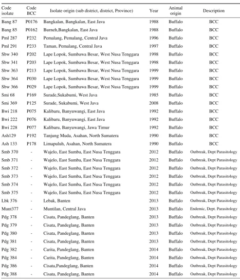
Table 1. T. evansi isolates used in this research
Code isolate
Code
BCC Isolate origin (sub district, district, Province) Year
Animal
origin Description
Bang 87 P0176 Bangkalan, Bangkalan, East Java 1988 Buffalo BCC
Bang 85 P0162 Burneh,Bangkalan, East Java 1988 Buffalo BCC
Pml 287 P232 Pemalang, Pemalang, Central Java 1996 Buffalo BCC
Pml 291 P233 Taman, Pemalang, Central Java 1997 Buffalo BCC
Sbw 340 P202 Lape Lopok, Sumbawa Besar, West Nusa Tenggara 1998 Buffalo BCC
Sbw 341 P203 Lape Lopok, Sumbawa Besar, West Nusa Tenggara 1998 Buffalo BCC
Sbw 363 P213 Lape Lopok, Sumbawa Besar, West Nusa Tenggara 1999 Buffalo BCC
Sbw 364 P030 Lape Lopok, Sumbawa Besar, West Nusa Tenggara 1999 Buffalo BCC
Sbw 366 P029 Lape Lopok, Sumbawa Besar, West Nusa Tenggara 1999 Buffalo BCC
Smi 68 P169 Surade,Sukabumi, West Java 1985 Buffalo BCC
Smi 369 P125 Surade, Sukabumi, West Java 2008 Buffalo BCC
Bwi 218 P075 Kalibaru, Banyuwangi, East Java 1992 Buffalo BCC
Bwi 222 P076 Kalibaru, Banyuwangi, East Java 1992 Buffalo BCC
Bwi 228 P077 Kalibaru, Banyuwangi, Jawa Timur 1992 Buffalo BCC
Ash129 P192 Tanjung Muda, Asahan, North Sumatera 1990 Buffalo BCC
Ash 133 P178 Limapuluh, Asahan, North Sumatera 1990 Buffalo BCC
Smb 370 - Wajelo, East Sumba, East Nusa Tenggara 2012 Buffalo Outbreak, Dept Parasitology
Smb 371 - Wajelo, East Sumba, East Nusa Tenggara 2012 Buffalo Outbreak, Dept Parasitology
Smb 372 - Wajelo, East Sumba, East Nusa Tenggara 2012 Buffalo Outbreak, Dept Parasitology
Smb 373 - Wajelo, East Sumba, East Nusa Tenggara 2012 Buffalo Outbreak, Dept Parasitology
Smb 374 - Wajelo, East Sumba, East Nusa Tenggara 2012 Buffalo Outbreak, Dept Parasitology
Smb 375 - Wajelo, East Sumba, East Nusa Tenggara 2012 Buffalo Outbreak, Dept Parasitology
Lbk 376 - Lebak, Banten 2013 Buffalo Outbreak, Dept Parasitology
Munt377 - Muntilan, Central Java 2013 Buffalo Endemic, Dept. Parasitology
Pdg 378 - Cisata, Pandeglang, Banten 2013 Buffalo Outbreak, Dept Parasitology
Pdg 379 - Cisata, Pandeglang, Banten 2013 Buffalo Outbreak, Dept Parasitology
Pdg 380 - Cisata, Pandeglang, Banten 2013 Buffalo Outbreak, Dept Parasitology
Pdg 381 - Cisata, Pandeglang, Banten 2013 Buffalo Outbreak, Dept Parasitology
Pdg 382 - Carita, Pandeglang, Banten 2014 Buffalo Outbreak, Dept Parasitology
Pdg 384 - Carita, Pandeglang, Banten 2014 Buffalo Outbreak, Dept Parasitology
Pdg 386 - Cisata,Pandeglang, Banten 2014 Buffalo Outbreak, Dept Parasitology
Table 2. Primers Sequence used for the amplification single and multiplex PCR
Primers Primer sequences Amplicon size (bp) Reference
ITS1
F 5`-CCGGAAGTTCACCGATATTG-3`
480 (Njiru et al. 2005) R 5`-TGCTGCGTTCTTCAACGAA-3`
RoTat 1.2 VSG F 5`-CTGAAGAGGTTGGAAATGGAGAAG-3` 151 (Salim et al. 2011)
R 5`-GTTTCGGTGGTTCTGTTGTTGTTA-3`
ESAG 6/7
F 5’-CATTCCAGCAGGAGTTGGAGG-3’
740 (Isobe et al. 2003) R 5’-TTGTTCACTCACTC TCTCTTTGACAG-3’
F = primer forward; R = primer reverse
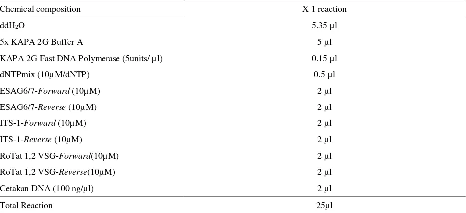
Table 3. Chemical Composition of mPCR optimalisation
Chemical composition X 1 reaction
ddH2O 5.35 µl
5x KAPA 2G Buffer A 5 µl
KAPA 2G Fast DNA Polymerase (5units/ µl) 0.15 µl
dNTPmix (10µM/dNTP) 0.5 µl
ESAG6/7-Forward (10µM) 2 µl
ESAG6/7-Reverse (10µM) 2 µl
ITS-1-Forward (10µM) 2 µl
ITS-1-Reverse (10µM) 2 µl
RoTat 1,2 VSG-Forward(10µM) 2 µl
RoTat 1,2 VSG-Reverse(10µM) 2 µl
Cetakan DNA (100 ng/µl) 2 µl
Total Reaction 25µl
DNA extraction
T. evansi from stabilates and buffalo blood was thawed at room temperature. Total genomic of 31 samples was extracted from 100 µl of stabilate/buffalo blood by using Genomic DNA Mini Kit (Geneaid, Taiwan) according to the manufacturer’s instructions. Purified DNA was stored at -20°C until further analysis. Samples T. evansi from stabilate were coded Mc (mice) and samples from buffalo blood were coded Buf (buffalo).
PCR Primers
Three primer pairs (1st BASE, Singapore) were used for single and multiplex PCR analysis (Table 1). ITS-1 primers was amplifying DNA at 480 bp fragment length, whereas RoTat 1.2 VSG and ESAG 6/7 primers were amplifying DNA at 151 bp and 740 bp fragment
length, respectively (Isobe et al. 2003; Njiru et al. 2005; Salim et al. 2011).
Bang87 T. evansi isolate purification as positive control
Single PCR analysis of ITS-1, Ro Tat 1,2 VSG and ESAG6/7 primers for T. evansi
First step of mPCR development was optimizing of each primer (Table 1) separately by using Bang87 T. evansi isolate as positive control. The PCR products should correspond to the size of the fragment gene of interest.
PCR amplification was performed using KAPA2G TM Fast PCR kit (KAPA BIOSYSTEMS, USA) on ABi GeneAmp thermal cycler 9700. Each reaction contained a final volume of 25µl, including 2 µl of 50-100 ng of genomic DNA; 5 µl of 5X KAPA 2G buffer A; 0.1 µl KAPA 2G Fast DNA Polymerase (5 unit/ µl); 0.5 µl dNTPmix (10µM/dNTP); 2 µl of each forward and reverse primer (10µM/µl) and 12,4 µl of sterile distilled water. PCR amplification was carried out the following conditions 35 cycles: one cycles initial denaturation step at 95°C for 1 minute; 35 cycles denaturation at 95°C for 10 second; 35 cycles annealing at 58°C for 15 second; 35 cycles extension at 72°C for 15 second and one cycle final extension at 72°C for 10 minutes. Multiplex PCR
Multiplex PCR was conducted by combining 3 primers on one PCR reaction. Multiplex PCR was carried out by using modified KAPA 2G TM Fast PCR kit (KAPA BIOSYSTEMS, USA). Composition of reagent, primer, and template was presented in Table 3. PCR condition was similar with previous step.
Visualization of PCR product
PCR products were resolved by electrophoresis at 100 volt in 1.5% (w/v) agarose gels stained with SYBR® Safe gel staining (InvitrogenTM).Visualization and analysis of fractionated DNA bands were carried out on GelDoc Transluminator (Cleaver). Diagnosis was considered positive when a specific product of each gene was amplified by PCR.
RESULT AND DISCUSSION
Identification of T. evansi by single PCR
Recent development in the molecular techniques have had considerable input into trypanosome identification, characterisation and diagnosis, accuracy and reliability at various taxonomic levels (Desquesnes & Davila 2002). PCR based methods was widely applied to detect trypanosome with high sensitivity and specificity (Gibson 2009). PCR use to detect DNA trypanosome was a reliable and accurate technique available to identify infected animal species naturally
for most species and subspecies of trypanosome (Welburn et al. 2001; Njiokou et al. 2004).
Product of single PCR amplification on Bang87 isolate as positive control with the three primers showed three DNA fragment with different sizes (Figure 1, 2, 3). The first DNA fragment at 480 bp (Figure 1) was an ITS-1 amplicon (Figure 1) (Salim et al. 2011) The second and the third DNA fragment at 151 bp (Figure 2) and 740 bp (Figure 3) was amplicon product of Ro Tat 1.2 (Njiru et al. 2005) dan ESAG6/7 (Isobe et al. 2003) respectively. Another 31 isolates used in this study both DNA template extracted from stabilate (Mc) or buffalo blood (Buf) also produced the same amplicon length (Figure 1, 2, 3). Thus all isolates used in this study was the T. evansi. Amplicons quality differences caused by differences in the quality of the DNA template.The three primers amplifies DNA target with the same PCR condition: one cycle of initial denaturation at 95°C for 3 minutes; 35 cycles of denaturation at 95°C for 10 second; 35 cycles of annealing at 58°C for 15 second; 35 cycles of DNA extension at 72°C for 15 second and one cycles of final DNA extension at 72°C for 10 minutes.
ITS-1 Primer which amplifying internal transcriber spacer-1 Ribosomal RNA (rRNA) gene was reported able to identify some trypanosome due to various length for specific species (Desquesnes & Dávila 2002). Internal Transcriber Spacers (ITS) was lied between repeated sequens at the core of 18S, 5.8S and 28S genes encoding the ribosomal RNA subunits, occurs in approximately 100-200 copies per genome of a trypanosome (Desquesnes et al. 2001). rRNA ITS-1 and ITS-2 sequence were separated by 5.8 S gene and connected by a small and large sub-unit rRNA gene in almost all eucaryotic organism (Hernandez et al. 1993). Internal transcribed spacer regions (ITS) which relatively short size and connected with highly conserved segment become the primer attachment site on PCR process (Desquesnes et al. 2001). The ITS1 is usually 300–800 bp in length, and has a variable length depending on the Kinetoplastida species, but is presumed to be constant within a species. Various ITS segment length between species and interspecies made region ITS a very useful molecular marker to identify mix infection of trypanosome species (Desquesnes & Dávila 2002).
500 bp 100 bp
(1) Bang 87 isolate (control-Mc) (4) Pdg 379 isolate (Buf) (7) Pdg 382 isolate (Buf) (2) Bang 85 isolate (Mc) (5) Pdg 380 isolate (Buf) (8) Pdg 384 isolate (Buf) (3) Pdg 378 isolate (Buf) (6) Pdg 381 isolate (Buf) (9) Pdg 386 isolate (Buf)
Figure 1. PCR amplification of 480 bp ITS-1 gene of T. evansi from stabilates (Mc) and buffalo blood (Buf)
(1) Bang 87 isolate (control-Mc) (4) Pdg 379 isolate (Buf) (7) Pdg 382 isolate (Buf) (2) Bang 85 isolate (Buf) (5) Pdg 380 isolate (Buf) (8) Pdg 384 isolate (Buf) (3) Pdg 378 isolate (Buf) (6) Pdg 381 isolate (Buf) (9) Pdg 386 isolate (Buf)
Figure 2. PCR amplification of 151 bp Ro Tat 1,2 VSG gene of T. evansi from stabilates (Mc) and buffalo blood (Buf)
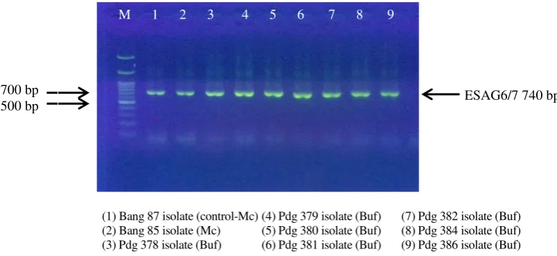
(1) Bang 87 isolate (control-Mc) (4) Pdg 379 isolate (Buf) (7) Pdg 382 isolate (Buf) (2) Bang 85 isolate (Mc) (5) Pdg 380 isolate (Buf) (8) Pdg 384 isolate (Buf) (3) Pdg 378 isolate (Buf) (6) Pdg 381 isolate (Buf) (9) Pdg 386 isolate (Buf)
Figure 3. PCR amplification of 740bp ESAG6/7 gene of T. evansi from stabilates (Mc) and buffalo blood (Buf)
M 1 2 3 4 5 6 7 8 9 M 1 2 3 4 5 6 7 8 9
M 1 2 3 4 5 6 7 8 9
ESAG6/7 740 bp ITS-1 480 bp
Ro Tat 1,2 VSG 151 bp 500 bp
700 bp 500 bp 100 bp
reported that the highest sensitivity against primer was gold standard for T. evansi.
Specific PCR product for T. evansi by using Rotat 1.2 VSG gene was 151 bp (Konnai et al. 2009). Molecular marker using this gene was able to distinguish T. evansi strain type A (Ro Tat) and type B (non Ro Tat) (Njiru et al. 2006). Bajyana & Hamers (1988) successfully isolated protein RoTat 1.2 VSG from Indonesian T. evansi isolate which futher developed into diagnostic CATT 1.2 VSG kit. Ro Tat 1.2 VSG antigen was the predominant Variable Antigen Type (VAT) to be expressed during early, middle and late stages of infection (Verloo et al. 2001). Therefore, in this study, primer RoTat 1.2 VSG was picked as one of primers used to identified T. evansi from Indonesia. Njiru et al. (2006) and Claes et al. (2004) reported that T. evansi was divided into type A (RoTat 1,2 VSG) ciculating in Asia, Africa, Shouth America, and Middle America and type B (non RoTat 1.2 VSG) which circulating in Africa, especially in Kenya. Amplicon product in this study showed that T. evansi Indonesian isolate was type A and no one isolate that including the type B.
Another primer used in this study was ESAG 6/7, a gene located in VSG. PCR product of ESAG 6/7 T. evansi length was 740 bp and was able to be expressed by T. evansi type A and B (Isobe et al. 2003; Mekata et al. 2009). ESAG 6/7 was a sensitive and specific primer against trypanosome due to its multi-copy gene ability encodes heterodimeric complex on transferrin receptor (Pruvot et al. 2010; Kabiri & Steverding 2001). According to Schell et al. (1991) and Kabiri & Steverding (2001) T. evansi use transferrin receptor in the host’s blood to obtain whole iron (Fe) serving in propagation phase. Transferrin receptor encoded by 2 expression-site-associated gen (ESAG6 and ESAG7) in
VSG region. Difference ESAG sequence was reported able to cause different transferrin affinity towards different host (Bitter et al. 1998; Salmon et al. 1994; Steverding et al. 1995).
Identification of T. evansi using multiplex PCR
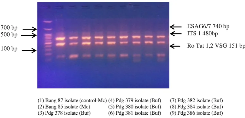
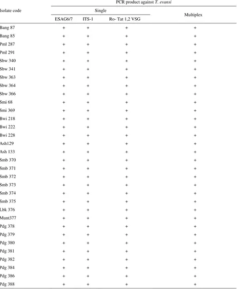
Multiplex polymerase chain reaction (mPCR) is a variant of PCR in which two or more target loci from one or more organisms are amplified using a mixture of locus-specific primer pairs in a single reaction (Markoulatos et al. 2002). Result of multiplex PCR amplification on agarose gel 1.5% visualization under UV light showed three DNA fragment with specific size for T. evansi. PCR product by primer Ro Tat 1.2 VSG produce 151 bp fragment length. Besides, ITS-1, and ESAG6/7 fragment length were 480 bp and 740 bp respectively (Figure 4). Multiplex PCR amplification product was same size with the single one (Table 4). The results of DNA amplification samples derived from stabilate (BCC) and buffalo blood (T. evansi circulating isolates in 2012-2014) are the same. Therefore, T. evansi identification by multiplex PCR against ITS-1, RoTat 1.2 VSG and ESAG6/7 also showed that 31 trypanosome isolates were T. evansi.
This result showed that multiplex PCR analysis by mixing three primer pairs in one reaction successfully marked with three DNA fragment in every column in the gel. During this time, identification of trypanosome species including T. evansi was carried out by single PCR (Sukanto et al. 2000; Njiru et al. 2005; Njiru et al. 2004). Single PCR reaction for T. evansi detection and identification in large number of samples was expensive and time consuming (Ahmed et al. 2013). It was required two or more primer pairs to identify one isolate
(1) Bang 87 isolate (control-Mc) (4) Pdg 379 isolate (Buf) (7) Pdg 382 isolate (Buf) (2) Bang 85 isolate (Mc) (5) Pdg 380 isolate (Buf) (8) Pdg 384 isolate (Buf) (3) Pdg 378 isolate (Buf) (6) Pdg 381 isolate (Buf) (9) Pdg 386 isolate (Buf)
Figure 4. Amplification product of multiplex PCR against ITS-1 (480 bp), Ro Tat 1.2 VSG (151 bp) and ESAG6/7 (740 bp) gene of T. evansi from stabilates (Mc) and buffalo blood (Buf)
M 1 2 3 4 5 6 7 8 9
ESAG6/7 740 bp ITS 1 480bp
Table 4. T. evansi isolate sample obtained from BCC and Departement of Parasitology, BBlitvet, Bogor
Isolate code
PCR product against T. evansi
Single
Multiplex ESAG6/7 ITS-1 Ro- Tat 1,2 VSG
Bang 87 + + + +
Bang 85 + + + +
Pml 287 + + + +
Pml 291 + + + +
Sbw 340 + + + +
Sbw 341 + + + +
Sbw 363 + + + +
Sbw 364 + + + +
Sbw 366 + + + +
Smi 68 + + + +
Smi 369 + + + +
Bwi 218 + + + +
Bwi 222 + + + +
Bwi 228 + + + +
Ash129 + + + +
Ash 133 + + + +
Smb 370 + + + +
Smb 371 + + + +
Smb 372 + + + +
Smb 373 + + + +
Smb 374 + + + +
Smb 375 + + + +
Lbk 376 + + + +
Munt377 + + + +
Pdg 378 + + + +
Pdg 379 + + + +
Pdg 380 + + + +
Pdg 381 + + + +
Pdg 382 + + + +
Pdg 384 + + + +
Pdg 386 + + + +
Pdg 388 + + + +
(Salim et al. 2011). Therefore, in this study multiplex PCR method was developed using more than 2 primer pairs in 1 PCR process. The multiplex PCR was cheaper because it only used one reaction in amplifying some
(Sharma et al. 2012). This study was the first report that used multiplex PCR for T. evansi detection.
Using mPCR to make a diagnosis is three to five times cheaper than using the classical species-specific primers, as the number of reactions required per sample is reduced to a single one. Njiru et al. 2005 and Davila et al. 2003 stated the use of many primers can also lead to the identification of multiple infection of unexpected trypanosome species, especially in wild hosts, vectors and field stocks. This test might indentify targeted trypanosome species without cross amplification between the targeted genes of different trypanosome species. This technique ensure a permanent screening of any unexpected trypanosome species that could grow in vivo or in vitro as a mixed infection.
CONCLUSION
Development of molecular detection technique of trypanosome DNA by mPCR using ITS-1, RoTat 1.2 VSG, and ESAG 6/7 primers has resulted in a considerable improvement of species-specificity in the diagnosis of these parasites to species level. mPCR success to amplify target gene from T. evansi sample from endemic and outbreak areas in Indonesia which was isolated since 1988-2014. Thirty one trypanosome isolates used in this study were T. evansi type A which circulating in Asia. This technique recommended to be used in field.
REFERENCES
Ahmed HA, Picozzi K, Welburn SC, Macleod ET. 2013. A Comparative Evaluation of PCR- Based Methods for species-specific determination of African animal Trypanosomes in Ugandan cattle. Parasites & Vectors 6:316-7. specific Multiplex PCR assay for simultaneous detection of Bacillus anthracis, Yersinia pestis, Burkholderia pseudomallei & Brucella species. Indian J Med Res. 138: 111–116
Bitter W, Gerrits H, Kieft R, Borst P. 1998. The role transferrin-reseptor variation in the host range of Trypanosoma brucei. Nature. 391:499-503.
Chamberlain JS, Gibbs RA, Ranier JE, Nguyen PN, Caskey CT. 1988. Deletion Screening of the Duchenne Muscular Dystrophy Locus via Multiplex DNA Amplification. Nucl Acids Res. 16:11141-11156.
Claes, Filip, Radwanska M, Urakawa T, Majiwa PAO, Goddeeris B, Büscher P. 2004. Kinetoplastid biology and disease diagnostic tool for the detection of Trypanosoma evansi infections. Kinetoplastid Biol Dis. 6:1-6. doi: 10.1186/1475-9292-3-3.
Davila AM, Herrera HM, Schlebinger T, Souza SS, Traub-Cseko YM. 2003. Using PCR for unraveling the cryptic epizootiology of livestock Trypanosomosis in the Pantanal. Braz Vet Parasitol. 117:1-13.
De Sá ARN, Steindel M, Demeu LMK, Lückemeyer DD, Grisard EC, Neto QADL, de Araújo SM, Toledo MJDO, Gomes ML. 2013. Cytochrome oxidase subunit 2 gene allows simultaneous detection and typing of Trypanosoma reangeli and Trypanosoma cruzi. Parasites & Vectors. 6:363. doi: 10.1186/1756-3305-6-363.
Desquesnes M, Dávila AMR. 2002. Applications of PCR-based tools for detection and identification of animal Trypanosomes: A Review and Perspectives. Vet Parasitol. 109:213-231. doi: 10.1016/S0304-4017(02)00270-4.
Desquesnes M, McLaughlin G, Zoungrana A, Davila AMR. 2001. Detection and identification of Trypanosoma of African livestock through a single PCR based on internal transcribed spacer 1 of rDNA. Int J Parasitol. 31:610-614.
[Ditjennak] Direktorat Jenderal Peternakan. 2012. Pedoman Pengendalian Dan Pemberantasan Penyakit Trypanosomiasis (Surra). Jakarta (Indones): Direktorat Kesehatan Hewan, Direktorat Jenderal Peternakan dan Kesehatan Hewan, Kementerian Pertanian.
Ekawasti F, Sawitri DH, Wardhana AH. 2014. Perbandingan metode penyimpanan darah vektor Surra (Lalat Haematophagus) untuk analisis Multiplex Polimerase Chain Reaction. Pamungkas D, Widiawati Y, Noor SM, Purwantari ND, Widiastuti R, Brahmantiyo B, Herawati T, Kusumaningsih A, Handiwirawan E, Puastuti W, editors. Prosiding Seminar Nasional Peternakan dan Veteriner. Bogor (Indones): Pusat Penelitian dan Pengembangan Peternakan. p. 209-217.
Fernandez D, Baradat B, Eleizalde M, Gonzalez-Marcano E, Perrone T, Mendoza M. 2009. Trypanosoma evansi: A comparison of PCR and parasitological diagnostic tests in experimentally infected mice. Trypanosoma Exp Parasitol. 121:1-7.
Gibson W. 2009. Species-specific probes for the identification of the African Tsetse-Transmitted Trypanosomes. Parasitol. 136:1501-1507.
Hernandez P, Martin-Parras L, Martinez-Robles ML, Schvartz-Man JB. 1993. Conserved features in the mode of replication of eukaryotic ribosomal RNA genes. EMBO J. 12:1475-1485.
Herrera HM, Dávila MR, Norek A, Abreu UG, Souza SS,
D’Andrea PS, Jansen AM. 2004. Enzootiology of Trypanosoma evansi in Pantanal, Brazil. Vet Parasitol. 125:263-275. doi:10.1016/j.vetpar.2004.07.013.
Hoare CA. 1972. The Trypanosomes of mammals. Oxford (UK): Blackwell Scientific Publications.
Holland WC, My LN, Dung TV, Thanh NG, Tam PT, Vercruysse J, Goddeeris BM. 2001. The influence of Trypanosoma evansi infection on the immunoresponsive- ness of experimentally infected water buffaloes. Vet Parasitol. 102:225-234.
Isobe T, Holmes EC, Rudenko G. 2003. The transferrin receptor genes of Trypanosoma equiperdum are less diverse in their transferrin binding site than those of the broad-host range Trypanosoma brucei. J Mol Evol. 56:377-386. doi: 10.1007/s00239-002-2408-z.
Jittapalapong, Sathaporn, Pinyopanuwat N, Inpankaew T, Sangvaranond A, Phasuk C, Chimnoi W, Kengradomkij C, Kamyingkird K, Sarataphan N, Desquesnes M and Arunvipas P. 2009. Prevalence of Trypanosoma evansi infection causing abortion in dairy cows in central Thailand. Kasetsart J Nat Sci. 43:53-57.
Kabiri M, Steverding D. 2001. Identification of a developmentally regulated iron superoxide dismutase of Trypanosoma brucei. Biochem J. 260:173-177.
Konnai, Satoru, Hirohisa Mekata, Claro N Mingala, Nancy S Abes, Charito a Gutierrez, Jesus Rommel V Herrera, Alan P Dargantes. 2009. Development and application of a quantitative real-time PCR for the diagnosis of Surra in water buffaloes. Infect Genet Evol. 9:449-452. doi: 10.1016/j.meegid.2009.01.006.
Markoulatos P, Siafakas N, Moncany M. 2002. Multiplex polymerase chain reaction: a practical approach. J Clin Lab Anal. 16:47-51.
Masake RA, Njuguna JT, Brown CC. 2002. The application of PCR-ELISA to the detection of Trypanosoma brucei and T. vivax infections in livestock. Vet Parasitol. 105:179-189.
Mekata, Hirohisa, Konnai S, Witola WH, Inoue N, Onuma M, Ohashi K. 2009. Molecular detection of Trypanosomes in cattle in South America and genetic diversity of Trypanosoma evansi based on expression-site-associated gene 6. Infect Genet Evol: J Mol Epidemiol Evol Genet Infect Dis. 9:1301-1305. doi: 10.1016/j.meegid.2009.07.009.
Mulumbu JM. 2006. The importance of trypanosoma congolense strain diversity in the epidemiology of bovine trypanosomiasis (Thesis). Faculteit Diergeneeskunde, Universiteit Gent.
Njiokou F, Simo G, Nkinin SW, Laveissi C. 2004. Infection rate of Trypanosoma brucei S.L., T. vivax, T. congolense ‘Forest Type’, and T. simiae in small wild vertebrates in South Cameroon. 92:139-146. doi: 10.1016/j.actatropica.2004.04.011.
Njiru ZK, Constantine CC, Masiga DK, Reid SA, Thompson RCA, Gibson WC. 2006. Characterization of Trypanosoma evansi type B §. Infect Genet Evol. 6:292-300. doi: 10.1016/j.meegid.2005.08.002.
Njiru NK, Constantine CC, Guya S, Crowther J, Kiragu JM, Thompson RC, Dávila AM. 2005. The use of ITS1
rDNA PCR in detecting pathogenic African Trypanosomes. Parasitol Res. 95:186-192.
Njiru ZK, Constantine CC, Ndung’u JM, Robertson I, Okaye S, Thompson RCA, Reid SA. 2004. Detection of Trypanosoma evansi in camels using PCR and CATT/T. evansi tests in Kenya. Vet Parasitol. 124:187-199. doi:10.1016/j.vetpar.2004.06.029.
[OIE] Office International des Epizooties Terrestrial Manual, 2012. Chapter 2.1.17. Trypanosoma evansi infection (surra).
Payne RC, Sukanto IP, Bazeley K, Jones TW. 1993. The effect of Trypanosoma evansi infection on the oestrous cycle of Friesian Holstein heifers. Vet Parasitol. 51:1-11. doi: 10.1016/0304-4017(93)90190-X.
Payne RC, Sukanto IP, Partoutomo S, Jones TW. 1994. Efficacy of Cymelarsan treatment of suramin resistant Trypanosoma evansi in cattle. Trop Anim Health Prod. 26:92-94.
Powers TO, Todd TC, Burnell AM, Murray PCB, Fleming CC, Szalanki AL, Adams BA, Harris TS. 1997. The internal transcribed spacer region as a taxonomic marker for nematodes. J Nematol. 229:441-450.
Pruvot M, Kamyingkird K, Desquesnes M, Sarataphan N. 2010. Veterinary parasitology a comparison of six primer sets for detection of Trypanosoma evansi by Polymerase Chain Reaction in Rodents and Thai Livestock. Vet Parasitol. 171:185-193. doi: 10.1016/j.vetpar.2010.04.001.
Reid SA. 2002. Trypanosoma evansi control and containment in Australasia. Trends Parasitol. 18:219-224. doi: 10.1016/S1471-4922(02)02250-X.
Salim B, Bakheit MA, Kamau J, Nakamura I, Sugimoto C. 2011. Molecular epidemiology of camel Trypanosomiasis based on ITS1 rDNA and RoTat 1 . 2 VSG gene in the Sudan. Parasites Vectors. 4. BioMed Central. Ltd: 31. doi: 10.1186/1756-3305-4-31.
Salmon D, Geuskens M, Hanocq F, Hanocq-Quertier J, Nolan D, Ruben L, Pays E. 1994. A novel heterodimeric transferrin receptor encoded by a pair of VSG expression site-associated genes in T. brucei. Cell. 78:75-86.
Sawitri DH. 2016. Studi virulensi T. evansi isolat Indonesia dengan penentuan marka molekuler DNA mikrosatelit dan analisis profil sitokin pada mencit (Thesis). [Jakarta (Indones)]: Faculty of Medicine, Universitas Indonesia.
Schell D, Borowy NK, Overath P. 1991. Transferrin in a growth factor for the bloodstream form of Trypanosoma brucei. Parasitol Res. 30:558-560.
Sharma P, Juyal PD, Singla LD, Chachra D, Pawar H. 2012. Comparative evaluation of Real Time PCR assay with conventional parasitological techniques for diagnosis of Trypanosoma evansi in cattle and buffaloes. Vet Parasitol. 190:375-382. doi: 10.1016/j.vetpar.2012. 07.005.
Steverding D, Stierhof YD, Fuchs H, Tauber R, Overath P. 1995. Transferrin-binding protein complex is the receptor for transferrin uptake in Trypanosoma brucei. J Cell Biol. 131:1173-1782.
Sukanto IP, Solihat L, Politedy F, Dachlan M, Wardhana AH, Satria E. 2000. Peran Diptera Hematofagus sebagai vektor Trypanosoma evansi. Haryanto B, Darminto, Hastiono S, Sutama I-K, Partoutomo S, Subandriyo, Sinurat AP, Darmono, Supar, Butar SOB, editors. Prosiding Seminar Nasional Teknologi Peternakan dan Veteriner. Bogor (Indones): Pusat Penelitian dan Pengembangan Peternakan. p. 481-487.
Uilenberg G. 1998. A field guide for the diagnosis, treatment and prevention of African animal Trypanosomes. In: FAO Corporate Document Repository. Agriculture Costumer Production http://www.fao.org/docrep/006 /x0413e/x0413e02.htm
Urakawa T, Verloo D, Moens L, Büscher P, Majiwa PAO. 2001. Trypanosoma evansi: Cloning and expression in Spodoptera frugiperda [correction of Fugiperda] insect cells of the diagnostic antigen RoTat1.2. Experiment Parasitol. 99:181-189. doi: 10.1006/expr.2001.4670.
Verloo D, Magnus E, Büscher P. 2001. General expression of RoTat 1. 2 variable antigen type in Trypanosoma evansi isolates from different origin. Vet Parasitol. 97:183-189.