Management of Infected Preauricular Sinus
Jacky Munilson, Effy Huriyati, Wahyu Triana
Otorhinolaryngology Head and Neck Surgery Departement Medical Faculty of Andalas University / Dr. M. Djamil Hospital
Abstract
Preauricular sinus is a congenital malformation of the external ear that manifest as a small dell adjacent to the external ear commonly at anterior margin of ascending limb of helix. This disease usually asymptomatic and isolated. The diagnosis is made clinically. Asymptomatic patient require no treatment. If the sinus is infected, the management is administration of antibiotic. If there is abscess formation, incision and drainage is required followed by surgery which aimed to complete dissection of sinus by using various technique like the simple sinectomy, sinectomy with supra-auricular approach and sinectomy with inside-out technique.
One case of infected right preauricular sinus in a 12 years old boy was reported which had been managed by antibiotic based on the cultured and had performed sinectomy with supra-auricular approach.
Key word : preauricular sinus, simple sinectomy, supraauricular approach
Abstrak
Sinus preaurikula merupakan suatu kelainan kongenital dari pembentukan telinga luar yang bermanifestasi sebagai suatu lobang kecil yang terdapat di sekitar telinga luar biasanya pada pinggir anterior dari heliks. Kelainan ini umumnya tidak menunjukkan gejala dan terisolasi. Diagnosis dapat ditegakkan berdasarkan klinis. Bila tidak menunjukkan gejala tidak membutuhkan pengobatan. Bila sinus mengalami infeksi, penanganannya dapat berupa pemberian antibiotika. Bila terjadi abses pada sinus dibutuhkan insisi dan drainase abses yang dilanjutkan dengan operasi yang bertujuan untuk mengangkat sinus secara keseluruhan dengan menggunakan berbagai teknik antara lain sinektomi sederhana, sinektomi dengan pendekatan supra-aurikula dan sinektomi dengan teknik “inside-out".
Dilaporkan suatu kasus sinus preaurikula kanan yang terinfeksi pada anak laki-laki usia 12 tahun yang telah dilakukan sinektomi dengan pendekatan supra-aurikula.
Kata kunci : sinus preaurikula, sinektomi sederhana, pendekatan supra-auricular
INTRODUCTION
Preauricular sinus is a common congenital anomaly in the children. The incidence approximately 0,3-0,9% among the pediatric population.1 Most of preauricular sinus
is manifest as small dells (width less than 3 mm) adjacent to external ear commonly at the anterior margin of the ascending limb of helix. This disease was first described by Van Heusinger in 1864.2,3
Preauricular sinus is often unilateral, only occasionally 25-50% of cases are bilateral and usually being inherited.4 The incidence is
varies globally. In United States is estimated at 0,1-0,9%, in England at 0,9%, in Taiwan at 1,6-2,5%, in Asian at 4-6% and in some areas of Africa at 4-10%.1,2
Preauricular sinus may be associated with other condition or syndrome in 3-10% of cases, primarily associated with deafness and branchio-oto-renal syndrome (BOR syndrome). When congenital anomaly coexist with this sinus,
auditory testing and renal ultrasound should be considered.5 The preauricular sinus was also
being inherited. Genetic linkage analysis study has reported that congenital preauricular sinus was localized at chromosome 8q11.1-q13.3.6,7
The majority of preauricular sinus is asymptomatic, isolated and require no treatment. When the sinus is infected, frequently become swollen, painful and produce discharge with foul
odor. The discharge came out from
desquamating epithelial debris or infection. The most common pathogen bacteria that cause infection is Staphylococcal species and less
commonly Proteus, Streptococcus and
Peptococcus species.3,8
The formation of preauricular sinus is
closely associated with embryological
branchial clefts externally and endodermal pharyngeal pouches internally. The first and second branchial arches each give rise to 3 hillocks that called the hillocks of His. Three hillocks arise from the caudal border of the first branchial arch will formed the tragus, helical crus and the helix, and three hillocks that arise from the cephalic border of the second branchial arch will form the antihelix, scapha and the lobule. These hillocks should unite during the next few weeks of embryogenesis. (figure 1) If the fusion of hillocks is incomplete, it can postulated source of the preauricular sinus. Another theory suggests that localized folding of ectoderm during auricular development is the cause of preauricular sinus formation.6,9,10
Figure 1 : Embriology origins of the auricle.11
The tract of preauricular sinus usually narrow and varies in length (usually short), tortuous paths and its orifices are very small. Preauricular sinus may leads to the formation of a subcutaneous cyst that intimately related to the tragal cartilage and the anterior crus of the helix. In almost cases, part of the tract connect to the perichondrium of the auricular cartilage. The preauricular sinus is usually found at lateral, superior, and posterior of the facial nerve and the parotid gland. The sinus also can extends into the parotid gland.11,12
There are 2 types of the preauricular sinus, the classical type and the variant type. The classical type is define as a preauricular sinus whose sacs are located on the anterior of the external auditory canal (EAC) and the variant type is the auricular sinus whose sacs are located at the postauricular area and sometimes presents the opening pit near the ascending limb of the helix. In distinguishing between the classical type and the variant type have been made the imaginary line that connect the tragal cartilage to the posterior margin of the ascending limb of helix. The variant type could be classified into
three types according to the location of dells : dell is located at the middle area on crus (type 1); dell is located at the superior area of the crus (type 2); and dell is located at the cymba concha (type 3). 1(figure 2)
Preauricular mass or cyst is usually considered as inflamed lymph node, an infected sebaceous cyst, furuncle and branchial cleft cyst.1,9
Figure 2 : The schematic location of fistular dells of the variant type and classical type of preauricular sinus on imaginary line that connecting between the tragal and the posterior margin of ascending limb of helix1
In the acute phase infection of the preauricular sinus, the intervention involves administration of appropriate antibiotic against the causative pathogen. If the abscess present, commonly need incision and drainage. Surgical excision of the sinus and its tract is required if the infection recurrent or persistent. Surgical excision is aimed to ensure complete dissection of the sinus. Incomplete dissection is believed to be the cause of recurrence of preauricular sinus. Recurrence rate after excision range from 0-42%.6,12
CASE REPORT
A 12 years old boy presented to the ENT out-patient clinic on April 4th 2012 with a
not healing. There was small dell at the right anterior part of ascending limb of helix from birth. From this small dell has ever came out the yellowish discharge and sometimes swelling. There was no fever. Mixturation was normal. There was no families have disease like that, no history of discharge from the right ear and no impairment of hearing. There was no families that have congenital impairment of hearing. There was no history of diabetic on his mother during pregnancy.
On physical examination, general
condition was moderately ill, composmentis cooperative and temperature was 36,80C. Pulse
rate 80x/minute and respiratory rate 18x/minute.
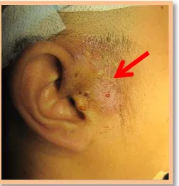
On ENT examination of the right ear, there was swelling at the anterior part of the auricle 3 x 1,5 x 0,5 cm in size, fluctuative, hyperemic and tenderness. (figure 3A) There was a small dell at the peak of the swelling, at the anterior of the ascending limb of helix. The ear canal was wide, tympanic membrane was intake and cone of light was positive. At the left ear, there was a small dell at the cymba concha and no infection surrounding the dell. (figure 3B) The ear canal was wide, tympanic membrane was intake and cone of light was positive. Nose and throat was normal.
A B
Figure 3 : A)The right infected preauricular sinus, B) A small dell at the cymba concha of left auricle.
The patient was diagnosed suspected infected right preauricular sinus in classic type and the left preauricular sinus in variant type. The patient was hospitalized to administer the intravenous antibiotic ceftriaxone 2 x 500 mg, metronidazole drip 3 x 500 mg and Tinoridine Hcl 2 x 50 mg. Before that, The swelling was
aspirated and found 1,5 cc serosanguineus
discharge. The discharge was sent to
microbiology laboratory for culture and sensitivity test.
On laboratory finding was found
hemoglobin 13,3g%, leukocyte 8.200/mm3 and
hematocrit 39%. On April 11th 2012, from culture
and sensitivity test was found the pathogen causing infection is Staphylococcus aureus and
sensitive with ampicillin sulbactam,
chloramphenicol, ciprofloxacin, erythromycin,
gentamycin, meropenem, netilmicin dan
sulfamethoxazole + trimethoprime while for cefotaxime dan cefoperazone was intermediate.
The intravenous antibiotic was changed with the antibiotic based on culture cefotaxime 2 x 1 gr and gentamycin 2 x 80 mg. After 3 days of administered of antibiotic, the swelling did not decrease and more fluctuative. The swelling was aspirated again and was found the bloody
purulent discharge. The patient was planned to perform incision and exploration of preauricular abcess followed by sinectomy in general anaesthesia. The preparation of operation was done in ENT out-patient clinic.
On laboratory finding was found
hemoglobin 13,5g%, leukocyte 7.600/mm3,
hematocrit 41%, trombosit 273.000/mm3, PT ,4 and APTT 44, . The patient was consulted to neurotology sub department for evaluation of facial nerve and hearing function. There was no facial nerve paralysis. Result of the tunning fork examination Rinne +/+, Weber no lateralization and Schwabach at the right and left ear was the same with the examiner. Pure tone audiometri can not be performed because of the swelling and pain at the anterior of auricle. One day before operation, the swelling was rupture and release the bloody purulent discharge and formed rupture wound at anterior of tragal 0,2 x 0,2 cm insize. (figure 4)
On April 18th 2012 has been performed
sinectomy that extended to supra-auricular
(supra-auricular approach) in general
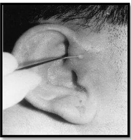
anaesthesia. Operating report, the patient was laying down on operating table in general anaesthesia and the head was tilted to the left side. Aseptic and antiseptic procedure was performed at the operating field. Tract of sinus was evaluated by intravenous canule no 22, the canule was entered through small dell approximately 1,5cm to inferior direction. Infitration with adrenalin 1 : 200.000 was done at surrounding the dell, supra-auricular and surounding the rupture wound. The tract of the sinus was demarcated with methylene blue which entered through the dell. (figure 5)
Figure 5 : Evaluated and demarcated the tract with methylene blue
An ellipse incision of skin around the dell was made. The tract of sinus was opened and separated with blunt and sharp dissection until the sacs was identified. Sac of the sinus was adhered to the superior part of tragal cartilage. Sac was removed together with superior part of tragal cartilage. Incision was extended to supraauricular around the ascending limb of helix. Pathologic tissue was removed. Rupture wound at the anterior of tragus was removed by ellipse incision and pathologic tissue at surrounding it was removed. The incision wound was washed by peroxide (H2O2 3%) and rinsed by Nacl 0,9%. The incision wound around the helix was sutured with chromic 4.0 and continued with prolene 5.0 whereas the rupture wound at anterior tragus was sutured with prolene 5.0. The incision was closed by compression dressing and operation was finished.
Figure 6 : Macroscopic view of preauricular sinus and its tracts
After operation, the patient was given cefotaxime 2 x 1 gr, gentamycin 2 x 80 mg, dexamethasone 3 x 5 mg tapering off and tramadol 1 ampul in 500 cc ringer lactat solution. Vital sign, bleeding from the incision wound and facial nerve paralysis was observed.
The first day after operation, there was no fever, bleeding from the incision wound and facial nerve paralysis. The therapy was continued.
On the third day after operation, the compression dressing was removed. The incision wound was seen swelling and pain. The wound was cleaned and pressed, serous discharge and clotting came out from the wound. A part of suture was loosed. Therapy was continued.
On the fifth day after operation the swelling at incision wound and pain was decrease. The patient was discharged with therapy cefixime 2 x 100mg and tinoridine hcl 2 x 50 mg.
On April 25th 2012 (7 days after
operation) patient was controlled to ENT out-patient clinic, the incision wound was not swelling again, no discharge surrounding the incision and no pain. A part of suture was still loosed. The incision wound was cleaned and the edge was harmed. The loosed suture was sutured again in local anaesthesia and closed with compression dressing. Therapy was continued.
On April 28th 2012 (3 days after
re-suture) patient was controlled, there was no infection surrounding the incision wound. The compression dressing was maintained and therapy was changed with roxytromisin 2 x 150 mg.
On 1st May 2012 (6 days after re-suture)
(9 days after re-suture) the incision wound was closed completely, no infection and formed thin scar. Whole of the suture was removed. Antibiotic was stopped and given antikeloid cream for the scar. (figure 7)
Figure 7 : Scar 2 weeks after surgery
DISCUSSION
A case of 12 years old boy with diagnosed infected right preauricular sinus in classical type was reported. The incidence of preauricular sinus was spread globally. In Asia and Africa was higher than Europe and American (4-10%).13
Jimoh et al14 have studied about prevalence of
preauricular sinus among Nigerians and found that the incidence was higher in age 1 to 45 years old and the commonest incidence in 1-18 years old, male is more affected than female with ratio 1,3 to 1,0, the left ear more often than the right ear and bilateral ear only 7%. Choi SJ et al1
report that incidence of variant type of preauricular sinus is 10,9%, and type 1 that has dell at the middle of the helical crus is the commonest one. These studies were similar with this case, the patient was male, 12 years old and has bilateral preauricular sinus. The right preauricular sinus is classical type while the left side is variant type and asymptomatic. In ENT out-patient clinic at Dr. M. Djamil hospital on January until December 2011 has been found 7 cases of preauricular sinus and 6 cases has undergone sinectomy surgery. On January until March 2012 only found 2 cases.
Preauricular sinuses arise in the antenatal period and usually present at birth, but part of them can become apparent later in life. Huang et al14 found only one quarter of
preauricular sinuses were became symptomatic
and almost one third of the symptom was appeared after 16 years old. Whereas Hanson et al9 said that most of people with preauricular
sinus is asymptomatic, only one third of patients aware of their malformation. The lesions became apparent at 9 years old. Goel AK et al15 from his
research found that the most common symptom of preauricular sinus is yellowish discharge come out from the sinus dell followed by sinus infection symptom like swelling, tenderness and abscess formation around the sinus. In these patients the symptoms initially were seen in the age of 12 years there was yellowish discharge from the sinus dell followed by swelling and pain around the sinus.
Preauricular sinus can be associated with hearing and renal problems and also associated with many syndromes. Some investigator recommended that auditory testing and renal ultrasonography should performed for all children with a preauricular sinus.2 Wang et al16
suggest that renal ultrasound should be performed on patients with a preauricular sinus accompanied by one or more of following item :
1. Another malformation or dysmorphic feature
2. A family history of deafness
3. An auricular and/or renal malformation 4. A maternal history of gestational
diabetes
This patient was not done the auditory testing and renal ultrasonography because there was no symptom, no malformation of ear and renal and no history of deafness in family and no history diabetic in his mother while pregnancy.
Firat et al17 in his study at 13.740
primary school children were found 36 children with preauricular sinus. The children were performed urinalysis, renal ultrasonography and otoacoustic emission (OAE). The result was prevalence of renal abnormality 2,7% and hearing impairment 2,7%. These was similar to the control group. The conclution is unnecessary to investigate hearing or urinary abnormality in patients with preauricular sinus, unless there is an association with a syndrome or history of hearing or renal impairment in the family.
Preauricular sinuses can be either inherited and sporadic. When inherited, they show an incomplete autosomal dominant pattern. Bilateral preauricular sinus was found in 25-50% of cases, and usually being inherited.2 In
The diagnosis of preauricular sinus was made clinically and may remain asymptomatic throughout life This disease usually found in ear, nose and throat examination. If asymptomatic require no treatment.3 Once infected can causes
pain, swelling and abscess formation. Treatment of acute infection or abscess in preauricular sinus is adequate drainage.17 Fluid drained from the
sinus should be cultured and given appropriate antibiotic.2 The antibiotics based on culture was
administered until all infection healed. When the symptoms are recurrent or persistent, sinus excision is a treatment of choice. The aim of surgery is to extirpate all squamous epithelium that lining of the sinus because incomplete remove of deep branching of the sinus can cause recurrence.18 Goel et al15 suggests that surgical
excision of preauricular sinus should be done in quiescent phase. The infection make difficulty to eradicate infection that make higher recurrence and bad surgical scar. In this study, the mean time that taken to achieve an infection-free preauricular sinus was 6 weeks. In this case, the patient had been treated with antibiotic for 6 weeks before surgery, but the infection was not healed. Therefore we choosed management by incision and drainage abscess then followed by sinus excision, but the abscess was rupture one day before the operation was done.
Standard technique for excision of preauricular sinus are simple sinectomy that provides an elliptical skin excision around the sinus opening and continued with dissection of sinus ramifications in the subcutaneous tissue under visual or palpatory guidance. For identification of sinus tract, there are many suggestions like use small lacrimal probe, injection of methylene blue intra-operative or pre-operative sonography that aimed grossly evaluating of sinus course.7
In preauricular sinus surgery, methylene blue is used to demarcat and differentiate between sinus and fistulae intra-operative. Dickson et al19, in his study assesses the utility
and safety of methylene blue in demarcating of preauricular sinus in children. The result was methylene blue demarcation provide minimally invasive and helps ensuring complete resection of preauricular sinus. Granizo et al20 suggest that
the combination of methylene blue staining and fistula probing may provide a suitable method for accurate preauricular sinus resection.
In 1990 Prasad et al4, for the first time
described a new surgical approach that named supra-auricular approach. This approach is made
based on the theory that preauricular sinus almost always included subcutaneous tissues
between the temporalis fascia and
perichondrium of the helix cartilage. (figure 8) Therefore the elliptical excision of the standard technique was extended upward to the pre and supra-auricular temporal without adverse aesthetic consequences. Lam et al8 compare two
excision techniques of the preauricular sinus between the simple sinectomy and auricular approach. The conclusion is supra-auricular approach statistically has lower recurrence rate than the simple sinectomy. This study was the same with the study whose done by Hassan et al5 that recommended the
supra-auricular approach for presupra-auricular sinus excision because statistically has significant lower recurrence rate than standard sinectomy technique. In this case also use the supra-auricular approach to facilitate removing of pathologic tissue around sinus infection.
Figure 8 : Incision of supra-auricular approach.8
J. Robert et al21 introduces a new surgical
Dunham et al11 found over 50% of
specimens reviewed, the sinocartilaginous distance was less than 0,5 mm and the epithelial tracts are continued with stromal tissue histologically that indistinguishable from the perichondrium
Figure 9 : Inside-out technique. A. a small skin island around the sinus is made, B. stay suture allow traction to facilitate dissection of tracts, C. the sinus is opened with sharp scissors, D. under magnified vision, the glistening lining (inside) and the outer wall of the tract (out) are dissected free of the surrounding tissue.21
If abscess has occurred, incision and drainage of the preauricular sinus was recommended. The incision and drainage can make disruption of sinus architecture that increased recurrence problem. Coatesworth et al22 described one technique in managing a
preauricular abscess that drainage of the pus from the abscess with little and no disturbance the underlying tissue around the sinus. This technique was done in local anesthesia. The blunt-ended lacrimal probe is inserted into the sinus dell then drainage the abscess without incision. The procedure may be repeated in subsequent days if necessary. If subsequent incision is required can be guided by the probe.22
(figure 10)
Figure 10 : lacrimal probe is entered to the infected preauricular sinus allowing the release of pus.22
If the preauricular sinus is rupture, can develop granulation tissue which difficult to heal. The epithelial-granular junction at the rupture area is not so clear. For moving the epithelium clearly used an operating microscope or magnifying glasses. Shu et al23 suggest that the
granulation tissue should be removed by curettage and continued with placement of small drain inside incision wound for 2 days to evacuate fluid accumulation. In this study did not report the recurrence and infection after surgery. Late recurrence is rare. When the recurrence has occured, mostly appeared within the first 2 weeks after surgery. The recurence often as result of residual cyst wall inside the wound. In this case did not used magnifying glasses or operating microscope during operation and also did not used drain after operation because the incision wound look clean and dry enough.
It is important to close the dead space after excision of preauricular sinus completely by mean closured layer by layer with or without drain and use compression dressing.18 Heo et
al24 introduce the new compression dressing
method by suture transfixion of silicone sheets. This technique can decrease the recurrence rate and haematoma formation after excision.
A preauricular sinus with a previous history of infection or active infection during the definitive surgery may have higher tendency of recurrence.3 recurrence of the preauricular sinus
often happened in patient whose post operative wound infection.17 Currie et al18 based on his
temporalis fascia to ensure complete removal of all epithelial components;(4) avoidance of sinus rupture; and (5) closure of wound dead space.17
Goel et al15 in his study found various factors
which resulted good surgical results are good surgical technique, infection-free period,and use of general anaesthesia. Yeo et al 25 report that
surgery which performed under local infiltrative anaesthesia has higher rate of recurrence than cases which used general anaesthesia.
REFERENCES
1. Choi SJ, Choung YH, Park K, Bae J, Park HY. The Variant Type of preauricular Sinus: Postauricular Sinus. The Laryngoscope 2007;117: 1798-1802
2. Scheinfeld NS, Silverberg Nb, Weinberg JM, Nozad V. The Preauricular Sinus: A review of its Clinical Presentation, Treatment, and Associations. Pediatric Dermatology
4. Leopardi G, Chiarella G, Conti S, Cassandro.
Surgical treatment of recurring
preauricular sinus: supra-auricular approach. Acta Otorhinolaryngologica Italica 2008: 28: 302-5
5. Hassan ME, Samir A. Pre-auricular sinus: Comparative Study of Two surgical Techniques. Annals of Pediatric Surgery 2007;3(3,4): 139-43 preauricular fistula maps to chromosome 8q11.1-q13.3. J Hum Genet 2003; 48:155-58
8. Lam HCK, Soo G, comparison of Two Surgical Techniques. The Laryngoscope 2001;111: 317-9
9. Scheinfeld NS. Preauricular Sinus. Update 2011 Aug 1 [cited 2012 Apr 28]. Available
from :
www.emedicine.medscape.com/article/11 18768-0verview.
10. Wilson CP. The Etiology of The Preauricular Sinuses. Actaoto-laryng 50: 226-32
11. Dunham B, Guttenberg M, Morrison W, Tom L. The Histologic Relationship of Preauricular Sinuses to Auricular cartilage. Arch otolaryngol Head Neck Surg 2009; 135(12): 1262-5
12. Tan T, Constantinides H, Mitchell TE. The preauricular sinus : A review of its aetiology, clinical presentation and management. International Journal of Pediatric Otorhinolaryngology 2005; 69: 1469-74
13. Jimoh OR, Alabi BS, adebayo SS. Prevalence of Preauricular Sinus Among Nigerians. Surgery Journal 2008;3(3): 61-3
14. Huang XY, Tay GS,nsaicheong GKL, Low WK. Preauricular Sinus. Arch Otolaryngol Head Neck Surg 2007;133:65-8
15. Goel AK, Slonia SC, Garg A, Rattan K. Preauricular sinus : When to operate?. Indian Journal of Otology 2011;17: 63-5 16. Wang RY, Earl DL, Ruder RO, Graham JM.
Syndromic Ear Anomalies and Renal Ultrasounds. Pediatrics 2001;108(2):1-8 17. FiratY , Sireci S, Yakinci C, Akarcay M,
Karakas HM et al. Isolated preauricular pits and tags: is it necessary to investigate renal abnormalities and hearing impairment?. Eur Arch Otorhinolaryngol 2008;265 : 1057-60
18. Currie AR, King WWK, Vilantis AC, Li AKC. Pitfall in the management of preauricular sinuses. British Journal of Surgery 1996;83: 1722-4
19. Dickson JM, Riding KH, Ludemann JP. Utility and Safety of Methylene Blue Demarcation of Preauricular Sinuses and Branchial Sinuses and Fistulae in Children. Journal of Otolaryngology Head &Neck Sursery 2009; 38(2): 302-10
20. Granizo RM, Perez-Herrero MC, Sanches-Cuellar A. Methylene blue staining and probing for fistula resection: application in a case of bilateral congenital preauricular fistulas. Int J. Oral Maxillofac Surg 2002;31 : 439-41
21. J Robert, Jong Baatenburg. A new surgical technique for treatment of preauricular sinus. Surgery 2005;137(5): 567-70 22. Coatesworth AP, Patmore H, jose J.
management of an infected preauricular sinus, using a lacrimal probe. The Journal of Laryngology & Otology 2003; 117: 983-4 23. Shu MT, Lin HC. Extirpation of Ruptured
24. Heo KW, Baek MJ, Park SK. Pressure dressing after excision of preauricular sinus: suture transfixion of silicone sheets. The Journal of laryngology & Otology 2009; 123: 1367-70