Identification of Plankton on Fish Pond of
Oreochromis niloticus
Infected
by
Viral Nervous Necrosis
Uun Yanuhar1), Muhammad Musa1), Dyah Tri Rahayu1), Diana Arfiati1) 1) Laboratory of Water and Biotechnology, Faculty of Fishery and Marine Science
University of Brawijaya, Indonesia.
Email: [email protected] / [email protected]
ABSTRACT
The purpose of study is to determine the type of plankton found in cultivation pond of nile tilapia infected by VNN. The method is to identify the plankton in the water pond and testing the VNN that infects nile tilapia related to plankton eaten using Polymerase Chain Reaction (PCR). VNN identification results on the nile tilapia digestive tract stomach and intestines as well as plankton found in the stomach and intestines indicate VNN positively. Plankton found in the digestive tract of fish is division of Chlorophyta, Bacillariophyta, phylum Rotifera, and Crustacea. It indicates that nile tilapia infected by VNN both on the gastrointestinal tract organs, stomatch or intestines and plankton were found with positive VNN digestive duct, while plankton found in water pond is VNN negative. The VNN infection depends on the nature of specifications VNN on nile tilapia and the plankton in the nile tilapia digestive duct. Keywords: Oreochromis niloticus, plankton, Viral Nervous Necrosis.
INTRODUCTION
Plankton is microscopic organism that, in general, is floating in the water and able to swim in very weak movement, because their movement is influenced by the movement of water. Phytoplankton is microscopic plant, and low-level classifications of plants, while each type of phytoplankton undergoes physiological and behavioral reactions to changes in
environmental quality either physically, chemically or biologically. Phytoplankton community acts as useful indicators of water quality (George, Nirmal Kumar, & Kumar, 2012). Zooplankton is microscopic animal and serves as the first consumer that eats phytoplankton. Zooplankton and phytoplankton is the food of most juvenile fish like nile tilapia (Orechromis niloticus) and they may serve as a vector of virus (Kitamura, Kamata, Nakano, & Suzuki, 2003); (Frada et al., 2014).
role of plankton in Nile tilapia cultivation pond contaminated water and other organisms such as water microorganisms. Betanodavirus can also be transmitted through natural food that is plankton (Yanong, 2013). For example, virus-infected plankton such as Artemia, Tigriopus japonicus (copepod), and Acetesinte medius (shrimp), or by organisms that are eaten. While vertically, betanodavirus can be transmitted via the mains or fish seeds obtained in advance to the offspring of fertilized egg (Gomez, Mori, Okinaka, Nakai, & Park, 2010).
Nile tilapia infected with VNN shows clinical symptoms of the Nile tilapia (Oreochromis niloticus) found at the site of research that has similarities with clinical symptoms of VNN-infected snapper and it is emphasizedby the advent of VNN genome with an analysis of 294 bp PCR with primary VNN. This study will examine any plankton found in water, plankton found in the digestive tract of nile tilapia, and VNN genome analysis as RNA viruses in the digestive tract organ of nile tilapia, plankton found in digestion tract of nile tilapia and plankton species identified, the condition water and environment and role of plankton in the spread of Viral Nervous Necrosis (VNN) nile tilapia cultivation pond.
MATERIALS AND METHODS
Sample.
The samples are Nile tilapia infected by Viral Nervous Necrosis (VNN) and the water pond that obtained from fisheries farm in Blitar, East Java, Indonesia.
Research design.
The research design is observational data using a laboratory analysis. Observations of Nile tilapia test on cultivation pond carried out
for 3 weeks which a week observation and repeated three times.
Water Sampling.
Water quality parameters observed are: brightness, temperature, pH, DO, CO2, NO3,
PO4, and TOM. Monitoring of water quality is
as much as 3 repetitions with a sampling interval is one week. Water sampling uses DO bottle and 600 ml bottle of mineral water.
Plankton sampling
c) The diversity index using Shannon-Wiener Index (Ghosh & Biswas, 2015).
H′ = − ∑s pi log pi
i =1 ... (3)
Where,
pi = the proportion of importance value of the i (pi = ni/N).
H' = Diversity Index.
Ni = the number of individual of i number.
N = Total Number of individuals. S = Number of species in the samples.
d) The domination Index using Simpson’s index (D) (Ghosh & Biswas, 2015).
D = Σ (pi)2 ... (4)
Where,
D = Domination Index. Pi = relative Abundance.
Polymerase Chain Reaction
After extraction of RNA, then the amplification process in the order of the PCR process revers-transcriptase is performed using the Quick Access AMV® RT-PCR (Promega) followed by nested PCR using Go Taq® Green Master Mix (Promega) and using the primary pair of VNN Forward 2 (F2): AGT GTC CGT GTG TCG CAT CT and Reverse 3 (R3): CGA GTC AAC GGT GAA ACG GA.
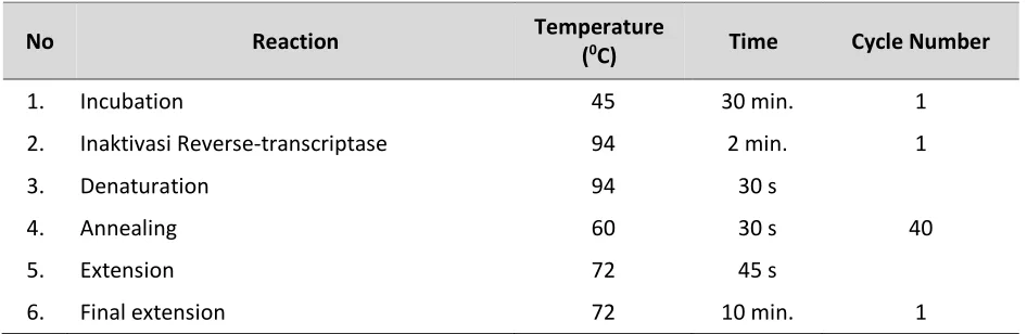
The reaction process is conducted in accordance with the protocol of each kit, to a final concentration of each primary 2.5 µl. Then, enter the template of RNA, a positive control and a negative control in the PCR machine (thermal cycler). Ensure that the program to be run is VNN. The setting of the temperature on a thermal cycler is as Table 1.
Table 1. The setting of the temperature on a thermal cycler.
No Reaction Temperature
(0C) Time Cycle Number
1. Incubation 45 30 min. 1
2. Inaktivasi Reverse-transcriptase 94 2 min. 1
3. Denaturation 94 30 s
40
4. Annealing 60 30 s
5. Extension 72 45 s
6. Final extension 72 10 min. 1
Electrophoresis Agarose
After the amplification process is complete, take 10 μL of each sample and add 2 μL of
staining solution (6x loading dye) on parafilm paper, then homogenized. Furthermore, around
10 μL of the sample and marker put into agarose gel and sink slowly. Then attach the
RESULTS AND DISCUSSION
Water quality
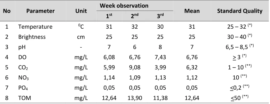
Quality of water in the Nile tilapia cultivation pond was shown in Table 2.
Table 2. The result of water quality parameter measurement in the cultivation pond.
No Parameter Unit Week observation Mean Standard Quality 1st 2nd 3rd
1 Temperature 0C 31 32 30 31 25 – 32 (*)
2 Brightness cm 25 25 25 25 30 – 40 (*)
3 pH - 7 6 8 7 6,5 – 8,5 (*)
4 DO mg/L 6,08 6,76 7,43 6,76 > 3 (*)
5 CO2 mg/L 5,99 9,08 3,99 6,32 1 – 10 (**)
6 NO3 mg/L 1,14 1,09 1,13 1,12 10 (**)
7 PO4 mg/L 0,05 0,05 0,05 0,05 <0,2 (**)
8 TOM mg/L 12,64 13,90 11,38 12,64 <50 (**)
Notes:
(*) SNI (Indonesia National Standard) (2009). (**) Marion (1998).
Plankton identification.
The results of the identification of plankton on the Nile tilapia cultivation pond of observations on week 1 to 3 obtained the kinds of phytoplankton and zooplankton. Phytoplankton consists of three divisions: division of Chlorophyta (2 genus), Cyanophyta (1 genus) and Bacillariophyta (7 genus). Zooplankton consists of three phyla which are phylum Arthropods (1 genus), crustaceans (1 genus) and Rotifera (1 genus).
Relative abundance
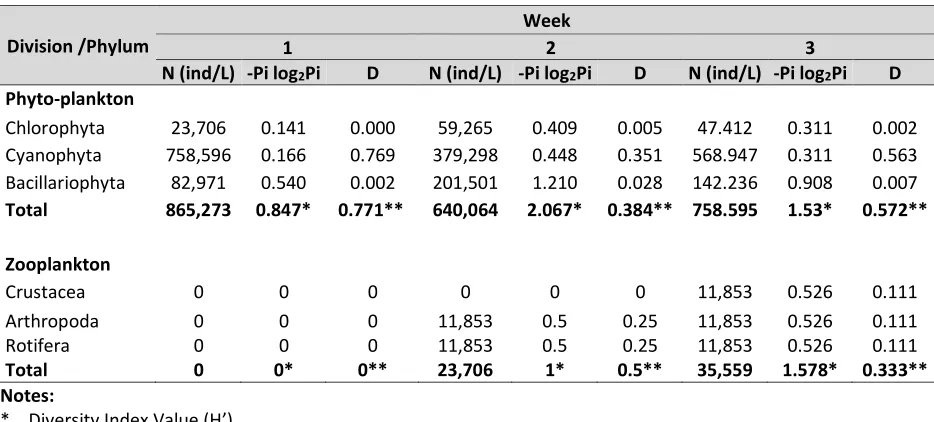
Based on the calculation of the relative abundance of phytoplankton found that overall (week 1 to 3) percentage of the value of the highest relative abundance is occupied by division of Cyanophyta (genus Merismopedia), followed by the division of Bacillariophyta then division of Chlorophyta. The result of the calculation of abundance (N) in Table 2 indicates that the division of Cyanophyta has high abundance. The results of relative abundance and abundance of division of Cyanophyta (Merismopedia sp.)
are high on every week due phytoplankton of the division was able to adapt to the aquatic environment and the availability of nutrients needed by the phytoplankton.
Abundance (N)
The result of the calculation of the abundance of plankton (Table 3) showed that phytoplankton abundance was highest in week 1 and then decreased in week 2 and increased its abundance in week 3. Overall, the highest contributor to the total value of abundance phytoplankton on each week are Cyanophyta division. Erdina et al. (2013), suggests that Cyanophyceae is able to survive in conditions without light and indicators of their high content of organic material in the water. Algae is capable of taking CO2 and
phosphorous in conditions of low concentrations in water. The Nile tilapia cultivation pond is included into the water with moderate fertility levels (mesotrofik) and the fertility of water is having plankton ranged from 104 to 107 ind/L (Goldman and
Table 3. The result of abundance, diversity and domination index.
Division /Phylum
Week
1 2 3
N (ind/L) -Pi log2Pi D N (ind/L) -Pi log2Pi D N (ind/L) -Pi log2Pi D
Phyto-plankton
Chlorophyta 23,706 0.141 0.000 59,265 0.409 0.005 47.412 0.311 0.002 Cyanophyta 758,596 0.166 0.769 379,298 0.448 0.351 568.947 0.311 0.563 Bacillariophyta 82,971 0.540 0.002 201,501 1.210 0.028 142.236 0.908 0.007
Total 865,273 0.847* 0.771** 640,064 2.067* 0.384** 758.595 1.53* 0.572**
Zooplankton
Crustacea 0 0 0 0 0 0 11,853 0.526 0.111
Arthropoda 0 0 0 11,853 0.5 0.25 11,853 0.526 0.111
Rotifera 0 0 0 11,853 0.5 0.25 11,853 0.526 0.111
Total 0 0* 0** 23,706 1* 0.5** 35,559 1.578* 0.333**
Notes:
* Diversity Index Value (H’). ** Domination Index Value (D).
Table 4. Plankton composition analysis result in digestion tract of Nile tilapia (Oreochromis niloticus).
Diversity Index (H')
Phytoplankton and zooplankton diversity index, the calculation results in Table 3, indicate that the nile tilapia cultivation pond included in the criteria low to moderate H'. According Utami (2001), the diversity index criteria are divided into three types of low diversity with H'<1, moderate diversity when 1<H'<3 and high diversity when H'>3. According to Krebs (1989), moderate diversity could mean that ecosystem is in fairly good condition, where the spread of the individual or the type of phytoplankton is almost evenly. Low diversity indicates the tendency of dominant species
Division/Philum Genus Total (ind/ml)
Week 1 Week 2 Week 3
a. Phytoplankton
Chlorophyta Netrium 1 1 2
Bacillariophyta Navicula 18 15 22
Nitzschia 5 4 8
Achnantes 2 1 6
Surirella 2 3 2
Tablelaria 4 6 2
Diatoma 2 3 5
Subtotal 34 33 47
b. Zooplankton
Crustacea Calanus 1 0 2
Rotifera Keratella 2 1 0
Subtotal 3 1 2
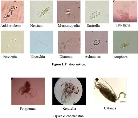
in an ecosystem due to the instability of environmental factors and population. The description of plankton observations with Olympus BX331 microscope is shown in Figure 1 and Figure 2.
Figure 1. Phytoplankton.
Figure 2. Zooplankton.
Domination Index (D).
Based on Table 3 on the dominance index of phytoplankton and zooplankton in week 1 and 2, there is a plankton species that dominates which are phytoplankton of division of Cyanophyta (genus Merismopedia) and in zooplankton, there is no species that dominate in the water. The kinds of phytoplankton have a different response to the ratio of nutrients dissolved in water. This condition causes phytoplankton community in one water has
a structure and dominance of different types with other water.
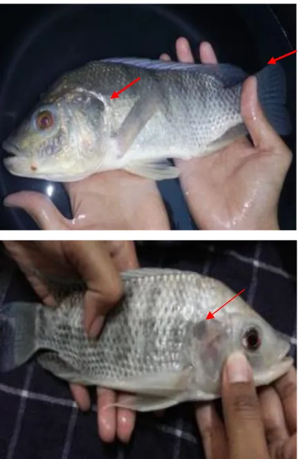
Analysis result of VNN-infected Nile tilapia. The observation of the morphology and behavior of the Nile tilapia showed the symptoms that fish infected with VNN. The visible symptoms are fish swimming at the surface, swim angled and loss of balance, weak movement, eyes bulging out and blackish-red, the body of mucus and body color is pale and some fish showed a darkening of the color of the body as shown in Figure 3.
Figure 3. VNN-Infected nile tilapia morphological condition.
OIE (2013) explains that there is a change in the habits of the VNN-infected fish like swimming round and round, upside down (whirling) or stomach position is above due to the swim bladder inflammation. Sometimes the VNN-infected fish swim vertically to the bottom of the pond or swimming round and round quickly or stomping the head to the surface of the water. VNN on Nile tilapia detection by PCR test on primary base of 294 bp showed that the intestine is positively infected by VNN.
Indication of the presence of plankton and VNN in Nile tilapia.
VNN genome amplified by PCR on Nile tilapia and plankton is shown in Figure 4 and 5.
Figure 4. Amplification of VNN Genome found in Intestine Nile tilapia: (1) Marker DNA lambda 1 bp; (2) Positive control; (3) Negative control; (4) Positive sample VNN 294 bp.
Figure 5. Amplification of VNN Genome in the plankton found in the digestive tract of Nile tilapia (Gastric and Intestine): (1) Marker DNA lambda 1 bp; (2) Positive control; (3) Negative control; (4) Positive sample VNN 294 bp.
However, brightness value of the water is low. This is caused by nile tilapia cultivation pond is included a shallow pond with a pond depth only about 25 cm. In addition, the bottom of the pool is still visible on the surface of the water so that it can be assumed that the brightness value of 100% at a depth of 25 cm.
The existence of VNN in water is affected by environmental conditions such as physico-chemical parameters of water. The most influencing water quality parameter on the existence of VNN is pH, temperature and TOM. The pH value, temperature and TOM range from 6-8, 30-32°C, and 11.376 to 13.904 mg/L (Table 2). VNN can survive in waters with environment pH ranging from 2-9. While at pH 11-12, it will cause the inactivation of the virus (Munday, 2003). At a temperature of 25°C or higher temperature, it can significantly affect VNN infection of the fish. The virus is able to survive the acidic pH conditions even at a temperature of 37°C (OIE, 2013). However, when the water temperature is more than 31°C, it will inhibit the proliferation of betanodavirus or VNN (Yuasa, Koesharyani, & Mahardika, 2007). While, the organic materials are associated with the build up of feces at the bottom of the water. If the feces is derived from the nile tilapia that were infected by VNN the more feces come out and piled up in the bottom of the pool, the more the virus found in the water and likely nile tilapia can be infected by VNN.
Merismopedia sp. is abundant in water temperatures ranging from 27 to 30°C. While at temperatures ranging from 27 to 31°C, Merismopedia species sp. is found abundantly in the waters of the freshwater reservoir. In addition, an increasing number of individuals Cyanophyta are due to an increase in nutrients in the water. Thus, the temperature obtained during observations
with a range between 31-32°C is a good temperature range for growth of Cyanophyta. Moreover, the fluctuations of the concentration of nutrients (nitrate and ortopfosfat) during the observation show the fluctuations of the number of cells in each week (Mahar, Larik, Narejo, & Jafri, 2010).
Based on Table 3, the abundance of zooplankton in week 1 is not found, while on week 2 and week 3, the total zooplankton abundance has increased. The existence of zooplankton is affected by several factors. The biotic factors such as the availability of food, the number of predators and competition are all factors that determine species composition of zooplankton. Zooplankton abundance has increased and decreased due to the factors of each zooplankton itself, such as growth, death, vertical distribution, and different migration and a change in water quality. Table 3 is based on analysis of the composition of plankton found in the digestive tract of the stomach and intestines of nile tilapia infected by VNN which is seen each type of plankton from division of Cyanophyta and bacillariophyta, and the type that is a type of crustacean, zooplankton and rotifera.
Granado-Lorencio, Toja, & León, 2008), respectively.
Proteins are composer of the viral capsid. Betanodavirus is utilizing the amino acid cysteine in forming the capsid. Meanwhile, aspartic acid plays important role in cutting of capsid protein on betanodavirus. The amino acids arginine and lysine also serve to bind the viral RNA genome into layers in the walls of the capsid (Costa & Thompson, 2016). Viruses require energy and amino acids contained in the body of a host cell to replicate (Wake and Morgan, 1986). The virus cannot synthesize their own proteins in the body because they do not have the ribosome. Besides viruses require amino acids, they also need nucleotides and lipids as the main material forming the body of the virus (Prasad & Schmid, 2012).
From the Figure 5, digestive tract of Nile tilapia infected by VNN with the target is the gastric and intestines showed positive VNN with ribbon length 294 bp. Similarly, plankton identified from the digestive tract of the type found plankton Chlorophyta (Netrium), and Bacillariophyta (Navicula, Nitzschia, and Achnantes) and the phylum Crustacea (Calanus) and Rotifera (Keratella). This shows that the amino acids contained in the host body (plankton) have compatibility amino acids required in the virus reproduction. So it can be indicated that the genus Nitzschia, Navicula, Achnantes, Keratella and Calanus can be a vector in the spread of VNN on nile tilapia body through the process of predation may also be referred to as a horizontal transmission.
CONCLUSIONS
It can conclude that the composition of the plankton identified consist of the division of Chlorophyta (Netrium), and Bacillariophyta (Navicula, Nitzschia, and
Achnantes) and the phylum Crustacea (Calanus) and Rotifera (Keratella). Both plankton in the digestive tract of nile tilapia and VNN in gastrointestinal tract of infected nile tilapia contains the amplified VNN with a length of 294 bp.
ACKNOWLEDGEMENT
My greatest gratitude is given for the BOPTN Higher Education Department that has supported this research with contract no. 033/SP2H/LT/DRPM/II/2016.
REFERENCES
Costa, J. Z., & Thompson, K. D. (2016). Understanding the interaction between Betanodavirus and its host for the development of prophylactic measures for viral encephalopathy and retinopathy. Fish & Shellfish Immunology, 53, 35–49.
http://doi.org/10.1016/j.fsi.2016.03.0 33
Cowey, C. B., & Corner, E. D. S. (1963). Amino acids and some other nitrogenous compounds in Calanus finmarchicus.
Journal of the Marine Biological Ossolinski, J. E., Sabanay, H., Ben-Dor,
S., … Vardi, A. (2014). Zooplankton
estuarine area of Gulf of Khambhat, India. The Egyptian Journal of Aquatic Research, 38(3), 157–170.
http://doi.org/10.1016/j.ejar.2012.12. 010
Ghosh, D., & Biswas, J. K. (2015). Macroinvertebrate diversity indices: A quantitative bioassessment of ecological health status of an oxbow lake in Eastern India, 3(2), 78–90. Gomez, D. K., Mori, K., Okinaka, Y., Nakai, T.,
& Park, S. C. (2010). Trash fish can be a source of betanodaviruses for cultured marine fish. Aquaculture, 302(3), 158– 163.
http://doi.org/10.1016/j.aquaculture. 2010.02.033
Guisande, C., Granado-Lorencio, C., Toja, J., & León, D. (2008). Identification of the main factors in structuring rotifer community assemblages in ponds of Doñana national park using the amino acid composition of the species.
Limnetica, 27 (2), 273–284.
Kitamura, S.-I., Kamata, S.-I., Nakano, S.-I., & Suzuki, S. (2003). Detection of marine birnavirus genome in zooplankton collected from the Uwa Sea, Japan.
Diseases of Aquatic Organisms, 54(1), 69–72.
http://doi.org/10.3354/dao054069 Mahar, M. A., Larik, Z. A., Narejo, N. T., &
Jafri, S. I. H. (2010). Limnological study
of fishponds and Kalribaghar lower canal at Chilya fish hatchery thatta, Sindh, Pakistan. Pakistan Journal of Zoology, 42 (4), 419–430.
Prasad, B. V. V., & Schmid, M. F. (2012). Principles of Virus Structural Organization. In Advances in experimental medicine and biology
(Vol. 726, pp. 17–47).
http://doi.org/10.1007/978-1-4614-0980-9_3
Rice, E. W., Baird, R. B., Eaton, A. D., & Clesceri, L. S. (2012). Standard Methods for the Examination of Water and Wastewater. American Public Health Association. Retrieved from https://www.awwa.org/store/produc tdetail.aspx?productid=28493774. Scholz, B., & Liebezeit, G. (2012). Compatible
solutes in three marine intertidal microphytobenthic Wadden Sea diatoms exposed to different salinities.
European Journal of Phycology, 47 (4), 393–407.
http://doi.org/10.1080/09670262.201 2.720714
Yuasa, K., Koesharyani, I., & Mahardika, K. (2007). Effect of High Water Temperature on Betanodavirus Infection of Fingerling Humpback Grouper Cromileptes altivelis. Fish Pathology, 42 (4), 219–221.