The effects of duration of mesenteric
artery ligation to the ratio of TNF-
α
/IL-10 in
a rat model of acute mesenteric ischemia
(AMI)
Adeodatus Yuda Handaya1, Vicky S. Budipramana2
1Digestive Surgery Division, Department of Surgery, Faculty of Medicine, Universitas
Gadjah Mada/Dr. Sardjito General Hospital, Yogyakarta, 2Digestive Surgery Division,
Department of Surgery, Faculty of Medicine, Airlangga University/Dr. Soetomo General Hospital, Surabaya, Indonesia
DOI: http://dx.doi.org/10.19106/JMedSci004904201702
ABSTRACT
The mortality rate ofacute mesenteric ischemia (AMI) is high due to the delay in diagnosis. Determination of potent biomarker for early AMI is the key in reducing the mortality. As a proinlammatory cytokine, the level of TNF-α might be affected during the ischemia and reperfusion, with the prediction duration of 60-120 min. High TNF-α level may stimulate the upregulation of IL-10 as an inhibitor of TNF-α. This provides a new opportunity for early diagnosis of AMI by measuring the ratio between those two cytokines. The purpose of this study was to investigate the effect of duration of the mesenteric artery to the ratio of TNF-α/IL-10 in a AMI rat model. This was an experimental study using Wistar rat. We performed mesenteric artery in 28 male rats to produce an AMI model, with ligation duration of 0, 30, 60, 90, 120, 150, and 180 minutes. At the end of ligation, blood samples were taken for measurement of TNF-α and IL-10 level using ELISA. For the microscopic examination of tissue necrosis, intestinal organ samples were taken and made into parafin blocks and stained using Haematoxylin-Eosin.TNF-α increased in minute 120 compared to other treatment groups (p<0.05). IL-10 increased in minute 180 compared to control group (p<0.05). Microscopic examination showed that the duration of ligation affects the structure and morphology of intestinal mucosa characterized by discoloration of organs along with increasing the ligation duration. Ligation of the superior mesenteric artery was found to be signiicantly increased the TNF-α level and to be compensated by increasing IL-10. It is assumed that when the IL-10 level, that has protective effect as an inhibitor, higher than TNF-α level as a proinlammatory cytokine on duration 150 minutes, it means no more inlammatory or cells is dead. Therefore TNF-α/IL-10 ratio can be used as a biomarker candidate of prognosic factor management of AMI.
ABSTRAK
peningkatan IL-10 yang merupakan inhibitor TNF-α. Hal ini memberikan peluang baru pada diagnostik awal kejadian IMA dengan mengetahui rasio antara kedua sitokin tersebut. Tujuan dari penelitian ini adalah untuk mengetahui pengaruh durasi ligasi arteri mesenterika superior terhadap rasio TNF-α/IL-10 pada tikus model IMA. Pada 28 ekor tikus Wistar jantan dilakukan ligasi arteri mesenterika superior untuk menghasilkan model IMA dengan durasi ligasi 0, 30, 60, 90, 120, 150, dan 180 menit. Pada akhir percobaan, dari masing-masing kelompok dilakukan pengambilan sampel darah untuk pemeriksaan kadar TNF-α dan IL-10 dengan ELISA. Pemeriksaan mikroskopik dilakukan untuk memeriksa jaringan nekrosis. Sampel jaringan disiapkan dari organ intestinal yang dibuat parafin blok dan dicat dengan Hematoxylin-Eosin. Hasil penelitian menunjukkan adanya perbedaan bermakna pada kenaikan kadar TNF-α pada menit ke-120 dibandingkan kelompok perlakuan lainnya (p<0,05). Kadar IL-10 juga meningkat secara bermakna pada kelompok perlakuan ligasi 180 menit dibandingkan kelompok kontrol (p<0,05). Hasil pemeriksaan mikroskopik menunjukkan bahwa lama ligasi mempengaruhi struktur morfologi mukosa. Dari penelitian dapat disimpulkan ligasi arteri mesenterika superior ditemukan secara peningkatan kadar TNF-α secara bermakna dan dikompensasi dengan meningkatkan IL-10. Hal ini diasumsikan bahwa ketika IL-10 yang memiliki efek perlindungan sebagai inhibitor lebih tinggi dari TNF-α sebagai sitokin proinlamasi pada menit ke 150, berarti tidak ada lagi inlamasi atau sel telah mati. Dengan demikian rasio TNF-α/IL-10 dapat digunakan sebagai calon biomarker factor proknosis penatalaksanaan AMI.
Keywords : acute mesenteric ischemia – mesenteric artery - TNF-α/IL-10 ratio – biomarker – early diagnosis
INTRODUCTION
Acute mesenteric ischemia (AMI) is a life-threatening syndrome of vascular emergency caused by inadequate blood low through the mesenteric vessels, resulting in ischemia and eventual gangrene of the bowel wall.1 Acute mesenteric ischemia has a high
mortality rate due to the delay in diagnosis and management in the hopsital. It requires early diagnosis and intervention to adequately restore mesenteric blood low and to prevent bowel necrosis and patient death. Clinical
presentation is nonspeciic in most cases and
can be characterized by an initial discrepancy
between severe abdominal pain and minimal clinical indings. Physical examination does not reliably differentiate between ischemic and infarcted bowel. Complications such as ileus,
peritonitis, pancreatitis, and gastrointestinal bleeding may also mask the initial signs and
symptoms of AMI.1,2
The incidence of AMI in the United States is 1 in 1000 patients and supported also by the high number of deaths due to the misdiagnosis of AMI patients, caused by the nonspeciic signs and symptoms.3 Necrosis
due to ischemia can trigger nearly 60-69% mortality in AMI patients and will take place in the period of J0-J90 post-surgery.4 Acute
mesenteric ischemia is an important factor associated with high mortality and morbidity in surgical and trauma patients ranges from 60-90% and the incidence continues to increase.5 Acute mesenteric ischemia is closely
associated with elevated level of TNF-α in the area of mesenteric vessels with a duration ranging from 60-120 min.6 This provides a
the incidence of intestinal infarction due to AMI that leads to increased mortality rate in patients.6 This, again, provides a new
opportunity in early diagnosis of AMI by measuring the ratio between TNF-α/IL-10.
Acute mesenteric ischemia comprises a group of pathophysiologic processes that have a common end point, which is bowel necrosis, speciically necrosis of the superior mesenteric artery (SMA).7,8 Intestinal
ischemia can quickly damage the tissue metabolism and resulted in damage of the intestinal mucosal barrier because intestine is a visceral organ containing unstable and vulnerable cells and can be damaged easily due to ischemia reperfusion injury.9 Besides, this damage will facilitate the invasion of bacteria, toxins and vasoactive mediators that will trigger myocardial depression, systemic inlammatory response syndrome (sepsis and septic shock), multisystem organ failure, and ended in the death of the patient. Mediators are released before the process of infarction is complete and known that necrosis can occur between 10 to 12 hours after the onset of AMI so that surgery can be performed before that timeframe.10 Intestinal ischemia with SMA occlusion followed by reperfusion will result in injury and apoptosis of jejunum and ileum. Death cells that caused by necrosis mechanism will also trigger varieties of extra and intra cellulars that will contribute to apoptosis.
Several factors that mediate injury and apoptosis such as reactive oxygen species (ROS), inlammatory leukocytes, mitochondrial dysfunction, and the release of cytochrome C from mitochondria into the cytosol.11 The inal stage of intestinal
necrosis will cause severe shock and excesses inlammatory response. Intestinal ischemia begins with supericial necrosis of the intestinal mucosa with or without involving the submucosa and muscularis focal area.
Increased inlammatory response caused by upregulation of speciic mediators such as platelet-activating factor and tumor necrosis factor produced by neutrophils, platelets, mast cells, and endothelial within the splanchnic circulation. These substances will increase the damage of the intestinal wall and contribute to the process of necrosis. As a consequence, the intestinal mucosal barrier is damaged and losing bacterial invasion resistance then lead to bacteremia and sepsis. Proteases, intraluminal bacteria and toxins, as well as free radicals produced during reperfusion of ischemic bowel segment are also thought to contribute to severe bowel necrosis.12
tissue injury and death. Balancing the levels of these two cytokines are a prime target for the prevention of ischemia-reperfusion that leads to the injury of intestine. Inhibition of NF-kB activation in intestinal epithelial cells is capable of preventing the increased concentration of systemic TNF-α after intestinal ischemia and reperfusion. Treating rat as animal model with anti-TNF antibodies and blocker with a soluble chimeric TNF-α receptor can prevent the inlux of neutrophil, tissue injury, and death after intestinal ischemia or reperfusion.6
In this study we reported the effects of duration of mesenteric artery ligation to the ratio of TNF-α/IL-10 in a rat model of AMI. We performed ligation of the superior mesenteric artery of the rats for various duration and determined their serum TNF-α and IL-10 levels. Macroscopic and histological examinations of intestinal tissues after ligation were also performed.
MATERIALS AND METHODS
Animal model of AMI
This was a true-experimental study with post-test only control group design using male Wistar rats (Rattus norvegicus) aged 2-3 months with body weight 150-200 g. The study was carried out in one step to compare serum TNF-α and IL-10 level of the rats between treatment groups of AMI model (intestinal hypoxia) and conducted at the Laboratory of Physiology, Faculty of Medicine, Airlangga University, Surabaya. Thirty ive rats divided into seven groups based on the duration of ligation were used in this study. The group 1 was control after superior mesenteric artery ligation for 0 min and group 2-7 were intervention groups after superior mesenteric artery ligation for 30, 60, 90, 120, 150 and 180 min, respectively. The rats were fasted for 12
h for excessive water intake and exposed to 2% isolurane to lose their relexes prior to the superior mesenteric artery ligation.
Examination of serum TNF-α and IL-10 level
Serum TNF-α and IL-10 levels were measured by using ELISA method. Serum was dissolved in buffer coating (1:50) and applied in 50 mL volume to ELISA microplate. TNF-α and IL-10 protein antigens were obtained from the isolation of rat model intestine cells. The ELISA microplate then were incubated at 4oC
overnight. Antigen suspension was discharged and the plate was washed with PBS-T twice for 5 min followed by incubation of 1% BSA for 45 min. Primary antibodies (anti TNF-α and IL-10) were incubated for 60 min followed with the incubation of secondary antibody anti-Rat was applied and incubated for 60 min. The coloring was initiated by incubation of SA-HRP for 60 min followed with the incubation of TMB substrate for 30 min. The reaction was stopped by adding 1 N HCl for 15 min and read on an ELISA reader at λ 492 nm. Washing were needed in every steps.
Macroscopic and histological examinaton of necrotic tissue
Statistical analysis
The data were presented as means ± standard error (SE) and veriied for the homogeneity using Kolmogorov-Smirnov test. The data were then analyzed using computerized one way Anova through SPSS 16 PS with signiicance level (p<0.05) to determine differences between the serum TNF-α and IL-10 levels between groups.
RESULTS
Serum TNF-α and IL-10 levels after
mesenteric artery ligation
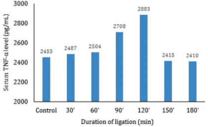
Mesenteric arterial ligation for 0, 30, 60, 90, 120, 150, 180 min was performed to evaluate the effect of duration of mesenteric artery ligation to the ratio of TNF-α/IL-10 in an AMI rat model. The TNF-α level was increased along with the duration of ligation in AMI rat model. The 120 min intervention group showed the highest level of TNF-α (FIGURE 1).
FIGURE 1. The effects of duration of mesenteric artery ligation to the serum TNF-α level
Serum TNF-α level after mesenteric artery ligation for 120 min was signiicantly different compared to that control and other mesenteric artery ligation groups (p<0.05). Whereas, the mesenteric artery ligation for 30, 60, 90, 150 and 180 min were not signiicantly different
compared to control (p>0.05) as presented in TABLE 1.
TABLE 1. Mean of serum TNF- α level between mesenteric artery ligation groups
Duration of ligation
(minutes) n Mean ± SE
0 (control) 5 2445 ± 33.16
30 5 2487 ± 43.85
60 5 2504 ± 14.93
90 5 2708 ± 68.23
120 5 2883 ± 108.50*
150 5 2415 ± 8.06
180 5 2410 ± 28.56
*) signiicantly different with p<0.05
The duration of mesenteric artery ligation affects the serum IL-10 level in AMI rat model. The 180 min mesenteric artery ligation group showed highest serum IL-10 level compared to the control and other mesenteric artery ligation groups. Other mesenteric artery ligation groups showed similar serum IL-10 level (FIGURE 2).
FIGURE 2. The effects of duration of mesenteric artery ligation to the serum IL-10 level
TABLE 2. Mean of serum IL-10 level between mesenteric artery ligation groups
Duration of ligation
(minutes) n Mean ± SE
0 (control) 5 1284 ± 15.60
30 5 1357 ± 25.57
60 5 1336 ± 41.69
90 5 1616 ± 31.84
120 5 1666 ± 55.46
150 5 1734 ± 89.31
180 5 3393 ± 281.86*
*) signiicantly different with p<0.05
and 150 min were not signiicantly different compared to control (p>0.05) as presented in TABLE 2.
Serum TNF-α and IL-10 levels ratio after
mesenteric artery ligation
Comparison between serum TNF-α and IL-10 levels in each mesenteric artery ligation is presented in FIGURE 5, whereas their serum TNF-α/IL-10 ratio is presented in TABLE 3. The serum TNF-α/IL-10 levels ratio decreased signiicantly after ligation for 90, 120, 150 and 180 min (TABLE 3). This study showed a balance between TNF-α/10, when TNF-α level increased, so did IL-10 level which shown in parallel line of those two cytokines.
FIGURE 3. Comparison between serum TNF-α and IL-10 levels in each mesenteric artery ligation
TABLE 3. Serum TNF-α/IL-10 level ratio after mesenteric artery ligation
Duration of ligation
(minutes) n TNF-α/IL-10 ratio
0 (control) 5 1.90
30 5 1.83
60 5 1.83
90 5 1.68*
120 5 1.73*
150 5 1.39*
180 5 0.71*
*) signiicantly different with p<0.05
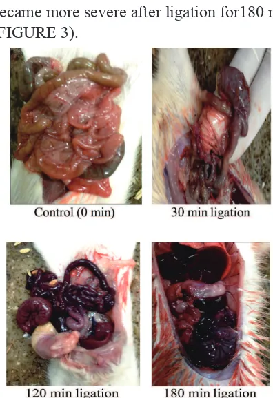
Intestinal tissue macroscopic examination after mesenteric artery ligation
Macroscopic examination showed control rats showed viable intestine. Whereas after mesenteric artery ligation for 30 min, intestine started to undergo edema and ischemia, after ligation 120 min intestine started to undergo necrosis and edema, and necrosis and edema became more severe after ligation for180 min (FIGURE 3).
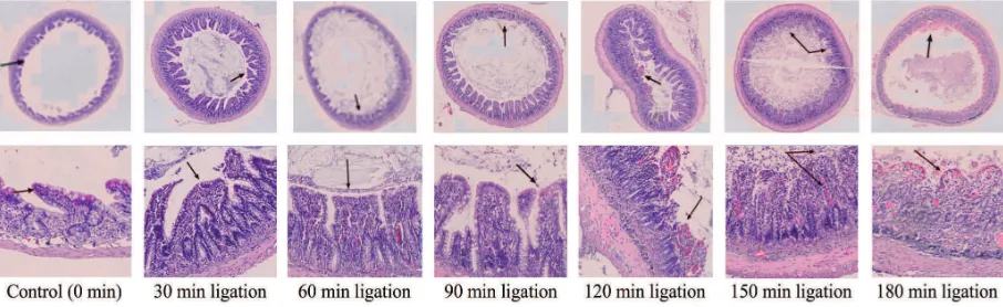
Histological examination of intestinal tissue after mesenteric artery ligation
Histological examination of intestinal tissue after mesenteric artery ligation showed normal intestinal villus, no lytic cell and normal vascularization in control rat (FIGURE 4).
Whereas after ligation for 30 min, edematous villus could was observed. Moreover, after ligation for 120 – 180 min edematous villus, damaged villus characterized by irregular shape, necrotic cell characterized by vascular rupture were observed (FIGURE 4).
FIGURE 4. Cellular necrotic level of intestinal tissue of AMI rats model after HE staining with 400x magniication
DISCUSSION
Acute mesenteric ischemia should be diagnosed earlier due to its high mortality rate. No speciic clinical inding and limitation of diagnostic test make early diagnose the most crucial part of this condition. Systemic inlammation response syndrome and sepsis is the main reason of AMI mortality rate. This study purpose to ind speciic serologic biochemical parameter as an early diagnose for AMI. In developing of early diagnose for AMI, several laboratory parameters that can be used for diagnosis of AMI such as ILs, TNF- α and CRP as a pro-inlammatory cytokine. An increasing level of those cytokines on AMI indicates systemic inlammatory response in early ischemia. This condition proves those potential cytokines not only can be used as a diagnosis, but more as a prognosis for patients with AMI.
This study found intestinal ischemia due to mesenteric artery ligation signiicantly
increased serum TNF- α level on 120 min post ligation compared to other intervention groups. Other study showed a similar result, elevating of serum TNF-α level on 60-120 min ligated mesenteric artery provided an early description for AMI prognosis. However, necrosis in intestinal tissue only can be seen 10-12 h after emersion of AMI symptoms.10
Previous study on rat model showed occlusion of superior mesenteric artery signiicantly elevates the serum TNF-α, IL-6, and IL-1 levels started from 2 h post ligation.
TNF-α level becomes important to prevent further post-ischemic damage to intestinal tissue. One of the main endogenous protective cytokines that can control TNF-α activity is IL-10. Previous study proved administration of phosphodiesterase type 4 (PDE4) might suppress chronic inlammation by inhibiting leukocyte recruitment, inhibiting increased level of TNF-α, and accelerating production of IL-10.15 TNF-α blockade and accelerating
production of IL-10 show capability of limiting injury due to inlammation followed by reperfusion of ischemic tissue. Anti TNF-α treatment may prevent cell death due to reperfusion injury. Predicted level of systemic TNF-α has major contribution to cell death due to ischemia as observed on animal model. PDE4 inhibitor is more potent to inhibit increased level of tissue TNF-α than systemic TNF-α.6
Inlammation due to post ischemia reperfusion eventually still becomes a focus in clinical studies to prevent further damage that leads to death caused by no speciic symptom and minimal clinical inding that leads to that condition.7 Prevention of AMI
should be done by measuring TNF-α/ IL-10 level ratio on certain duration. Alteration of those two cytokines is started since 120 min post-ischemia due to ligation of the superior mesenteric artery as observed in animal model. Endogenous control from intestinal cell shows reperfusion post intestinal ischemia cause tissue dysfunction. Ischemia may lead to elevation of free radical, activation of PAF, pro-inlammatory cytokine and chemokine, Kallikrein system that contribute to severity in post-reperfusion injury. It may be caused by increasing of neutrophil migration to the ischemic area. Another factor that may contribute to post-reperfusion injury severity is chronic inlammation due to activation of NFkB. Increasing in that kind of molecular
regulation will trigger elevation of TNF-α, which is a pro-inlammatory cytokine. This cytokine will stimulate post-reperfusion injury. Another theory suggests that IL-10 plays a role in inlammation homeostasis control. Previous study showed a balance between TNF-α/IL-10 level from the beginning of reperfusion process might prevent further tissue injury and cellular death. This may be a contradiction to a clinical condition where it is not only necessary to balance TNF-α/ IL-10 level in a post-reperfusion setting, furthermore, it should be used to suppress mortality rate due to IMA by early diagnose using TNF-α/IL-10 level ratio predictor.
This study showed a signiicant increasing TNF-α level was detected on 60-120 min post ligation. The data showed imbalance level of pro-inlammatory cytokine in ischemic area and systemic circulation. It could increase intestinal injury severity before reperfusion procedure, and decrease success rate of reperfusion procedure. This study found that before underwent reperfusion, IL-10 level, as an anti-inlammatory cytokine, elevated to prevent further TNF-α activity. TNF-α gradually increased on 60 min, and increased signiicantly on 120 min, while IL-10 started to increase on 60 min and signiicantly increased on 180 min. A new hypothesis for AMI prognosis is emerging, if the measurement for AMI is done earlier, obtained TNF-α/IL-10 levels ratio can be used as a baseline for further management. If the gap between TNF-α and IL-10 levels is increasing, tissue viability becomes worse, and so does reperfusion success rate. This study also showed that early diagnose on irst 120 min post-ischemia was a potential time to establish prognosis and tissue damage severity in order to establish the right management for AMI.
inlammation rate, which was important to avoid further tissue injury. This is expected to reduce the rate of cellular injury and death when undergoing reperfusion procedure to help restore homeostasis in ischemic area. Therapy mechanism by injecting exogenous IL-10 or anti-inlammatory drugs is providing a potential chance to suppress ischemic tissue cellular death rate, in a way of suppressing TNF-α level and stimulating elevation of IL-10 level as a negative feedback mechanism.
CONCLUSION
Mesenteric artery ligation can signiicantly increase serum TNF-α and IL-10 levels in a rat model of AMI. It proved that the increase of the TNF-α level is compensated by the increase the IL-10 level in rat model of acute mesenteric ischemia. It is assumed that when IL-10 level, that has protective effect as an inhibitor, higher than TNF-α as a proinlammatory cytokine on duration 150 min, means no more inlammatory or cells is dead. It is indicated that serum TNF-α/IL-10 levels ratio can be used as a biomarker candidate of prognosic factor management of AMI.
ACKNOWLEDGEMENTS
We would like to thank all technicians for their valuable assistance during laboratory works.
REFERENCES
1. Acosta S. Mesentric ischemia. Curr Opin Crit Care 2015; 21:171-8.
2. Clair DG, Beach JM. Mesentric ischemia. N Engl J Med 2016; 374:959-68.
3. Hussain D, Sarfraz SL, Baliga SK, Hartung R. Acute mesenteric ischemia: experience in tertiary care hospital. J Ayub Med Coll
4. Aouini F, Bouhaffa A, Baazaoui J, Khelii
S, Ben Maamer A, Hous N, et al. Acute
mesenteric ischemia: Study of predictive
factors of mortality. Tunis Med 2012; 90(7):533-6.
5. Huang HH, Chang YC, Yen DH, Kao WF, Chen JD, Wang LM, et al. Clinical factors and outcomes in patients with acute mesenteric ischemia in the emergency department. J
Chin Med Assoc 2005;68(7):299-306. http://
dx.doi.org/10.1016/S1726-4901(09)70165-0
6. Souza DG & Teixeira MM. The balance between the production of tumor necrosis factor-α and interleukin-10 determines
tissue injury and lethality during intestinal
ischemia and reperfusion. Mem Inst Oswaldo Cruz, Rio de Janeiro 2005; 100 (suppl. 1): Med 2004; 164(10):1054-62. http://dx.doi. org/10.1001/archinte.164.10.1054
8. Park SW, Chen SW, Kim M, Brown KM,
Kolls JK, D’Agati VD, et al. Cytokines
induce small intestine and liver injury after
renal ischemia or nephrectomy. Lab Invest
2011; 91(1):63-84. http://dx.doi.org/10.1038/
labinvest.2010.151
9. Mallick IH, Yang W, Winslet MC, Seifalian AM.
Ischemia-reperfusion injury of the intestine
and protective strategies against injury. Dig
Dis Sci 2004; 49(9):1359-77. http://dx.doi.
org/10.1023/B:DDAS.0000042232.98927.91 10. Ansari, P. Acute mesenteric ischemia. The
Merck Manual for Health Care Professional. New Jersey: USA. Merck and Sharp Dohme Corp, 2007.
11. Wu B, Ootani A, Iwakiri R, Fujise T, Tsunada
S, Toda S, et al. Ischemic preconditioning
mitochondria-dependent pathway in rat small intestine. Am J Physiol Gastrointest Liver Physiol 2004; 286(4):580-7. http://dx.doi. org/10.1152/ ajpgi.00335.2003
12. Wiesner W, Khurana B, Ji H, Ros PR. CT of acute bowel ischemia. Radiology 2003; 226(3):635-50. http://dx.doi.org/10.1148/ radiol.2263011540
13. Zhang Y, Leng YF, Xue X, Zhang Y, Wan T, Kang YQ. Effects of penehyclidine
hydrochloride in small intestinal damage
caused by limb ischemia-reperfusion. World
J Gastroenterol 2011; 17(2):254-9. http:// dx.doi.org/10.3748/wjg.v17.i2.254
14. Wullaert A, Bonnet MC, Pasparakis M. NF-Κβ in the regulation of epithelial homeostasis
and inlammation. Cell Res 2011; 21:146-58. http://dx.doi.org/10.1038/cr.2010.175