Biosaintifika 10 (3) (2018) XXX-XXX
Biosaintifika
Journal of Biology & Biology Educationhttp://journal.unnes.ac.id/nju/index.php/biosaintifika
Plantlets Regeneration from Crown Bud Slicing of Pineapple (
Ananas
comosus
)
Zulkarnain, Neliyati, Eliyanti
DOI: http://dx.doi.org/10.15294/biosaintifika.v10i3.8079
Faculty of Agricultural, Universitas Jambi, Indonesia
Abstract
Pineapple propagation by lateral shoots, suckers or crowns is often confronted with limited number of regenerated seedlings and high diversity in flowering and fruit formation. In order to solve this problem, this study offer an alternative method by using tissue culture techniques. This study aimed to determine the effect of growth regulators on plantlet regeneration from bud slicing of pineapple cv. Tangkit. Four levels of 2.4-D (0.0, 0.001, 0.01 and 0.1 ppm) in combination with BA (0.0, 0.1, 1.0 and 10.0 ppm) were tested on solid MS medium.Cultures were incubated in total darkness for a week followed by transfer to 16-hour photoperiod. Results showed that explants treated with 2,4-D and/or BA succeeded in regenerating adventitious shoots. Average leaf number did not differ significantly among treatments (P = 0.60). Highest leaf number (2.99 ± 0.23) was obtained on medium with 0.01 ppm 2,4-D without BA, followed by 0.1 ppm 2,4-D without BA (2.85 ± 0.33). Mean-while, roots were only formed on medium with 0.1 ppm 2.4-D without BA (4.2 ± 0.37 per shoot). Thus, complete plantlets were regenerated only on medium sup-plemented with 0.1 ppm 2,4-D without BA. The growth of plantlets was relatively uniform, and plantlet acclimatization succeeded 100% on Jiffy pots. The finding of optimum concentration of 2.4-D and BA in this study is important to develop standard protocol for in vitro propagation of pineapple cv. Tangkit. Thus, the benefit of producing seeds in large quantities and relatively uniform in growth is made pos-sible through tissue culture technique.
How to Cite
Zulkarnain, Z., Neliyati, N., & Eliyanti, E. (2018). Plantlets Regeneration from Crown Bud Slicing of Pineapple (Ananas comosus). Biosaintifika: Journal of Biology & Biology Education, 10(3), XXX-XXX.
History Article
Received 29 November 2016 Approved 19 September 2018 Published 31 December 2018
Keywords 2.4-dichlorophenoxy-acetic acid; benzyladenine;
Ananas comosus; in vitro
culture; micropropagation.
Correspondence Author:
Kampus Pinang Masak, Mendalo Indah, Jambi 36361 E-mail: [email protected]
p-ISSN 2085-191X e-ISSN 2338-7610
Zulkarnain et al. / Biosaintifika 10 (3) (2018) XXX-XXX
karnain & Neliyati, 2017, Zuraidaet al., 2011). In an earlier study of pineapple tissue culture, Mapes (1973) was successfully regene-rated plants from shoot culture, while Sitaet al. (1974) obtained plantlet from meristem culture of Smooth Cayenne cultivar. Regeneration of pineapple plants through somatic embryogene-sis has also been reported by a number of rese-archers (Daquinta et al., 1996, Sripaorayaet al., 2003). Soneji et al. (2002) used leaf segments as explant materials. Most protocols, however, used dormant axillary buds from fruit crowns as planting materials (Atawiaet al., 2016, Rahman et al., 2001, Sonejiet al., 2001, Sripaorayaet al., 2003). These dormant buds, however, need to be stimulated to grow into maturation stage in order to regenerate plants. For this reason, auxin and cytokinin should play an important role in exp-lant organogenic response during the culture. The dichlorophenoxyacetic acid (2,4-D) and benzyla-denine (BA) are two widely used auxin and cy-tokinin in tissue culture system of many plants (Kristantiet al., 2013, Sariet al., 2014, Wahyuni et al., 2017).
This paper describes a simple in vitro pro-cedure for obtaining pineapple cv Tangkit plants using axillary buds from crown slicing as initial explant materials. This procedure will facilitate clonal propagation techniques of this pineapple cultivar in order to provide uniform quality seeds.
METHODS
Preparation of plant material
Fruit crowns were isolated from pineapp-le cv Tangkit obtained from farmers’ gardens in Tangkit Baru village, Muaro Jambi district. All leaves attached to crowns were removed to obtain a “naked crown” with clearly visible dormant axillary buds. The crowns were then sliced hori-zontally into three parts to obtain a section of the base, the middle and the tip pieces. The middle pieces were used as the source of the explant ma-terial, while the other two parts were discarded. The pieces were then sterilized by immersion in 0.02 % HgCl2 solution for 5 minutes, then rinsed three times with sterile distilled water. After the sterilization process was completed, the pieces were cut into small cube block of about 0.5 × 0.5 × 0.5 cm with a dormant axillary bud. The blocks were then used as planting material and ready to be cultured in the medium within culture flasks.
Medium composition and environmental conditions
Culture medium used was MS composi-tion (Murashige & Skoog, 1962), supplemented
INTRODUCTION
Pineapple (Ananas comosus (L.) Merr.) is one of important fruit commodities from Brome-liaceae family cultivated in Jambi Province. The cultivation of pineapple in Jambi has become a priority since 1983, with Tangkit Baru Village, Muaro Jambi Regency as the production cent-re (Zulkarnain, 2017). The Tangkit cultivar has been registered in the Ministry of Agriculture Re-public of Indonesia as a local variety of Jambi Province, with registration No. 40/PVL/2009. The fruit has soft aroma, sweet and fine texture with little fibre. The shelf life of this fruit may last up to seven days after harvest. Another advanta-ge of pineapple cv Tangkit is that their ability to grow on peat soils with pH below 5.0.
In general, pineapple fruits are high in fib-re, an important component of a healthy diet that improve the digestion system. They also contain a good array of vitamins and minerals including vitamins A and C, folic acid, calcium, and man-ganese (Hossainet al., 2015). Another benefit of pineapple fruit is to improve skin and hair health, increase energy and reduce body weight. Many studies have suggested that consuming pineapp-les may relieve osteoarthritis and diarrhea (Pavan et al., 2012), decrease the risk of obesity and its associated cardiovascular dysfunction (Ahmed, 2016), besides it also has a strong antioxidant capacity (Luet al., 2014). These benefits, along with the sweet taste, fresh aroma and attractive shape make pineapple attracts many people, both for fresh consumption or processing industries such as jam, syrup and canned pineapple.
Pineapples cv Tangkit are routinely pro-pagated vegetatively by means of lateral shoots, basal suckers or crowns as they are parthenocar-py (do not producing seeds). However, vegetati-ve propagation using lateral shoots, suckers and crowns are confronted with the limited number of propagules produced. Crowns are always car-ried along with the fruit at the time of marketing, while suckers are often available in limited num-ber. The size of the seedling often varies great-ly, giving rise to high diversity in flowering time and fruit formation (Sripaoraya et al., 2003). In addition, being vegetatively propagated, conven-tional hybridization for better pineapple varieties are cumbersome and time consuming (Mhatre, 2007). Therefore, an alternative of pineapple pro-pagation method that can be used is by tissue cul-ture techniques. Many authors have reported suc-cessful production of pineapple via tissue culture system during the last few years (Atawiaet al., 2016, Danso et al., 2008, Yapoet al., 2011,
Zulkarnain et al. / Biosaintifika 10 (3) (2018) XXX-XXX
with vitamins (myo-inositol, glycine, nicotinic acid, pyridoxine-HCl and thiamine-HCl) and 3% sucrose (w/v). Plant growth regulators 2,4-dichlo-rophenoxyacetic acid (2,4-D) (0.0; 0.001; 0.01 and 0.1 ppm) and benzyl adenine (BA) (0.0; 0.1; 1.0 and 10 ppm) were added to the medium, pro-ducing 16 treatment combinations. The pH of the medium was set to 5.8 ± 0.02 before compacting with 0.8% (w/v) agar (Difco Bacto) and distri-buted into culture flasks each of 10 mL. The me-dium was then autoclaved at a pressure of 1.06 kg cm-2 and temperature of 121 °C for 15 minutes.
One explant was cultured on medium in culture flask of each treatment combinations. The cultures were then incubated in total dark-ness for 7 days prior to shifted on shelves within the culture room at 25 ± 1 oC and photoperiod
of 16 hours per day under a light intensity of 50 μmol m-2 s-1 from white TL lamps (Phillips
Indo-nesia).
Acclimatization
The regenerated plantlets (buds with shoots and roots) were removed from culture flasks and transplanted in Jiffy pots (Jiffy-7 ™) which had been soaked in water for 15 minutes. Each pot was planted one plantlet. The plantlets were then maintained in a shade house with light intensity ranging from 100 to 170 μmol m-2 s-1, natural
pho-toperiod and day/night temperature of 25/18 oC
and relative humidity of approximately 80%.
Design and Data analysis
The experiment was arranged in a simple randomized design with five replications. Para-meters suchs as percentage of explants regenera-ting adventitious shoots, root formation, number of leaves and callus proliferation were observed every week for 16 weeks. Data obtained were cal-culated using decriptive statictics (Microsoft Cor-poration, 2016) and standard errors (SEs) were determined.
RESULTS AND DISCUSSION
While explants cultured in medium wit-hout growth regulator did not show any progress until the end of the trial period (Table 1), all ex-plants cultured in medium equipped with 2,4-D or BA or combination of both regenerated adven-titious shoots (Figure 1). This proves that the re-generation of adventitious shoots from dormant axillary buds of Tangkit pineapple requires the presence of auxin and/or cytokinins in culture medium, with 2,4-D as a critical factor for plant-let growth. Some earlier researchers also reported
the need for auxin and cytokinin involvement in inducing morphogenetic responses in a number of Bromeliaceae plants. For example Dyckia dis-tachya (Pompelli & Guerra, 2005), Tillandsia eizii
(Pickens et al., 2006), Vriesea scalaris (da Silvaet al., 2009), Vriesea reitzii (Guerra & Dal Vesco, 2010), Aechmea blanchetiana and A. distichantha
(Santa-Rosaet al., 2013) were successfully rege-nerated by adventitious shoot formation under in vitro system. In all of these successes, the presen-ce of cytokinin (BA) was proven to be crucial in the formation of adventitious shoots.
Figure 1. Adventitious shoots formation on
crown slicing explants of pineapple cv Tangkit. A = 0.001 ppm 2,4-D + 0.01 ppm BA; B = 0.01 ppm 2,4-D + 10.0 ppm BA; C = 0.001 ppm 2,4-D + 1.0 ppm BA; D = 0.01 ppm 2,4-D + 0.1 ppm BA.
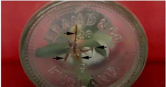
Root formation with an average number of 4.2 ± 0.37 per shoot occured only in the addi-tion of 0.1 ppm 2.4-D without BA, and they are yellowish-white in colour (Figure 2). This is in ac-cordance with report of Sripaorayaet al. (2003) regarding tissue culture of pineapple cv Phuket (belongs to Queen group similar to cv Tangkit). Therefore, in our study, complete plantlets regen-eration occured only from explants cultured on medium with 2.4-D alone.
Figure 2. Roots (indicated by arrowheads)
formed on adventitious shoot grown from crown bud slicing of pineapple cv Tangkit treated with 0.1 ppm 2,4-D alone.
In general, the use of synthetic auxin in tis-sue culture system was aimed at accelerating root
Zulkarnain et al. / Biosaintifika 10 (3) (2018) XXX-XXX
formation and increasing rooting rate. Rovereet al. (2013) and Yuet al. (2017) suggested that root formation in tissue culture will take place when auxin is present in culture medium. The presence of auxin, either alone or in combination with a low concentration of cytokinin is a critical factor for the proliferation of root primordia on cultu-red explants (Zulkarnain, 2009). This is probably due to their participation in the regulation of cell cycling, division, and differentiation (Tapingkae et al., 2012, Zulkarnainet al., 2015).
The effect of 2,4-D and BA in the range of concentrations tested did not give any significant difference in leaves formation (P = 0.60). The ave-rage number of leaves ranged from 2.57 ± 0.50 to 2.99 ± 0.23. These results indicate that plantlet growth does not show high diversity, so, this in
vitro propagation method is very appropriate
ef-fort to obtain pineapple with a uniform growth. This is apparently more advantageous compared to conventional method by using suckers. Sripa-orayaet al. (2003) claimed that propagation using suckers will result in non-uniform growth and flo-wering times among progenies due to differences in suckers age at planting time.
All cultured explants did not produce
cal-lus, though 2,4-D is known as a strong auxin that frequently used to induce callus formation in a number of plant species such as sweet po-tato (Oggemaet al., 2007), wheat (Rashidet al., 2009), sugar cane (Tahiret al., 2011), gendarussa (Wahyuni et al., 2017) and orchid (Budisantosoet al., 2017). This finding, however, is in accordance with report by (Sonejiet al., 2002) where MS me-dium supplemented with 2,4-D was not conduc-tive to induce callus growth on leaf explants of pineapple. This probably due to the concentration of 2,4-D was still too low to induce callus prolife-ration. Ikeuchiet al. (2013) suggested that in ge-neral, callus proliferation will occur when auxin and cytokinin present in intermediate rasio. High rasio of auxin-to-cytokinin or cytokinin-to-auxin will lead to root and shoot regeneration, respec-tively. In pineapple cv Phuket, Sripaoraya et al. (2003) found that callus was formed on leaf ex-plants with the presence of 2,4-D concentrations of 0.5 to 2.0 ppm, while in our experiment the highest concentration of 2.4-D was only 0.1 ppm. This result confimed the report of De Silva et al. (2008) who found that 2,4-D application of up to 54.3 ppm failed to induce calli from meris-temic globular bodies of pineapple cv Moris
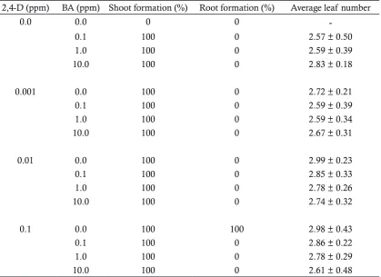
Table 1. The effect of 2,4-D and BA on shoots, roots and leaves formation on crown slicing explants
of pineapple cv Tangkit.
2,4-D (ppm) BA (ppm) Shoot formation (%) Root formation (%) Average leaf number
0.0 0.0 0 0
-0.1 100 0 2.57 ± 0.50
1.0 100 0 2.59 ± 0.39
10.0 100 0 2.83 ± 0.18
0.001 0.0 100 0 2.72 ± 0.21
0.1 100 0 2.59 ± 0.39
1.0 100 0 2.59 ± 0.34
10.0 100 0 2.67 ± 0.31
0.01 0.0 100 0 2.99 ± 0.23
0.1 100 0 2.85 ± 0.33
1.0 100 0 2.78 ± 0.26
10.0 100 0 2.74 ± 0.32
0.1 0.0 100 100 2.98 ± 0.43
0.1 100 0 2.86 ± 0.22
1.0 100 0 2.78 ± 0.29
10.0 100 0 2.61 ± 0.48
± Standard Error. Each treatment consists of 5 replications.
Zulkarnain et al. / Biosaintifika 10 (3) (2018) XXX-XXX
and Josapine. Thus, we believe that the very low concentration of 2,4-D employed in culture me-dium was the key factor causing the absence of callus formation in this trial.
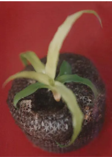
Figure 3. In vitro plantlet of pineapple cv Tangkit
transplanted onto Jiffy pot during acclimatiza-tion process.
Acclimatization was done after the plant-lets were large enough (at 16 weeks after culture initiation). Only plantlets generated on medium supplemented with 0.1 ppm 2,4-D alone were subjected to acclimatization as they have root system. They were transplanted onto Jiffy pots ready-made medium (Figure 3). All explants ac-climatized on Jiffy pot showed good growth and development, so that they can be transferred to polybag with soil for further planting in the field. The acclimatization rate of 100% indicated that both environmental conditions and substrate used to support plantlet growth have met their requirements.
The finding of optimum concentration of 2,4-D and BAP in this study is useful for in vitro shoots formation from crown bud slicing of pine-apple. The shoots can be subcultured to new me-dium to develop multiple shoots, or transferred to rooting medium to grow into complete plantlets which can be acclimatisized for field evaluation. Through this study of in vitro propagation techni-que, a standard protocol for mass propagation of pineapple cv. Tangkit can be developed. The
research is very important to provide the needs of quality seeds, because normal vegetative pro-pagation of pineapple is hampered by the limited availability of propagation materials.
CONCLUSION
Propagation of pineapple cv Tangkit is made possible through tissue culture techniques using crown bud slicing as explants source. This is a new approach to the effort of multiplying pineapple plants in large quantities. In addition, plants generated through tissue culture were pro-ven to be relatively uniform in growth, making it very profitable in cultivation.
REFERENCES
Ahmed, M. M. (2016). Pineapple Juice Ameliorates the High Fat Diet-induced Alterations in Car-diac Gene Expression Pattern in Male Rats.
International Journal of Biochemistry Research & Review, 15 (4), 1-11.
Atawia, A. R., F. M. A. EL-Latif, S. F. EL-Gioushy, S. S. Sherif & O. M. Kotb. (2016). Studies on Mi-cropropagation of Pineapple (Ananas comosus
L.). Middle East Journal of Agriculture Research,
5 (2), 224-232.
Budisantoso, I., N. Amalia & K. Kamsinah. (2017). In Vitro Callus Induction from Leaf Explants of Vanda sp. Stimulated by 2,4-D. Biosaintifika: Journal of Biology & Biology Education, 9 (3), 492-497.
da Silva, A. L. L., E. T. H. Franco, E. B. Dornelles, C. L. R. Bortoli & M. Quoirin. (2009). In Vi-tro Multiplication of Vriesea scalaris E. Morren (Bromeliaceae). Iheringia Série Botânica, 64 (2), 151-156.
Danso, K. E., K. O. Ayeh, V. Oduro, S. Amiteye & H. M. Amoatey. (2008). Effect of 6-Benzylamino-purine and Naphthalene Acetic Acid on In Vitro Production of MD2 Pineapple Planting Materials. World Applied Sciences Journal, 3 (4), 614-619.
Daquinta, M. A., A. Cisneros, Y. Rodríguez, M. Es-calona, M. Pérez, I. Luna & C. G. Borroto. (1996). Embríogénesis somática en pina ( Anan-as comosus (L.) Merr.). Acta Horticulturae, 425, 243-246.
De Silva, M. A. Kadir, M. A. Aziz & S. Kadzimin. (2008). Callus Induction in Pineapple (Ananas comosus L.) cv. Moris and Josapine. International Journal of Agricultural Research, 3 (4), 261-267. Guerra, M. & L. Dal Vesco. (2010). Strategies for the
Micropropagation of Bromeliads. In S. M. Jain & S. J. Ochatt [eds.], Protocols for In Vitro
Propagation of Ornamental Plants, 47-66. Hu-mana Press, New York.
Hossain, M. F., S. Akhtar & M. Anwar. (2015). Nu-tritional Value and Medicinal Benefits of
Zulkarnain et al. / Biosaintifika 10 (3) (2018) XXX-XXX
apple. International Journal of Nutrition and Food Sciences, 4 (1), 84-88.
Ikeuchi, M., K. Sugimoto & A. Iwase. (2013). Plant Callus: Mechanisms of Induction and Repres-sion. The Plant Cell, 25, 3159-3173.
Kristanti, I., N. A. Habibah & L. Herlina. (2013). Op-timization of 2,4-D Concentration, BA, and Long Irradiation to Promote Regeneration Buds from Callus of Soybean. Biosaintifika: Journal of Biology & Biology Education, 5 (1), 50-57.
Lu, X.-H., D.-Q. Sun, Q.-S. Wu, S.-H. Liu & G.-M. Sun. (2014). Physico-Chemical Properties, Antioxidant Activity and Mineral Contents of Pineapple Genotypes Grown in China. Mol-ecules, 19, 8518-8532.
Mapes, M. O. (1973). Tissue Culture of Bromeliads. Proceedings of International Plant Propagator Society, 23, 47-55. . Tissue culture of bromeli-ads. Proceedings of International Plant Propa-gator Society, 23, 47-55.
Mhatre, M. (2007). Micropropagation of Pineapple,
Ananas comosus (L.) Merr. In S. M. Jain & H. Häggman [eds.], Protocols for Micropropa-gation of Woody Trees and Fruits, 499–508. Springer, Dordrecht, The Netherland.
Microsoft Corporation. (2016). Microsoft Office 365 ProPlus Version 1708. Microsoft Corporation, New York, USA.
Murashige, T. & F. Skoog. (1962). A revised medium for rapid growth and bio assays with tobacco tissue cultures. Physiologia Plantarum, 15, 473-497.
Oggema, J. N., M. G. Kinyua & J. P. Ouma. (2007). Optimum 2,4-D Concentration Suitable for Embryogeneic Callus Induction in Local Ke-nyan Sweet Potato Cultivars. Asian Journal of Plant Sciences, 6 (3), 484-489.
Pavan, R., S. Jain, Shraddha & A. Kumar. (2012). Properties and Therapeutic Application of Bromelain: A Review. Biotechnology Research In-ternational, 2012, 1-6.
Pickens, K. A., J. Wolf, J. M. Affolter & H. Y. Wetz-stein. (2006). Adventitious Bud Development and Regeneration in Tillandsia eizii. In Vitro Cellular & Developmental Biology - Plant, 42 (4), 348-353.
Pompelli, M. F. & M. P. Guerra. (2005). Microprop-agation Enables the Mass PropMicroprop-agation and Conservation of Dyckia distachya Hassler. Crop Breeding and Applied Biotechnology, 5, 117-126. Rahman, K. W., M. N. Amin & M. A. K. Azad.
(2001). In Vitro Rapid Clonal Propagation of Pineapple, Ananas comosus (L.) Merr. Plant Tis-sue Culture, 11, 47-53.
Rashid, U., S. Ali, G. M. Ali, N. Ayub & M. S. Masood. (2009). Establishment of an Efficient Callus Induction and Plant Regeneration System in Pakistani Wheat (Triticum aestivum) Cultivars.
Electronic Journal of Biotechnology, 12 (3), 1-12. Rovere, F. D., L. Fattorini, S. D’Angeli, A. Veloccia, G.
Falasca & M. M. Altamura. (2013). Auxin and
Cytokinin Control Formation of the Quiescent Centre in the Adventitious Root Apex of Ara-bidopsis. Annals of Botany, 112 (7), 1395-1407. Santa-Rosa, S., F. V. D. Souza, Á. M. Vidal, C. A. da
S Ledo & J. R. F. de Santana. (2013). Micro-propagation of the Ornamental Vulnerable Bromeliads Aechmea blanchetiana and Aechmea distichantha. Horticultura Brasileira, 31, 112-118. Sari, N., E. S. R & Sumadi. (2014). Optimization of
Type and Concentration of PGR in Embryo-genic Callus Induction and Regeneration into plantlets on Carica pubescens (Lenne & K.Koch).
Biosaintifika: Journal of Biology & Biology Educa-tion, 6 (1), 52-59.
Sita, G. L., R. Singh & C. P. A. Layer. (1974). Plantlets through shoot tip culture in pineapple. Current Science, 43, 724.
Soneji, J. R., P. S. Rao & M. Mhatre. (2001). Soma-clonal Variation in Micropropagated Dormant Axillary Buds of Pineapple (Ananas comosus L., Merr.). The Journal of Horticultural Science and Biotechnology 77 (1), 28-32.
Soneji, J. R., P. S. Rao & M. Mhatre. (2002). In Vitro
Regeneration from Leaf Explants of Pineapple (Ananas comosus L., Merr.). Journal of Plant Bio-chemistry and Biotechnology, 11 (2), 117-119. Sripaoraya, S., R. Marchant, J. B. Power & M. R.
Dav-ey. (2003). Plant regeneration by somatic em-bryogenesis and organogenesis in commercial pineapple (Ananas comosus L.). In Vitro Cellular and Developmental Biology - Plant, 39, 450-454. Tahir, S. M., K. Victor & S. Abdulkadir. (2011). The
Effect of 2, 4-Dichlorophenoxy Acetic Acid (2,4-D) Concentration on Callus Induction in Sugarcane (Saccharum officinarum). Nigerian Journal of Basic and Applied Science, 19 (2), 213-217.
Tapingkae, T., Z. Zulkarnain, M. Kawaguchi, T. Ikeda & A. Taji. (2012). Somatic (Asexual) Proce-dures (Haploids, Protoplasts, Cell Selection) and Their Applications. In A. Altman & P. M. Hasegawa [eds.], Plant biotechnology and agri-culture: Prospects for the 21st century, 139-162.
Academic Press, Oxford.
Wahyuni, D. K., P. Andriani, A. N. M. Ansori & E. S. W. Utami. (2017). Callus Induction of Gen-darussa (Justicia gendarussa) by Various Con-centration of 2,4-D, IBA, and BAP. Biosainti-fika: Journal of Biology & Biology Education, 9 (3), 402-408.
Yapo, E. S., T. H. Kouakou, M. Kone, J. Y. Kouadio, P. Kouame & J.-M. Merillon. (2011). Regen-eration of Pineapple (Ananas comosus L.) Plant through Somatic Embryogenesis. Journal of Plant Biochemistry and Biotechnology, 20 (2), 196-204.
Yu, J., W. Liu, J. Liu, P. Qin & L. Xu. (2017). Auxin Control of Root Organogenesis from Callus in Tissue Culture. Frontiers in Plant Science, 8, 1385.
Zulkarnain, Z. (2009). Kultur Jaringan Tanaman: Solusi Perbanyakan Tanaman Budidaya. Jakarta, IDN:
Zulkarnain et al. / Biosaintifika 10 (3) (2018) XXX-XXX
PT Bumi Aksara.
Zulkarnain, Z. (2017). Budidaya Buah-Buahan Tropis. Yogyakarta, IDN: Deepublish.
Zulkarnain, Z. & N. Neliyati. (2017). The Effect of NAA and BAP on Tissue Culture of Tangkit Pineapple (Ananas comosus (L.) Merr. cv. Tang-kit). Biospesies, 10 (1), 1-10.
Zulkarnain, Z., T. Tapingkae & A. Taji. (2015). Ap-plications of In Vitro Techniques in Plant Breeding. In J. M. Al-Khayri, S. M. Jain & D. V. Johnson [eds.], Advances in Plant Breeding
Strategies: Breeding, Biotechnology and Mo-lecular Tools, 293-328. Springer International Publishing Switzerland, Switzerland.
Zuraida, A. R., A. H. Nurul Shahnadz, A. Harteeni, S. Roowi, C. M. Z. Che Radziah & S. Sreeraman-an. (2011). A Novel Approach for Rapid Mi-cropropagation of Maspine Pineapple (Ananas comosus L.) Shoots using Liquid Shake Culture System. African Journal of Biotechnology, 10 (19), 3859-3866.