저작자표시-비영리-변경금지 2.0 대한민국 이용자는 아래의 조건을 따르는 경우에 한하여 자유롭게
l 이 저작물을 복제, 배포, 전송, 전시, 공연 및 방송할 수 있습니다. 다음과 같은 조건을 따라야 합니다:
l 귀하는, 이 저작물의 재이용이나 배포의 경우, 이 저작물에 적용된 이용허락조건 을 명확하게 나타내어야 합니다.
l 저작권자로부터 별도의 허가를 받으면 이러한 조건들은 적용되지 않습니다.
저작권법에 따른 이용자의 권리는 위의 내용에 의하여 영향을 받지 않습니다. 이것은 이용허락규약(Legal Code)을 이해하기 쉽게 요약한 것입니다.
Disclaimer
저작자표시. 귀하는 원저작자를 표시하여야 합니다.
비영리. 귀하는 이 저작물을 영리 목적으로 이용할 수 없습니다.
변경금지. 귀하는 이 저작물을 개작, 변형 또는 가공할 수 없습니다.
수의학박사학위논문
Generation of transgenic cloned dogs using adipose-derived mesenchymal
stem cells
지방줄기세포를 이용한 형질전환 복제개의 생산
2016 년 2 월
서울대학교 대학원
수의학과 임상수의학 전공
오 현 주
Generation of transgenic cloned dogs using adipose-derived mesenchymal
stem cells
지방줄기세포를 이용한 형질전환 복제개의 생산
지도교수 이 병 천
이 논문을 수의학 박사학위논문으로 제출함
2015 년 10 월 서울대학교 대학원 수의학과 임상수의학 전공
오 현 주
오현주의 박사학위논문을 인준함 2015 년 12 월
위 원 장 (인)
부위원장 (인)
위 원 (인)
위 원 (인)
위 원 (인)
Generation of transgenic cloned dogs using adipose-derived mesenchymal
stem cells
by Hyun Ju Oh
A THESIS SUBMITTED IN PARTIAL
FULFILLMENT OF THE REQUIREMENT FOR THE DEGREE OF DOCTOR OF PHILOSOPHY
in
Veterinary Clinical Sciences
Department of Veterinary Medicine, Graduate School Seoul National University
We accept this thesis as confirming to the required standard
Seoul National University December 2015 © Hyun Ju Oh
Declaration
This thesis is submitted by the undersigned for examination for the degree of Doctor of Philosophy to the Seoul National University. This thesis has not been submitted for the purposes of obtaining any other degree or qualification from any other academic institution.
I hereby declare that the composition and experiment of this thesis and the work presented in it are entirely my own.
Hyun Ju Oh
i
Generation of transgenic cloned dogs using adipose-derived mesenchymal
stem cells
Hyun Ju Oh
(Supervisor: Byeong Chun Lee, D.V.M., Ph.D.) Veterinary Clinical Sciences
Department of Veterinary Medicine, Graduate School Seoul National University
ABSTRACT
Since the birth of the first cloned dog ‘Snuppy’, valuable canids were produced by SCNT using adult somatic cells. In addition, genetically modified dogs were generated by SCNT using fetal fibroblasts. Fetal fibroblasts are preferred as nucleus donors for SCNT used in producing transgenic dogs because they have excellent proliferative ability, are capable of being genetically modified, and have the ability to produce live offspring. However, the donor transgenic cells become senescent and
ii
unusable because stable transgene-expression, homologous recombination or multiple transfections require a long time for in vitro culture. As an alternative to fetal cells, recent reports indicate that some mesenchymal stem cells (MSCs) lines can be maintained sufficiently long enough for homologous recombination events to take place. MSCs can proliferate for many passages in culture and show constant growth.
Furthermore, MSCs have the ability to give rise several differentiated cell types. Thus the object of this study was to determine whether canine adipose–derived mesenchymal stem cells (cASCs) can be a suitable donor cell for producing transgenic cloned dogs. In several laboratory animals and humans, ASCs are of considerable interest because they are easy to harvest and can generate a huge number of cells from a small quantity of adipose tissue. ASCs have applications in various research areas, such as cell therapy and tissue engineering especially in bone reconstruction. In order to cASCs in SCNT, this study compared cellular proliferation rate, viability, cellular size and expression patterns of genes related to pluripotency and epigenetic modification between canine fetal fibroblasts (cFFs) and cASCs. The cFFs were established from fetuses of pregnant beagle at the 28th day. The cASCs were isolated from subcutaneous adipose tissue collected from the inguinal region of a healthy dog.
The cASCs were characterized through flow cytometry to be positive for CD29, CD44, CD73, CD90 and CD105, but negative for CD31, CD34 and CD45. Proliferation pattern, cellular viability as well as cell size at each passage of cFF and cASC were compared when the culture reached confluence. In addition, real time-PCR was performed to investigate different mRNA transcripts expression in both cell lines.
iii
Moreover, the cASCs were evaluated as a potential donor cell using interspecies SCNT (iSCNT); cASCs were cultured in two different culture media (RCMEP or DMEM) and used for iSCNT. Next, to generate transgenic cloned dog, cASCs were established from a transgenic cloned beagle produced by nuclear transfer of canine fetal fibroblasts modified genetically with a red fluorescent protein (RFP) gene. The cASCs expressed RFP gene and cell-surface marker characteristics of MSCs, including CD29, CD44 and thy1.1. Furthermore, the cASCs underwent osteogenic, adipogenic, myogenic, neurogenic and chondrogenic differentiation when exposed to specific differentiation- inducing conditions.
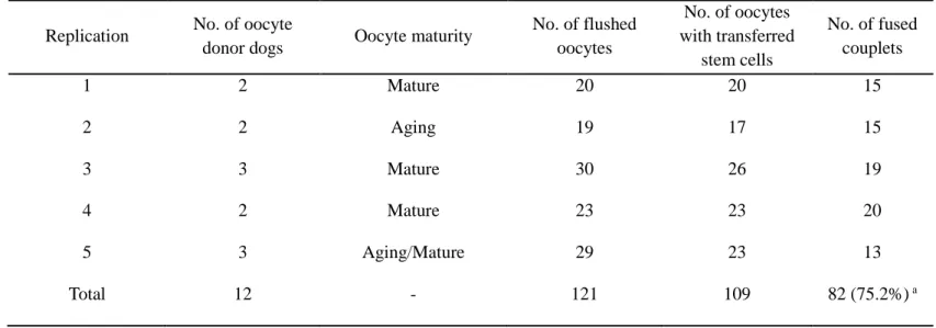
Isolated cASCs were used for SCNT and after embryo transfer into recipient, RFP-expressing transgenic recloned beagle pups (Magic) were produced by nuclear transfer of cASCs derived from a transgenic cloned beagle (Ruppy1). Another purpose of this study is to determine the degree of genetic identity between the cloned (Ruppy1) and recloned (Magic) dogs and evaluated whether the RFP expression and CMV promoter methylation of these two transgenic dogs are age-dependent. To produce a transgenic dog that expresses neuron specific transgene, human synapsin 1 promoter as primarily neuron selective was chosen. Synapsin 1-RFP (SYN1-RFP) was introduced into cASCs via lentiviral vector infection. The SYN1-RFP cells were injected into enucleated in vivo matured dog oocytes and fused by electric stimulation. The fused- couplets were transferred into the uterine tube of five naturally estrus-synchronized surrogates.
iv
As results, the cFFs and cASCs differed in the number of generation but not in doubling times at all passages. The mean cell size of cASCs was significantly smaller than that of cFFs. Cellular viability and apoptosis were significantly lower in cASCs when compared to passage-matched cFFs. The level of HDAC1 transcript in cASCs was significantly higher than in cFFs, but expression of DNMT1 was not different between the two groups. OCT4 and SOX2 transcripts showed significantly higher expression in cASCs than in cFFs. Thus, canine adipose-derived stem cells (cASCs) are promising as donor cells for SCNT. With this in mind, cASCs were evaluated as a potential donor cell using interspecies SCNT (iSCNT). RCMEP cultured cells contained significantly higher amount of SOX2, NANOG, OCT4, DNMT1 and MeCP2 than DMEM cultured cells (P <0.05). However, there was no significant difference in the rate of development to blastocysts between the two groups. Thus, these results showed that altering gene expression levels in donor cells by changing the culture medium did not influence subsequent in vitro development of cloned embryos.
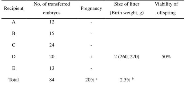
In SCNT for generating transgenic cloned dog, one dog among five (20%) maintained pregnancy and subsequently gave birth of two healthy cloned pups. The present study demonstrated for the first time the successful production of transgenic cloned beagles by nuclear transfer of cASCs derived from a transgenic cloned beagle. Moreover, the degree of genetic identity between the cloned and recloned dogs showed that both cloned dogs carried a single copy and same integration site of the RFP gene. The transgene protein quantity of both transgenic dogs, showed no significant difference in the relative RFP expression between the 1-year-old Ruppy1 and the 1-year-old Magic.
v
Also, transgene protein levels increased with aging of the two dogs, while promoter methylation status decreased with age. Gene expression and promoter methylation showed similar opposite profiles during growth of the two transgenic dogs. Lastly, neuron specific transgene-expressed dogs were generated by SCNT and three cloned pups (SYN1-RFP A, SYN1-RFP B, and SYN1-RFP C) were produced by natural delivery or C-sec. One of them is still alive, healthy and does not show any abnormalities.
In this thesis, cASCs have superior proliferation patterns, epigenetic modification and pluripotency ability compared to cFFs and as well as capable of producing transgenic dogs by SCNT. Furthermore, cASCs can become a valuable resource to provide an unlimited supply of identical nuclei and to produce a neuronal degenerative disease model dog.
Key words: somatic cell nuclear transfer, canine adipose-derived stem cells, transgenic cloned dog, recloning, synapsin 1 promoter
Student number: 2014-30552
vi
TABLE OF CONTENTS
ABSTRACT ... i
TABLE OF CONTENTS ... vi
LIST OF TABLES ... ix
LIST OF FIGURES ... x
LIST OF ABBREVIATIONS ... xvii
PUBLICATION LISTS ... xviii PART I. GENERAL INTRODUCTION ... 1 1. Literature review ... 2 2. General objective ... 16 PART II. GENERAL METHODOLOGY ... 17 1. Chemicals and materials ... 18 2. Care and use of animals ... 18 3. Preparation of donor fibroblasts and somatic cell nuclear transfer .... 18 4. Embryo transfer and pregnancy diagnosis ... 19 5. Microsatellite and mitochondrial DNA analysis of cloned puppies ... 19 PART III. Analysis of canine adipose-derived mesenchymal stem cells21
Chapter I. Comparison of cell proliferation and epigenetic modification of gene expression patterns in canine fetal fibroblasts and adipose tissue-derived mesenchymal stem cells. ... 22
vii
1. Introduction... 22 2. Materials and methods ... 24 3. Results ... 30 4. Discussion ... 37 Chapter II. Effect of culture medium type on canine adipose-derived mesenchymal stem cells and developmental competence of interspecies cloned embryos. ... 40
1. Introduction... 40 2. Materials and methods ... 42 4. Discussion ... 54
PART IV. Generation of transgenic cloned dog using canine adipose- derived mesenchymal stem cells ... 58
Chapter I. Recloned dogs derived from adipose-derived mesenchymal stem cells of a transgenic cloned beagle. ... 59
1. Introduction... 59 2. Materials and methods ... 61 3. Results ... 69 4. Discussion ... 81 Chapter II. Age-dependent alteration of transgene expression and cytomegalovirus promoter methylation in transgenic cloned and recloned dogs.
... 85 1. Introduction... 85 2. Materials and methods ... 87 3. Results ... 93 4. Discussion ... 99
viii
Chapter III. Neuron-specific expression of the red fluorescence protein in cloned dogs. ... 101
1. Introduction... 101 2. Materials and methods ... 103 3. Results ... 107 4. Discussion ... 113
PART V. FINAL CONCLUSION ... 114 REFERENCES ... 117 국문초록 ... 136
ix
LIST OF TABLES
Table 1. Primer sequences used for quantitative PCR ...29 Table 2. List of primers used for Real-Time PCR...47 Table 3. Developmental competence of bovine interspecies somatic cell nuclear
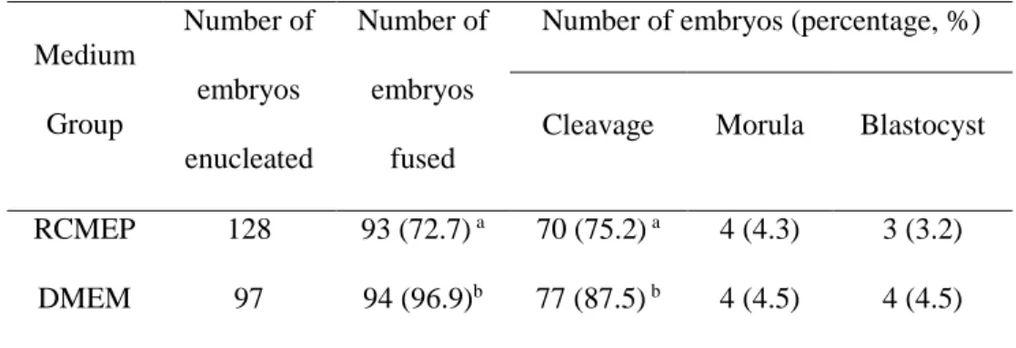
transfer embryos derived from donor cells cultured in two medium51 Table 4. Somatic cell nuclear transfer using canine adipose-derived mesenchymal
stem cells ...72 Table 5. In vivo developmental ability of cloned embryos derived from canine
adipose-derived mesenchymal stem cells ...73 Table 6. Microsatellite genotyping of recloned beagles. ...74 Table 7. Target CpG islands and primers for pyrosequencing ...92 Table 8. In vivo developmental ability of embryos cloned from SYN1-RFP cells
... 109
x
LIST OF FIGURES
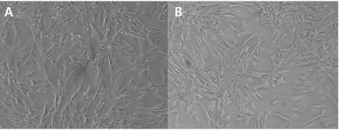
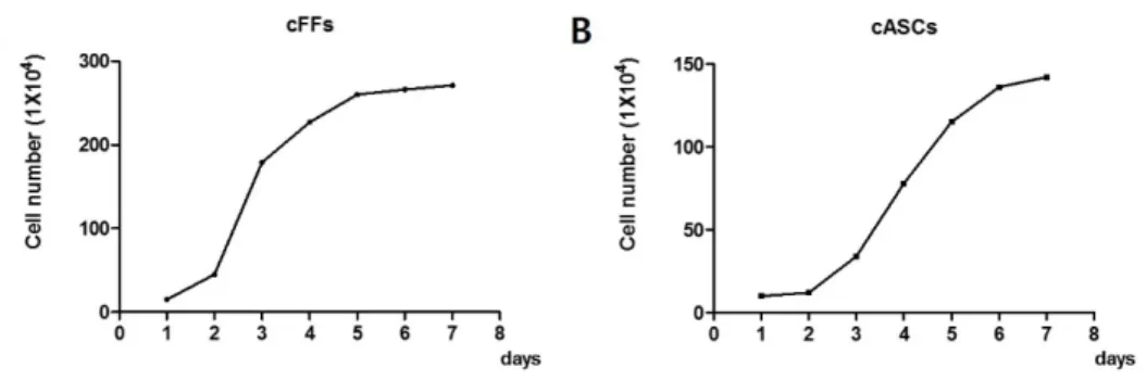
Figure 1. Photographs of cell growth in fourth passage. Morphology of (A) canine fetal fibroblasts (cFFs) and (B) adipose tissue-derived mesenchymal stem cells (cASCs) (magnification 100x). ...32 Figure 2. Growth curve of cFFs and cASCs. The sigmoidal curve (lag phase, log
phase, plateau) growth pattern observed in cFFs and cASCs at the 4th passage. Equal numbers of cells (1 X 10⁵) were seeded in triplicate and aliquots were counted daily during a period of 8 days. (A) Growth curve of cFFs by two fetal cell lines; (B) Growth curve of cASCs by two cASC lines. ...33
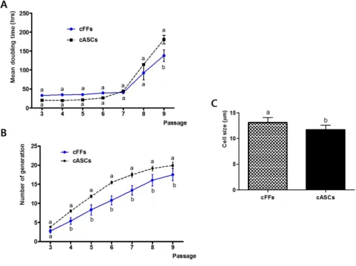
Figure 3. Cellular proliferation pattern and cell size. (A) Doubling time of cFFs and cASCs; (B) The generation number of cFFs and cASCs; (C) Cell size of cFFs and cASCs. Different superscripts (a, b) represent significant differences between groups (P < 0.05). Each experiment was performed at least five times using cFFs (pooled data from two fetal cell lines) and cASCs (pooled data from two cASC lines) during the 3–9th passages.
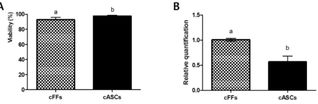
Data show mean ± SEM of the two cell lines in each group. ...34 Figure 4. Cellular viability of cFFs and cASCs. (A) Average viability of cFFs and
cASCs; (B) Expression profiles of BAX/BCL2 in cFFs and cASCs.
Different superscripts (a, b) represent significant differences between groups (P < 0.05). Each experiment was performed at least five times
xi
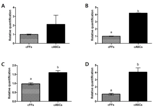
using cFFs (pooled data from two fetal cell lines) and cASCs (pooled data from two cASC lines) during the 3–9th passages. Data show mean ± SEM of the two cell lines from each group. ...35 Figure 5. Expression level of genes related to epigenetic modification and
pluripotency. Expression profiles of (A) DNMT1 and (B) HDAC1 related to epigenetic modification; Expression profiles of (B) OCT4 and (D) SOX2 related to pluripotency in cFFs and cASCs. Different superscripts (a, b) represent significant differences between groups (P < 0.05). Each experiment was performed at least five times. Data show mean ± SEM of the two cell lines from each group. ...36
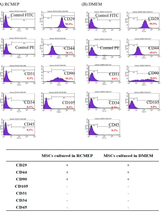
Figure 6. Cell surface staining of ASCs. FACS analysis detecting CD29, CD44, CD90, CD105, CD31, CD34 and CD45 antigen expression of ASCs cultured in (A) RCMEP (B) DMEM....50
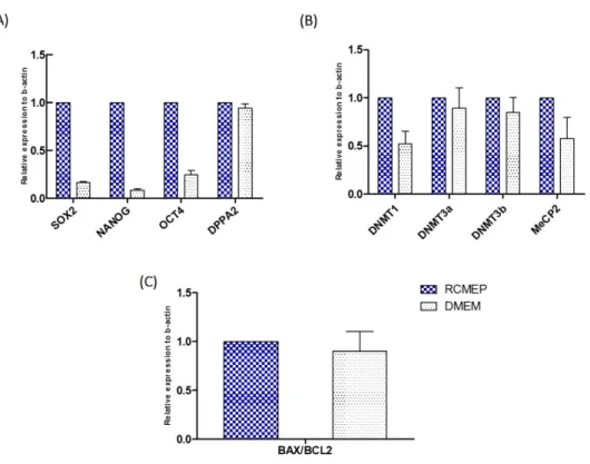
Figure 7. Real-time PCR analysis of ASCs. Relative abundance of transcripts of (A) SOX2, NANOG, OCT4, DPPA2; (B) DNMT1, DNMT3a, DMNT3b, MeCp2; (C) BAX, and BCL2 apoptotic genes in donor cells cultured in RCMEP media and DMEM media. Data presented as mean ± SEM of at least five replicates. Asterisk (*) superscripts indicate a significant difference (P<0.05). ...52 Figure 8. Real-time PCR analysis of iSCNT blastocyst. Gene expression pattern of
(A) NANOG, SOX2, OCT4, DPPA2; (B) DNMT1, DNMT3a, DNMT3b, MeCP2; (C) Na/K ATPase, Glut-1, E-cad, BAX/BCL2 expression pattern
xii
in iSCNT blastocysts. The figure shows the average relative abundance in cloned embryos. The transcripts of all genes were normalized to that of b- actin. Data presented as mean ± SEM of at least five replicates. Different superscripts indicate a significant difference (P<0.05). ...53 Figure 9. Morphology of canine adipose-derived mesenchymal stem cells derived
from an RFP transgenic cloned beagle. (A) Visible light images; (B) Fluorescence images (x 200). ...75
Figure 10. FACS analysis detecting CD29, CD44, Thy1.1, CD31, CD73, CD105 and CD34 antigen expression. The percentage of cells shows fluorescence intensity with specific antibody staining, as compared to nonspecific fluorescence (control). ...76
Figure 11. In vitro osteogenic, myogenic and chondrogenic differentiation of RFP transgenic dog derived adipose-derived mesenchymal stem cells by immunofluoresence at passage two. The cells transfected with RFP showed (A1) morphology of osteogenic differentiation and (A2) morphology of RFP labeling (x 100). (A3) osteogenic control cultured in normal adipose mesenchymal stem cell medium during 14 days showing negative Alizarin red s staining (x 100). (A4) osteogenic induction during 14 days culture showed morphological changes and mineralized deposits as indicated by positive Alizarin red S staining (x 100). (B1) cells showing morphology of myogenic differentiation and (B2) morphology of RFP labeling (x 200). (B3) the negative control of the myosin immunostaining
xiii
(x 200). (B4) myogenic differentiation showing expression of myosin as positive immunostaining with myosin antibody (green color) (x 200). (C1) cells showing morphology of chondrogenic differentiation and (C2) morphology of RFP labeling (x 100). (C3) chondrogenic differentiation 21 days after induction showing lacunae with extracellular proteoglycan formation as evidenced by positive staining with toluidine blue O (x 100).
(C4) lacunae as indicated by arrows (x 200). Every experiment was repeated three times. ...77
Figure 12. In vitro adipogenic and neurogenic differentiation of transgenic dog derived adipose-derived mesenchymal stem cells by immunofluoresence staining at passage two. (A1) morphology of adipogenic differentiation and (A2) morphology of RFP labeling (x 100) is shown. (A3) adipogenic control cultured in normal adipose mesenchymal stem cell medium during 21 days showed negative staining with Oil red O (x 100). (A4) adipogenic induction during 21 days showed morphological changes and accumulation of lipid-rich vacuoles in the cytoplasm as evidenced by positive Oil red O staining (x 100). (A5) lipid droplet deposition (vacuoles) demonstrating positive adipogenic induction. Morphology of neurogenic differentiation 10 days after induction showing large central bodies and neurites (x 400 of a quadrangle in A4). Neurogenic differentiation showing (B1) control, (B2) NSE antibody (green color), (B3) MAP-2 antibody (green color), (B4) TUJ1 antibody (green color) and (B5) GFAP
xiv
antibody (green color) expression by positive immunostaining (x 400).
Every experiment was repeated three times. ...79 Figure 13. The first dogs recloned by nuclear transfer of adipose-derived
mesenchymal stem cells derived from a transgenic cloned dog. (A) The transgenic recloned dog at 2 days after birth. They are named Magic and Stem. (B) Recloned Magic which carries the RFP gene (left) and a non- transgenic puppy (control, right). Notice that the claws and pads of Magic are tinged with red even in bright field illumination; (C) Visible light image; (D) Fluorescence image. ...80
Figure 14. Analysis of transgene integration in Ruppy1 and Magic. (A) Southern blot analysis of Ruppy1 and Magic was performed using a Hind III restriction enzyme. (B) The RFP transgene was detected by Southern blot.
M, marker; N, negative control; R1, Ruppy1; MG, Magic. (C) The integration position of the transgene. The RFP gene was inserted on chromosome 25 in both transgenic cloned dog. ...95
Figure 15. Age-dependent expression of RFP in tissue samples from Ruppy1 and Magic. (A) Western-blot analysis for RFP abundance in each dog at ages 1 and 4 years. (B) RFP expression, normalized to Beta-actin level in each sample. Analysis was replicated three times. Data are presented as the meanstandard deviation. Statistical analysis was performed with.
Statistical significance was determined by one-way analysis of variance (ANOVA) followed by Bonferroni posthoc testing, using GraphPad Prism
xv
version 5 (Graphpad Incorporation, San Diego, USA). a, b indicates P≤0.05. R1-1y, 1-year-old Ruppy1; R1-4y, 4-year-old Ruppy1; M-1y, 1- year-old Magic; M-4y, 4-year-old Magic. ...96 Figure 16. Age-dependent expression of CMV promoter methylation in tissue
samples from Ruppy1 and Magic. (A) Representative pyrograms obtained from samples for 4 select cytosine-phosphate-guanine (CpG) sites in the CMV promoter. Each gray shaded column indicates the assayed CpG dinucleotide, and the percentage of methylation at that CpG dinucleotide is indicated above. The yellow shaded boxes are internal bisulfite-modification control assessments. (B) The average methylation percentages of cytosine-phosphate-guanine (CpG) in the CMV promoter of Ruppy1 and Magic, measured at ages 1 and 4 years.
R1-1y, 1-year-old Ruppy1; R1-4y, 4-year-old Ruppy1; M-1y, 1-year-old Magic; M-4y, 4- year-old Magic. ...97
Figure 17. Generation of SYN1-RFP puppies by SCNT using cASCs. (A) Transgene integration of SYN1-RFP puppies by Southern blot analysis;
(B) SYN1-RFP C dog with RFP gene under human synapsin promoter by SCNT at 1 month after birth, and named Tung-B. M, marker; P, positive control; N, negative control; SYN-A, SYN1-RFP A puppy; SYN-B, SYN1-RFP B puppy; SYN-C, SYN1-RFP C puppy. ... 110 Figure 18. RPF protein using ELISA analysis. Heart, kidney, liver, lung, spleen,
cerebrum, cerebrum, cerebellum, midbrain, hippocampus, peripheral
xvi
nerves, skin and spinal cord of SYN1-RFP C was analyzed. And the skin of Ruppy1 was used as positive control. ... 111 Figure 19. RFP expression pattern in the body and organs of SYN1-RFP C dog.
(A, A′) Claws of Ruppy1 which carries the RFP gene under CMV promoter (left) and 1-week-old SYN1-RFP C which carries the RFP gene under synapsin promoter (right); (B, B′) ventral view of SYN1-RFP C spinal cord at 4 years of age; (C, C′) dorsal view of SYN1-RFP C brain at 4 years of age. (A–C) visible light image. (A′–C′) fluorescence image.
... 112
xvii
LIST OF ABBREVIATIONS
ASC Adipose derived mesenchymal stem cell MSC Mesenchymal stem cell
BSA Bovine serum albumin cDNA Complementary DNA CMV Cytomegalovirus
DMEM Dulbecco’s modified eagle’s medium DNA Deoxyribonucleic acid
mtDNA Mitochondrial DNA
MII Metaphase II
PB Polar body
PCR Polymerase chain reaction SCNT Somatic cell nuclear transfer
DPBS Dulbecco’s phosphate buffered saline
ET Embryo transfer
FBS Fetal bovine serum IVC In vitro culture
P4 Progesterone
RFP Red fluorescent protein SYN Synapsin 1
RT Reverse transcript
xviii
PUBLICATION LISTS
PUBLICATION PAPERS
1. Oh, H. J., Fibrianto, Y. H., Kim, M. K., Jang, G., Hossein, M. S., Kim, H. J., Kang, S. K., Lee, B. C., and Hwang, W. S. (2005) Effects of canine serum collected from dogs at different estrous cycle stages on in vitro nuclear maturation of canine oocytes. Zygote 13, 227-232
2. Oh, H. J., Kim, M. K., Jang, G., Kim, H. J., Hong, S. G., Park, J. E., Park, K., Park, C., Sohn, S. H., Kim, D. Y., Shin, N. S., and Lee, B. C. (2008) Cloning endangered gray wolves (Canis lupus) from somatic cells collected postmortem.
Theriogenology 70, 638-647
3. Oh, H. J., Hong, S. G., Park, J. E., Kang, J. T., Kim, M. J., Kim, M. K., Kang, S.
K., Kim, D. Y., Jang, G., and Lee, B. C. (2009) Improved efficiency of canine nucleus transfer using roscovitine-treated canine fibroblasts. Theriogenology 72, 461-470
4. Oh, H. J., Park, J. E., Kim, M. J., Hong, S. G., Ra, J. C., Jo, J. Y., Kang, S. K., Jang, G., and Lee, B. C. (2011) Recloned dogs derived from adipose stem cells of a transgenic cloned beagle. Theriogenology 75, 1221-1231
5. Oh, H. J., Park, E. J., Lee, S. Y., Soh, J. W., Kong, I. S., Choi, S. W., Ra, J. C., Kang, S. K., and Lee, B. C. (2012) Comparison of cell proliferation and epigenetic modification of gene expression patterns in canine foetal fibroblasts and adipose
xix
tissue-derived mesenchymal stem cells. Cell proliferation 45, 438-444
6. Hong, S. G., Oh, H. J., Park, J. E., Kim, M. J., Kim, G. A., Park, E. J., Koo, O. J., Kang, S. K., Jang, G., and Lee, B. C. (2011) Production of offspring from cloned transgenic RFP female dogs and stable generational transmission of the RFP gene.
Genesis 49, 835-840*
7. Oh, H. J., Lee, T. H., Lee, J. H., and Lee, B. C. (2012) Trichostatin a improves preimplantation development of bovine cloned embryos and alters expression of epigenetic and pluripotency genes in cloned blastocysts. The Journal of veterinary medical science / the Japanese Society of Veterinary Science 74, 1409-1415 8. Oh, H. J., Park, J. E., Park, E. J., Kim, M. J., Kim, G. A., Rhee, S. H., Lim, S. H.,
Kang, S. K., and Lee, B. C. (2014) Analysis of cell growth and gene expression of porcine adipose tissue-derived mesenchymal stem cells as nuclear donor cell.
Development, growth & differentiation 56, 595-604*
9. Kim, G. A., Oh, H. J., Lee, T. H., Lee, J. H., Oh, S. H., Lee, J. H., Kim, J. W., Kim, S. W., and Lee, B. C. (2014) Effect of culture medium type on canine adipose-derived mesenchymal stem cells and developmental competence of interspecies cloned embryos. Theriogenology 81, 243-249*
10. Oh, H. J., Kim, G. A., Rhee, S. H., Kim, M. J., Saadeldin, I. M., and Lee, B. C.
(2015) Age-dependent alteration of transgene expression and cytomegalovirus promoter methylation in transgenic cloned and recloned dogs. Molecular reproduction and development 82, 330-331
11. Oh, H. J., Choi, J., Kim, M. J., Kim, G. A., Jo, Y. K., Choi, Y. B., and Lee, B. C.
xx
(2016) Propagation of elite rescue dogs by somatic cell nuclear transfer. Animal science journal 87, 21-6.
12. Kim, M. K., Fibrianto, Y. H., Oh, H. J., Jang, G., Kim, H. J., Lee, K. S., Kang, S.
K., Lee, B. C., and Hwang, W. S. (2004) Effect of beta-mercaptoethanol or epidermal growth factor supplementation on in vitro maturation of canine oocytes collected from dogs with different stages of the estrus cycle. Journal of veterinary science 5, 253-258
13. Kim, M. K., Fibrianto, Y. H., Oh, H. J., Jang, G., Kim, H. J., Lee, K. S., Kang, S.
K., Lee, B. C., and Hwang, W. S. (2005) Effects of estradiol-17beta and progesterone supplementation on in vitro nuclear maturation of canine oocytes.
Theriogenology 63, 1342-1353
14. Lee, B. C., Kim, M. K., Jang, G., Oh, H. J., Yuda, F., Kim, H. J., Hossein, M. S., Kim, J. J., Kang, S. K., Schatten, G., and Hwang, W. S. (2005) Dogs cloned from adult somatic cells. Nature 436, 641
15. Hossein, M. S., Kim, M. K., Jang, G., Fibrianto, H. Y., Oh, H. J., Kim, H. J., Kang, S. K., and Lee, B. C. (2007) Influence of season and parity on the recovery of in vivo canine oocytes by flushing fallopian tubes. Animal reproduction science 99, 330-341
16. Hossein, M. S., Kim, M. K., Jang, G., Oh, H. J., Koo, O., Kim, J. J., Kang, S. K., Lee, B. C., and Hwang, W. S. (2007) Effects of thiol compounds on in vitro maturation of canine oocytes collected from different reproductive stages.
Molecular reproduction and development 74, 1213-1220
xxi
17. Jang, G., Kim, M. K., Oh, H. J., Hossein, M. S., Fibrianto, Y. H., Hong, S. G., Park, J. E., Kim, J. J., Kim, H. J., Kang, S. K., Kim, D. Y., and Lee, B. C. (2007) Birth of viable female dogs produced by somatic cell nuclear transfer.
Theriogenology 67, 941-947
18. Kim, M. K., Hossein, M. S., Oh, H. J., Fibrianto, H. Y., Jang, G., Kim, H. J., Hong, S. G., Park, J. E., Kang, S. K., and Lee, B. C. (2007) Glutathione content of in vivo and in vitro matured canine oocytes collected from different reproductive stages. The Journal of veterinary medical science / the Japanese Society of Veterinary Science 69, 627-632
19. Kim, M. K., Jang, G., Oh, H. J., Yuda, F., Kim, H. J., Hwang, W. S., Hossein, M.
S., Kim, J. J., Shin, N. S., Kang, S. K., and Lee, B. C. (2007) Endangered wolves cloned from adult somatic cells. Cloning and stem cells 9, 130-137
20. Jang, G., Hong, S. G., Oh, H. J., Kim, M. K., Park, J. E., Kim, H. J., Kim, D. Y., and Lee, B. C. (2008) A cloned toy poodle produced from somatic cells derived from an aged female dog. Theriogenology 69, 556-563
21. Jang, G., Oh, H. J., Kim, M. K., Fibrianto, Y. H., Hossein, M. S., Kim, H. J., Kim, J. J., Hong, S. G., Park, J. E., Kang, S. K., and Lee, B. C. (2008) Improvement of canine somatic cell nuclear transfer procedure. Theriogenology 69, 146-154 22. Choresca, C. H., Jr., Koo, O. J., Oh, H. J., Hong, S. G., Gomez, D. K., Kim, J. H.,
Lee, B. C., and Park, S. C. (2009) Different culture conditions used for arresting the G0/G1 phase of the cell cycle in goldfish (Carassius auratus) caudal fin- derived fibroblasts. Cell biology international 33, 65-70
xxii
23. Hong, S. G., Jang, G., Kim, M. K., Oh, H. J., Park, J. E., Kang, J. T., Koo, O. J., Kim, D. Y., and Lee, B. C. (2009) Dogs cloned from fetal fibroblasts by nuclear transfer. Animal reproduction science 115, 334-339
24. Hong, S. G., Jang, G., Oh, H. J., Koo, O. J., Park, J. E., Park, H. J., Kang, S. K., and Lee, B. C. (2009) The effects of brain-derived neurotrophic factor and metformin on in vitro developmental competence of bovine oocytes. Zygote 17, 187-193
25. Hong, S. G., Kim, M. K., Jang, G., Oh, H. J., Park, J. E., Kang, J. T., Koo, O. J., Kim, T., Kwon, M. S., Koo, B. C., Ra, J. C., Kim, D. Y., Ko, C., and Lee, B. C.
(2009) Generation of red fluorescent protein transgenic dogs. Genesis 47, 314-322 26. Park, J. E., Hong, S. G., Kang, J. T., Oh, H. J., Kim, M. K., Kim, M. J., Kim, H.
J., Kim, D. Y., Jang, G., and Lee, B. C. (2009) Birth of viable puppies derived from breeding cloned female dogs with a cloned male. Theriogenology 72, 721- 730
27. Choresca, C. H., Jr., Koo, O. J., Hong, S. G., Oh, H. J., Gomez, D. K., Kim, J. H., Lee, B. C., and Park, S. C. (2010) Effect of dimethyl sulfoxide on cell cycle synchronization of goldfish caudal fin derived fibroblasts cells. Reproduction in domestic animals 45, e73-77
28. Hong, S. G., Oh, H. J., Park, J. E., Kang, J. T., Kim, M. J., Yoon, J. H., Chang, J.
H., Kim, M. K., Jang, G., and Lee, B. C. (2010) Serum levels of reproductive hormones and ultrasonographic monitoring of ovarian follicles in female cloned dogs. The Journal of veterinary medical science 72, 89-92
xxiii
29. Kim, M. J., Oh, H. J., Park, J. E., Hong, S. G., Kang, J. T., Koo, O. J., Kang, S.
K., Jang, G., and Lee, B. C. (2010) Influence of oocyte donor and embryo recipient conditions on cloning efficiency in dogs. Theriogenology 74, 473-478 30. Park, J. E., Kim, M. K., Kang, J. T., Oh, H. J., Hong, S. G., Kim, D. Y., Jang, G.,
and Lee, B. C. (2010) Growth and hematologic characteristics of cloned dogs derived from adult somatic cell nuclear transfer. Cellular reprogramming 12, 141- 150
31. Park, J. E., Oh, H. J., Hong, S. G., Jang, G., Kim, M. K., and Lee, B. C. (2010) Effects of activin A on the in vitro development and mRNA expression of bovine embryos cultured in chemically-defined two-step culture medium. Reproduction in domestic animals 45, 585-593
32. Atikuzzaman, M., Koo, O. J., Kang, J. T., Kwon, D. K., Park, S. J., Kim, S. J., Gomez, M. N., Oh, H. J., Hong, S. G., Jang, G., and Lee, B. C. (2011) The 9-cis retinoic acid signaling pathway and its regulation of prostaglandin-endoperoxide synthase 2 during in vitro maturation of pig cumulus cell-oocyte complexes and effects on parthenogenetic embryo production. Biology of reproduction 84, 1272- 1281
33. Han, J. E., Choresca, C. H., Koo, O. J., Oh, H. J., Hong, S. G., Kim, J. H., Shin, S.
P., Jun, J. W., Lee, B. C., and Park, S. C. (2011) Establishment of glass catfish (Kryptopterus bicirrhis) fin-derived cells. Cell biology international reports 18, e00008
34. Hong, S. G., Koo, O. J., Oh, H. J., Park, J. E., Kim, M., Kim, G. A., Park, E. J.,
xxiv
Jang, G., and Lee, B. C. (2011) Post-mortem re-cloning of a transgenic red fluorescent protein dog. Journal of veterinary science 12, 405-40733.
35. Kim, M. J., Oh, H. J., Park, J. E., Kim, G. A., Hong, S. G., Jang, G., Kwon, M. S., Koo, B. C., Kim, T., Kang, S. K., Ra, J. C., Ko, C., and Lee, B. C. (2011) Generation of transgenic dogs that conditionally express green fluorescent protein.
Genesis 49, 472-478
36. Hong, S. G., Oh, H. J., Park, J. E., Kim, M. J., Kim, G. A., Koo, O. J., Jang, G., and Lee, B. C. (2012) Production of transgenic canine embryos using interspecies somatic cell nuclear transfer. Zygote 20, 67-72
37. Kang, J. T., Kim, H. J., Oh, H. J., Hong, S. G., Park, J. E., Kim, M. J., Kim, M.
K., Jang, G., Kim, D. Y., and Lee, B. C. (2012) SRY-positive 78, XY ovotesticular disorder of sex development in a wolf cloned by nuclear transfer.
Journal of veterinary science 13, 211-213
38. Kim, G. A., Oh, H. J., Park, J. E., Kim, M. J., Park, E. J., Jo, Y. K., Jang, G., Kim, M. K., Kim, H. J., and Lee, B. C. (2012) Species-specific challenges in dog cloning. Reproduction in domestic animals 47 Suppl 6, 80-83
39. Kim, M. J., Oh, H. J., Kim, G. A., Park, J. E., Park, E. J., Jang, G., Ra, J. C., Kang, S. K., and Lee, B. C. (2012) Lessons learned from cloning dogs.
Reproduction in domestic animals 47 Suppl 4, 115-119
40. Kim, M. J., Oh, H. J., Park, J. E., Kim, G. A., Park, E. J., Jang, G., and Lee, B. C.
(2012) Effects of mineral supplements on ovulation and maturation of dog oocytes.
Theriogenology 78, 110-115
xxv
41. Park, J. E., Kim, M. J., Ha, S. K., Hong, S. G., Oh, H. J., Kim, G. A., Park, E. J., Kang, J. T., Saadeldin, I. M., Jang, G., and Lee, B. C. (2012) Altered cell cycle gene expression and apoptosis in post-implantation dog parthenotes. PloS one 7, e41256
42. Saadeldin, I. M., Koo, O. J., Kang, J. T., Kwon, D. K., Park, S. J., Kim, S. J., Moon, J. H., Oh, H. J., Jang, G., and Lee, B. C. (2012) Paradoxical effects of kisspeptin: it enhances oocyte in vitro maturation but has an adverse impact on hatched blastocysts during in vitro culture. Reproduction, fertility, and development 24, 656-668
43. Jung, S. H., Yim, S. H., Oh, H. J., Park, J. E., Kim, M. J., Kim, G. A., Kim, T. M., Kim, J. S., Lee, B. C., and Chung, Y. J. (2013) De novo copy number variations in cloned dogs from the same nuclear donor. BMC genomics 14, 863
44. Kim, G. A., Oh, H. J., Park, J. E., Kim, M. J., Park, E. J., Lim, S. H., Kang, S. K., Jang, G., and Lee, B. C. (2013) Employing mated females as recipients for transfer of cloned dog embryos. Reproduction, fertility, and development 25, 700- 706
45. Kim, M. J., Oh, H. J., Park, J. E., Kim, G. A., Park, E. J., Jo, Y. K., and Lee, B. C.
(2013) Duration of gestation in pregnant dogs carrying cloned fetuses.
Theriogenology 79, 257-260
46. Choi, J., Lee, J. H., Oh, H. J., Kim, M. J., Kim, G. A., Park, E. J., Jo, Y. K., Lee, S. I., Hong do, G., and Lee, B. C. (2014) Behavioral analysis of cloned puppies derived from an elite drug-detection dog. Behavior genetics 44, 68-76
xxvi
47. Kim, G. A., Oh, H. J., Kim, M. J., Jo, Y. K., Choi, J., Park, J. E., Park, E. J., Lim, S. H., Yoon, B. I., Kang, S. K., Jang, G., and Lee, B. C. (2014) Survival of skin graft between transgenic cloned dogs and non-transgenic cloned dogs. PloS one 9, e10833048.
48. Kim, M. J., Oh, H. J., Kim, G. A., Jo, Y. K., Choi, J., Kim, H. J., Choi, H. Y., Kim, H. W., Choi, M. C., and Lee, B. C. (2014) Reduced birth weight, cleft palate and preputial abnormalities in a cloned dog. Acta veterinaria Scandinavica 56, 18 49. Moon, J. H., Kim, J. H., Im, H. J., Lee, D. S., Park, E. J., Song, K., Oh, H. J.,
Hyun, S. B., Kang, S. C., Kim, H., Moon, H. E., Park, H. W., Lee, H. J., Kim, E.
J., Kim, S., Lee, B. C., and Paek, S. H. (2014) Proposed Motor Scoring System in a Porcine Model of Parkinson's Disease induced by Chronic Subcutaneous Injection of MPTP. Experimental neurobiology 23, 258-265
50. Choi, Y. B., Kim, G. A., Oh, H. J., Kim, M. J., Jo, Y. K., Setyawan, E. M., Lee, S.
H., and Lee, B. C. (2015) Cloning of the short-tailed Gyeongju Donggyeong dog via SCNT: conserving phenotypic inheritance. The Journal of veterinary medical science. Epup
51. Kim, G. A., Oh, H. J., Kim, M. J., Jo, Y. K., Choi, J., Kim, J. W., Lee, T. H., and Lee, B. C. (2015) Effect of primary culture medium type for culture of canine fibroblasts on production of cloned dogs. Theriogenology 84, 524-530
52. Kim, M. J., Kang, S. C., Kim, J. H., Oh, H. J., Kim, G. A., Jo, Y. K., Choi, J., Kim, H., Lee, Y. H., Yoo, J. M., Eom, K. D., and Lee, B. C. (2015) Ectopic liver and gallbladder in a cloned dog: Possible nonheritable anomaly. Theriogenology
xxvii 84, 995-1002
53. Kim, M. J., Oh, H. J., Kim, G. A., Suh, H. N., Jo, Y. K., Choi, Y. B., Kim, D. H., Han, H. J., and Lee, B. C. (2015) Altering histone acetylation status in donor cells with suberoylanilide hydroxamic acid does not affect dog cloning efficiency.
Theriogenology 84, 1256-1261
54. Lee, J. H., Kim, G. A., Kim, R. S., Lee, J. S., Oh, H. J., Kim, M. J., Hong, D. K., and Lee, B. C. (2015) Reproductive ability of a cloned male detector dog and behavioral traits of its offspring. Journal of veterinary science Epub
55. Saadeldin, I. M., Oh, H. J., and Lee, B. C. (2015) Embryonic-maternal cross-talk via exosomes: potential implications. Stem cells and cloning : advances and applications 8, 103-107
56. Setyawan, E. M., Kim, M. J., Oh, H. J., Kim, G. A., Jo, Y. K., Lee, S. H., Choi, Y.
B., and Lee, B. C. (2015) Maintaining canine sperm function and osmolyte content with multistep freezing protocol and different cryoprotective agents.
Cryobiology 71, 344-349
* Both authors contributed equally
1
PART I
GENERAL INTRODUCTION
2
1. Literature review
1.1. Canine mesenchymal stem cells
The application of stem cells has rapidly emerged for regenerative medicine and tissue engineering in veterinary medicine. A tissue or organ can renew itself because undifferentiated stem cells can differentiate to yield the major specialized cell types of the tissue or organ [1]. Stem cells can be classified as embryonic or adult stem cells, according to a developmental stage be recovered [2-4]. Embryonic stem cells are derived from the inner cell mass of the blastocyst and adult mesenchymal stem cells (MSCs) originate from particular three germ layers [2-4]. Although embryonic stem cells are theoretically highly beneficial, there are various limitations as cell regulations, ethical considerations, and genetic manipulation to their use imposed. In contrast, MSCs are more easily accessible, with neither ethical considerations nor immunoreactivity. Thus MSCs as adipose-derived mesenchymal stem cells (ASCs) or bone marrow-derived stem cells (BMSCs), are getting the spotlight in the fields of stem cell research and regenerative medicine [1, 5].
1.1.1. Sources of canine mesenchymal stem cells
Adult stem cells can be obtained from mesodermal tissues (bone marrow, trabecular bone, synovium, adipose tissue, muscles and cartilage), endodermal tissues (thymus and liver) and ectoderm tissues (skin, hair follicle and dental pulp) [6]. Many
3
canine research and clinical articles have been published using ASCs or BMSCs, because these are well established and characterized [7-9]. BMSCs are not only be obtained easily and non-invasively, but also be considered the best standard for use in tissue regeneration among MSCs [7-10]. Nonetheless, ASCs derived from adipose tissue have been recently considered a highly attractive option compared to BMSCs, because of the ease of tissue collection and the availability of adipose tissue in the body [11]. In addition to, the fact that stem cell yields are greater from adipose tissue than from other stem cell reservoirs is a significant factor in their suitability for use in regenerative medicine [11].
1.1.2. Characterization of canine mesenchymal stem cells
Human MSCs are characterized by minimally three criteria according to the International Society for Cellular Therapy: (1) plastic adherence when maintained in standard culture conditions, (2) expression of a specific cell surface antigen marker (3) at least tri-lineage differentiation potential [4, 12]. However, unlike human MSCs, there are no available uniform characterization criteria in specific for MSCs from animal origin and especially from canine origin [4, 13]. Nevertheless, some criteria as for human are available for the dog [14, 15]. Recently, undifferentiated canine MSCs can be characterized morphologically, immune-phenotypically and by their gene expression.
Morphologically, MSCs are identified as spindle-shaped cells that grow on plastic dishes in a monolayer [16]. Immunophenotyping has not been universally well
4
characterized, but this have performed using specific cross-reaction antibodies to indicate surface antigen expression [4, 12]. Several studies have demonstrated the simultaneous expression of several cell surface positive markers (CD29, CD73, CD105 and CD44, Thy-1 CD90 glycoprotein and MHC-I), and negative markers (CD34, CD45, CD14, CD117, SSEA-3, SSEA-4, TRA-1-60, TRA-1-81, and CD146) either in BMSC or in ASC [4, 16, 17]. In human MSC research, gene expression is frequently used to characterize undifferentiated MSCs and the most commonly studied genes with positive expression are the pluripotency-associated transcription factors NanoG, OCT4 and SOX2 [18]. In line with human MSCs, canine MSCs also appear to be positive for these three gene expression markers [17, 19]. Characterization of these MSCs at the mRNA level is a valuable alternative when no monoclonal antibodies are available to characterize them at the protein level [4].
Lastly, the ability of MSCs to differentiate into multiple lineages through osteogeneis, chondrogenesis and adipogenesis has also been demonstrated for characterization of MSCs [4, 12]. Osteogenesis is verified by the polygonal appearance of the differentiated osteoblast containing nodular aggregates that stain positively with von Kossa and by the mRNA expression of osteoblast markers (Runx2, Osteonectin, Osterix and BSP) [15, 17]. Chondrogenesis is characterized by the presence of three- dimensional aggregates, which stain positive for alcian blue indicating the presence of sulphated proteoglycans. Expression of COL2A, Aggrecan, COMP, COL10A and Sox9 can be detected in the differentiated cells [16, 20]. Adipogenesis is characterized
5
by the appearance of round-shaped cells with Oil Red O staining of lipid droplets [15].
Adipose-induced cells express PPARg2, CEBPa, FABP4 and LPL [15, 20].
1.1.3. Therapeutic applications of canine mesenchymal stem cells
The main purpose of canine MSC is the conduction of experimental animal studies in which the dog served as a preclinical model for humans. In 1998, the cultured autologous BMSCs were examined the effect on the healing of generated defects in canine femurs. The group treated with a carrier loaded with MSCs showed substantial new bone formation, while the untreated femurs demonstrated atrophic non-union. This result hold potential to provide an alternative strategy to autologous bone-grafting [9]. MSC-based therapies in dogs have mainly been focusing on bone and cartilage repair and inflammatory diseases such as osteoarthritis. Nevertheless, an important share of musculoskeletal problems in dogs consists of tendon/ligament injuries, which are comparable to the human. Since natural repair mechanisms often do not deliver functional recovery or current therapeutic strategies show minimal effectiveness, MSC-based therapies can be considered as potentially revolutionary in this field. Therefore, a challenging future prospect involves the research on MSC- based therapy for these canine tendon/ligament injuries. Since dogs are considered to be a superior animal model for humans, so not only dogs but also human can benefit from state-of-the-art research in this exciting field.
6
1.2. Canine somatic cell nuclear transfer
1.2.1. Somatic cell nuclear transfer
SCNT in mammals is an assisted reproductive technique to produce an animal from a single cell nucleus using an enucleated oocyte. Nuclear transfer (NT) only became technically feasible in amphibians around 1950 [21] and in mammals about 30 years later [22]. Finally, the first successful mammals cloning was reported that a sheep named Dolly was obtained by cultured somatic cells from an adult ewe in 1997 [23]. Since then, a number of mammals have been produced by SCNT [24-29]. In addition, because somatic cells can be proliferated and genetic modified in vitro, this technique has been applied to different fields of biomedicine including xenotransplantation, production of bioreactor, cell therapy, and production of human disease model animals [30, 31].
1.2.2. Dog cloning
The cloning of canids was succeeded in 2005, several years after the birth of Dolly the sheep and also after the cloning of numerous other laboratory and farm animal species. The delay of successful canine SCNT was due to the unique reproductive characteristics of the female dog in comparison to other domestic mammals, such as ovulation of immature canine oocyte and a requirement of 2–5 days for the completion of meiosis within the oviduct [32, 33]. When the technology for the recovery of in vivo matured oocyte was established, the application of cloning also
7
became possible. Finally, the world’s first cloned dog was produced in 2005 from adult skin cell of male Afghan hound, named ‘Snuppy’ which stands for Seoul National University puppy [34]. ‘Snuppy’ was genetically identical to the donor dog, and his mitochondrial DNA was originated from their oocyte donor dog. From that birth onwards, canine SCNT technique has been developed for companion dog cloning [34, 35], expanding the elite pool of canids[36], conservation of endangered canine species [37, 38] and generation of transgenic cloned dog for animal model research [39].
Nonetheless, the low efficiency of canine SCNT was a major obstacle to extensive use of this technology. In order to overcome the problem, numerous factors affecting the NT have been studied; cell cycle of donor cell [40], status of oocyte maturation [35], fusion condition [41], condition of oocyte donor and recipient dog [42].
1.2.3. Donor cell and SCNT
Most of efforts to improve the efficiency of SCNT have been focused on donor cells. First, G0 or G1 phase of donor cell cycle is a major factor because it facilitate nuclear reprogramming in cloned embryo [43]. The cell cycle synchronization of donor cells can be induced by serum starvation, contact inhibition, and some chemical treatment including roscovitine [43, 44]. In dogs, contact inhibition and chemical treatment are available, and roscovitine treatment (15μg/ml for 24 h) improved cell cycle synchronization and cloning efficiency [40]. Another important factor related to donor cell is cell type; until now, various cell types, such as fetal fibroblasts [40, 45, 46]
and adult fibroblasts [35, 40, 41, 47-49], have been used as nuclear donors for
8
producing cloned dog. The general purpose of canine SCNT is to conserve the companion dog, the elite working dog and the endangered canine species, adult fibroblasts are used mainly as donor cell. On the other hand, fetal fibroblasts are preferred as nucleus donors for SCNT used in producing transgenic animals because they have excellent proliferative ability, are capable of being genetically modified, and have the ability to produce live offspring [26, 27, 40, 50] [26, 27, 40, 50]. Recently, transgenic puppies were born following nuclear transfer from canine fetal fibroblasts transfected with the exogenous genes [40, 46]. Although there is difference in SCNT efficiency between canine fetal fibroblasts and adult fibroblasts, they can be reprogrammed and selected as a donor nucleus for the SCNT [51].
1.3. Transgenic cloned dogs using SCNT
The dog has been proposed for human disease model because they have many common genetic diseases. Moreover, dog exhibit 359 genetic diseases similar to those experienced by humans (http://omia.angis.org.au, accessed April 2015), making them one of the important models for various human hereditary diseases. In addition, dogs 1) have organ sizes comparable to those of humans, unlike the traditional rodent models, 2) generally cohabitate with human beings, minimizing different environmental effects and 3) receive exceptional medical care [40, 52, 53]. For producing transgenic dog, the use of pronuclear injection or homologous recombination are needed. However, these methods are highly inefficient in large animals because of the limitation of random
9
integration of highly variable expression and unavailable of embryonic stem cells [54].
Recently, SCNT technique using transgenic donor cells generated transgenic large animals [24, 25], this technique would be feasible to produce genetically modified dog.
1.3.1. Transgenic dogs by cloning
Two kinds of transgenic dogs were born following nuclear transfer from fetal canine fibroblasts transfected with the Red fluorescent protein (RFP) gene [40] or Green fluorescent protein (GFP) conditionally expressed by doxycycline administration (Teton) [46]. In addition, transgenic cloned dogs produced exhibited normal reproductive ability, and the foreign gene was successfully inherited to their offspring as well as stable insertion of the transgene into the genome [46, 55].
Therefore, canine SCNT technique could be applied to produce human genetic disease model.
1.3.2. Transgenic dogs by recloning
The production of transgenic animals through a combination of genetic engineering and SCNT has grown, however, there are limitations in the use of these technique [56]. First, to establish transgenic donor cells for SCNT, transfection and selection procedures must be performed. During this procedures, the donor cells easily become senescent and the number of transgenic cells that can be used for SCNT are limited [57]. Second, mosaic pattern of transgene integration or a heterogeneous number of transgene copies integrated at different chromosome locations disturb
10
homogeneity among transgenic animals and increase variegation of transgene expression with aging which complicate interpretation of the developmental regulation of transgenes [58]. The strategy to overcome these obstacles is to reclone the first generation of transgenic animals. Several studies have demonstrated that the rejuvenated cells from cloned animals can be used to produce recloned offspring in several species including cattle [26, 59], pig [60] and cat [61]. These reports indicated that life-span of the donor cells can be elongated infinitely using recloning technique and transgenic animals can be produced as much as desired. In dog, recloning of the world’s first transgenic cloned dogs expressing RFP was tried to overcome cellular senescence and to produce more advanced transgenic dogs [62]. To establish a cloned transgenic cell line, donor cell from the male dogs who was died at 11 weeks after birth was harvested. Embryos reconstructed by nuclear transfer using the transgenic cell were transferred into recipients, one puppy was produced and confirmed as a recloned dog [62]. Although the recloned puppy was stillborn, recloning technique can be used for establishing a rejuvenated fibroblast cell line and propagating the recloned transgenic dog.
1.4. Canines as Human Disease model
Domesticated dogs are increasingly being identified as good models for a variety of biomedical research fields as they have a number of unique advantages over other commonly used experimental animals. The most interesting thing is that dog share
11
their habitat with their owner and they generally receive a good level care including highly-trained healthcare. This modeling of the human condition offers valuable opportunities for researchers to examine complex problems such as environmental contributions to diseases, aging and its effect on disease susceptibility and progression, and the effects of long-term treatment therapy protocols [63-65].
Another advantage is that over the past several centuries, domestication and selective breeding of dogs has resulted in nearly 400 distinct populations and thus the most naturally occurring genetic diversity in any one species besides humans. Careful breeding for trait selection has inadvertently resulted in breed-specific disease susceptibilities and approximately 400 naturally-occurring, inherited diseases have been identified in dogs. Most predicted canine genes have known as human homologs and many of these heritable canine diseases have been associated with mutations in canine and human homologous genes. The enormous genetic diversity of canine breeds (many of which have extensive pedigree information) and the broad range of spontaneously-occurring canine diseases afford researchers opportunities to examine genetic etiologies and explore the possibility of gene therapies. Unfortunately, this genetic diversity is also the main disadvantage of canine models as there are breed- specific differences in physiology and metabolism, especially idiosyncrasies in pharmacodynamics and pharmacokinetics, which can introduce complications when interpreting or translating the results. However, increasing research in this area is expanding on our current understanding of these issues [63-65].
Finally, due to the rapid aging of dogs, there is a shorter duration for disease
12
development and progression. This is an enormous advantage in the context of drug development as clinical trial study times are significantly reduced. Potential for rapid study times along with reduced regulatory guidelines and the increasing acceptance of canine models of human diseases by regulatory bodies is resulting in the growing use of dogs as models for translational medicine. Canine models have served to advance human medicine in a number of areas and have been instrumental in some, such as narcolepsy, hemophilia, retinal degeneration, and muscular dystrophy [63-65].
1.4.1. The dog as Alzheimer’s disease model
There are significant breed-specific differences in canine longevity; smaller breeds having longer lifespans and larger breeds having shorter lifespans. Generally, the beagle has been selected as the main breed used for aging studies as its median lifespan is 12 to 14 years, and beagles over 9 years of age are considered “old”
representing humans aged 66 to 96 years. Both cognitive and neuropathological changes in aged Beagles have been well documented and seem to closely approximate human clinical observations in many aspects. These features make dogs, and in particular beagles, well-matched for studies of human aging and age-related conditions such as Alzheimer’s disease (AD) [66].
Like humans, aged dogs naturally suffer from AD that is characterized by the deposition of significant amounts of Amyloid β (Aβ) protein and the development of diffuse plaques, the extent of which quantifiably correlate with cognitive decline [67].
However, AD-afflicted dogs do not appear to naturally develop neurofibrillary tangles
13
as observed in humans. This may be explained by the fact that while the Aβ amino acid sequence is identical in dogs and humans, the Tau amino acid sequence is appreciably different in the two species. It has been suggested that presence of Aβ deposits and plaques and lack of neurofibrillary tangles represents early stages of AD, which may uniquely position the canine AD model for investigations into the possibilities of preventative measures and early interventions. Therapeutic strategies under investigation using canine AD models include antioxidant diets and behavioral enrichment. Both of these regimens have been shown to improve AD pathology when delivered individually and even more so when administered in combination. Other treatment strategies being pursued in dogs include disruption of Aβ processing by anti- inflammatory or statin drugs and immunization against Aβ peptide [66, 68].
1.4.2. Human synapsin 1 promoter
The widespread phenotype diversity within the central nervous system underscores the importance of restricting transgene expression to a specified target cell type [69-71]. Failure to do so results in gene expression in non-target cells that confounds data interpretation and may lead to undesirable side effects. Restricting gene expression to a specified cell population within the brain by using cell-selective promoters remains an attractive approach [72, 73]. In addition, cell-type-specific promoters are advantageous since they are less likely to activate host cell defense machinery and are less sensitive to cytokine-induced promoter inactivation than viral
14
promoters [73]. As such, improved stability and longevity of gene expression can be expected.
Synapsins are a family of neuronal phosphoproteins that coat the cytoplasmic surface of small synaptic vesicles [74]. This family consists of four proteins, synapsin Ia and synapsin Ib and synapsin IIa and synapsin IIb. Synapsins I and II are generated via alternative splicing from two different genes [74]. Molecular cloning of bovine,
human, and rat synapsins revealed striking homologies in the amino-terminal 420 amino acids of all four synapsins. The major difference between synapsins I and II lies in the C-terminal domain of the synapsin I isoforms. This domain contains clusters of basic amino acids as well as two recognition sites for Ca /calmodulin-dependent protein kinase II [74]. Synapsin I has been postulated to link synaptic vesicles to the cytoskeleton, thus regulating the availability of synaptic vesicles for exocytosis [75]. In addition, a role for synapsin I in the regulation of short term plasticity has been suggested [76]. Mice lacking synapsin I or both synapsins I and II are viable and fertile with no gross anatomical abnormalities. These mice, however, frequently experience seizures, indicating the essential functions of the synapsins in synaptic vesicle regulation [76]. Virtually all neurons express the synapsins [74] and there are no non- neuronal counterparts known for the synapsins, in contrast to the synaptic vesicle proteins synaptobrevin, synaptophysin, and synaptotagmin [77, 78]. In transgenesis, direction and limitation of gene expression to neurons have been demonstrated with the Synapsin I promoter [79-82]. Though being highly specific, transgene expression cassette exhibits retained neuronal specificity and a considerably elevated level of
15
expression of two- to three- fold [83, 84]. Thus, restricted expression of synapsins I and II in the nervous system suggests as good tool for investigating the neuron-specific gene expression.
16
2. General objective
The purpose of this study is to generate transgenic dogs using cASCs which has multi-lineage differentiation potential for further applications to human disease models.
This thesis is composed of 5 parts. In part I; as a general introduction, it was explained why I have designed and performed this study. In part II; general methodology used in this study was described. In part III; several properties of cASCs as donor cell were analyzed. Then, iSCNT was performed using cASCs for evaluating its potential as donor cell. Finally, transgenic dogs were generated by canine SCNT using cASCs in part IV. In part V; a final conclusion of this study was described.
17
PART II
GENERAL METHODOLOGY
18
1. Chemicals and materials
All chemicals were obtained from Sigma-Aldrich Co. LLC. (Missouri, USA) unless otherwise stated.
2. Care and use of animals
In this study, mixed-breed female dogs (Canis familiaris) between 1 and 5 years of age were used as oocyte donors and embryo transfer recipients. The study was conducted in accordance with recommendations described in ‘‘The Guide for the Care and Use of Laboratory Animals’’ published by Institutional Animal Care and Use Committee (IACUC) of Seoul National University. In that regard, facilities for dog care and all procedures met or exceeded the standards established by the Committee for Accreditation of Laboratory Animal Care at Seoul National University.
3. Preparation of donor fibroblasts and somatic cell nuclear transfer
Canine fibroblasts were obtained by skin biopsy cultures from cell donor dogs.
After establishment of fibroblast monolayer derived from the tissue explants, the cells were maintained in culture, passaged, cryopreserved in 10% DMSO and stored in liquid nitrogen. The cells from passage numbers 2 to 6 were used as nuclear donor cells for SCNT. Collection of in vivo dog oocytes was performed approximately 72 h after ovulation and enucleation was done as described in previous reports [13,14]. A single
19
fibroblast was introduced into the perivitelline space of an enucleated oocyte. Couplets were then placed in a solution of 0.26 M mannitol, 0.1 mM MgSO4, 0.5 mM Hepes and 0.05% (w/v) BSA and fusion was induced using two pulses of direct current of 72 V for 15 µsec with an Electro-Cell Fusion apparatus (NEPA GENE Co., Chiba, Japan).
The fused couplets were activated by a 4 min incubation with 10 μM calcium ionophore, followed by 4 h of culture in 1.9 mM 6-dimethylaminopurine (Sigma- Aldrich Corp.).[15].
4. Embryo transfer and pregnancy diagnosis
Within 4 h after reconstruction, activated embryos were surgically transferred into the oviducts of the surrogate mothers. Recipients synchronized in natural estrus were used. Reconstructed embryos were placed in the ampulla using a 3.5 F Tom Cat Catheter (Sherwood, St. Louis, MO, USA). Pregnancies were detected around 23 days post ET using a SONOACE 9900 (Medison Co. LTD, Seoul, Korea) ultrasound scanner with an attached 7.0 MHZ linear probe. Pregnancy was monitored by ultrasound every 2 weeks after initial confirmation.
5. Microsatellite and mitochondrial DNA analysis of cloned puppies
Parentage analysis was performed to confirm the genetic identity of the offspring.
Genomic DNA was extracted from blood samples of the surrogate mothers, cloned pups and trypsinized nuclear donor cells. The isolated genomic DNA samples were
20
used for microsatellite assay with nine canine microsatellite markers and for mitochondrial (mt) DNA analysis. Microsatellite length variations were assayed by polymerase chain reaction (PCR) amplification with fluorescently labeled locus- specific primers and PAGE on an automated DNA sequencer (ABI 373: Applied Biosystems, Foster City, CA). Proprietary software (GeneScan and Genotyper; Applied Biosystems) was used to estimate the PCR product size in nucleotides. For the mtDNA analysis, oligonucleotide primers were synthesized based on the complete nucleotide sequence of canine mtDNA (GenBank accession no. U96639) [25]: forward, 5´- CCTAAGACTTCAAGGAAGAAGC-3´; reverse, 5´- TTGACTGAATAGCACCTTGA-3´. PCR amplifications were conducted and the products were purified using a Power Gel Extraction Kit (Qiagen, Hilden, Germany).
The purified PCR products were sequenced with an ABI3100 instrument (Applied Biosystems), and their identities with mtDNA were confirmed by BLAST search.
21
PART III
Analysis of canine adipose-
derived mesenchymal stem cells
22
Chapter I. Comparison of cell proliferation and epigenetic modification of gene expression patterns in canine fetal fibroblasts and adipose tissue-derived mesenchymal stem cells.
1. Introduction
Dogs have high potential as animal models for human genetic diseases because they not only exhibit similarities in longevity and physiology but also share a large number of disease types with humans (http://omia.angis.org.au, accessed Feb 2012) [85, 86]. In addition, it has become possible to generate transgenic model dogs that exhibit specific human disease traits [86]. For this purpose, SCNT has emerged as the most suitable technique, because alternatives such as pronucleus injection or germline- transmissible embryonic stem cells are not available, unlike in other species [40, 46] .
Fetal fibroblasts are preferred as nucleus donors for SCNT used in producing transgenic dogs because they have excellent proliferative ability, are capable of being genetically modified, and have the ability to produce live offspring [26, 27, 40, 50].
Using virus-driven, gene-inserted fetal fibroblasts as donor cells, cloned dogs were produced that continuously expressed a red fluorescent protein gene and conditionally expressed a green fluorescent protein [40, 46]. However, the donor cells become senescent and unusable because stable transgene-expression, homologous recombination or multiple transfections require a long time for in vitro culture [26, 87]. In addition,
23
using dog fetal fibroblasts raises an unresolved issue that causes much emotional reaction and heated ethical debate, mainly because of the need to destroy fetuses.
As an alternative to fetal cells, recent reports indicate that some mesenchymal stem cell (MSC) lines can be maintained sufficiently long enough for homologous recombination events to take place [88, 89]. MSCs can proliferate for many passages in culture and show constant growth; furthermore, MSCs have the ability to give rise to several differentiated cell types [90, 91]. In this study, I chose MSCs derived from adipose tissue. The adipose tissue presents some advantages with respect to other investigated stem cell sources because they are easily obtained from lipoaspiration or minimally invasive surgery and can be readily expanded to generate hundreds of millions of cells from a small quantity of fat [92]. Because of these properties, canine adipose derived-stem cells (cASCs) have considerable therapeutic potential for use in several disease processes including treatment of human malignancies. In addition, a recent report demonstrated that cASCs can generate cloned pups when used as donor cells in SCNT [93].
This study comprised a basic experiment to establish criteria for selecting suitable nuclear donor cells for generating transgenic cloned dogs, by comparison analysis of various aspects of ASCs and fetal fibroblasts. The aims of the study were to compare canine fetal fibroblast (cFFs) and cASCs for 1) proliferation rates, viability and cellular size; 2) expression patterns of HDAC1 and DNMT1 genes associated with epigenetic modification and of OCT4 and SOX2 associated with pluripotency and early embryonic development.
24