• Tidak ada hasil yang ditemukan

Directory UMM :Data Elmu:jurnal:B:Biological Psichatry:Vol48.Issue12.2000:

N/A
N/A
Protected

Academic year: 2017

Membagikan "Directory UMM :Data Elmu:jurnal:B:Biological Psichatry:Vol48.Issue12.2000:"

Copied!
7
0
0

Teks penuh

Loading

Gambar

Figure 1. Serotonin 1A receptor knockout mice show increasedlatency to feed in the novelty-suppressed feeding task
Figure 3. Increased tachycardia in serotonin 1A receptor knock-out mice exposed to foot shock
Figure 4. Decreased hypothalamic–pituitary–adrenal axis activ-ity in serotonin 1A receptor knockout mice.in 5-HT (A) Serum cortico-sterone levels after open field exposure
Figure 5. Expression of the serotonin 1A receptor (5-HTLevels of 5-HTdorsal raphe (DR) and caudal linear nuclei (CLN), and lowerlevels in the median raphe nucleus (MR) of the brainstem

Referensi

Dokumen terkait

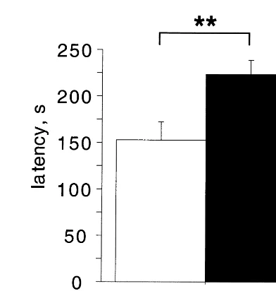
When animals received three rather than one conditioning trial, significant FLI was seen not only in the iNTS but also in the parabrachial nucleus (PBN), and the central nucleus of

Among the thalamic neurons were inhibited by acute immobilization, orexigenic agents, a potent and selective 5-HT 1A receptor which was significantly antagonized by preinjection of