Evidence for a Link Between Pathogenicity and the Role of Imp
Bacterial Transport Effector Proteins in Soybean Infection by
Xanthomonas axonopodis
pv.
glycines
ANY FITRIANI1‡, ANTONIUS SUWANTO1∗, ARIS TRI WAHYUDI1,
AND BUDI TJAHJONO2
1Department of Biology, 2Department of Plant Protection, Institut Pertanian Bogor, Darmaga Campus, Bogor 16680
Xanthomonas axonopodis pv. glycines (Xag) is the causal agent of bacterial pustule disease of soybeans. A non-pathogenic mutant of Xag (M715) was constructed employing transposon mutagenesis which showed similar epiphytic survival inplanta to its wild type strain (YR32). The objective of this work was to identify and to analyze genes involved in pathogenicity in Xag YR32. Inverse Polymerase Chain Reaction (IPCR) was used to isolate the DNA flanking transposon insertion. A 1.3 kb flanking DNA fragment was sequenced and analyzed employing BLAST program to study homology, the position of transposon insertion and to predict the structure and function of the gene. One of the Open Reading Frames (ORFs) shared homology with inner membrane proteins (imps) of
Xanthomonas axonopodis pv. citri (GenBank accession No. NC 003919). Northern blot analysis revealed that an imps
gene was monocistronic and the size of imps mRNA in YR32 was slightly longer than in M715. Reverse Transcriptase-PCR analysis demonstrated that the imps transcript in M715 was much less abundant than in the wild type YR32. Transposon (mini-Tn5-Kmr-Tpr) was determined to be inserted close to the end of C-terminal region of imps gene and might be sufficient to destabilize the imps transcript in M715 and so influence effectors transportation from Xag to plant cell.
Key words: Xanthomonas axonopodis, transposon insertion, non-pathogenic mutant, imps
_____________________________________________
_________________
‡Present address: Department of Biology Education, Universitas
Pendidikan Indonesia, Jalan Dr. Setiabudhi 229, Bandung 40154
∗Corresponding author, Phone/Fax: +62-251-315107,
E-mail: [email protected]
Bacterial pustule disease of soybeans could decrease soybean productivity. A number of researchers have tried to study the mechanism of pathogenicity in Xanthomonas axonopodis pv. glycines (Xag). In our laboratory, this research was initiated by Mesak et al. (1994) thorough the establishment of a modified soybean cotyledon bioassay (Hwang et al. 1992). Rukayadi et al. (2000) obtained a non-pathogenic mutant (M715) which failed to cause disease in planta but with survival rate of fitness on the soybean phyllosphere similiar to that of wild type. This mutant was generated from Tn5 mutagenesis of wild type strain YR32 (Rukayadi 1998). Akhdiya (2000) amplified DNA flanking the transposon in a recombinant plasmid with primer Km-Tn903 and M13F. An amplicon with size 0.7 kb was obtained by PCR. Pratiwi (2004) has been identified as a flanking DNA transposon with a size of 1.8 kb. The DNA fragment was sequenced and analyzed with bioinformatics. There are three frames, (i) frame 1, 68 nucleotides resembles the C-terminal region of type II secretion system protein in Xanthomonas axonopodis pv. citri str. 306, (ii) frame 2, resembles the end sequence of AEO11699 (gene of type II secretion system protein) in X.axonopodis pv. citri str. 306, (iii) frame 3, resembles the gene of iroN that encoded the TonB dependent receptor in X. axonopodis pv. citri str. 306. All of these studies have not revealed the genes involved in the lost of pathogenicity in M715. The objectives of this study were to identify the gene disrupted by the transposon in M715 and analyse the transcript of the disrupted gene in M715 and its wild type (YR32).
MATERIALS AND METHODS
Bacterial Strains and Plasmid. Bacteria used were E. coli strain DH5α (F- lacZrM15 recA hsdR17 gyrA thi) (Sambrook and Russell 2001) and X. axonopodis pv. glycines strain YR32 (Wild type, Rifr ) (Rukayadi 1998) and strain M715 (Rifr Kmr, pat) (Rukayadi 1998; Widjaja et al. 1999). The plasmid used was pFT3551 (Ampr, imp-cp-Xag, pGEM-T Easy).
Growth Conditions and Media. X. axonopodis pv. glycines YR32 and M715 were grown routinely in Luria Bertani broth (LB) at pH 7.0 or on YDCA (10 g of yeast extract, 5 g of dextrose, 20 g of CaCO3, and 20 g of agar added to 1 litre) at 30 oC. Escherichia coli strains were cultured at 37 oC in LB. Antibiotics were supplemented when appropriate at concentrations of 25 (kanamycin = Km), and 100 (ampicillin = Amp, rifampicin = Rif, ) µg ml-1.
Total Genomic DNA and Plasmid Isolation. Total genomic DNA isolation was carried out as described by Lazo et al. (1987). Plasmid isolation and digestion were carried out as described by Sambrook and Russell (2001).
Inverse Polymerase Chain Reaction (IPCR). The strategy for inverse PCR was as far Wahyudi et al. (2001). DNA templates for inverse PCR were prepared from approximately 1 µg of X. axonopodis pv. glycines genomic DNA digested with EcoRV. The digested DNA was further purified by ethanol precipitation. DNA pellets were diluted in 10 µl ddH2O and ligated by ligase (New England Biolabs, Beverly, USA). The ligation reaction was incubated overnight at 16 oC and the DNA was then precipitated with ethanol, and the resultant DNA pellets diluted in 10 µl ddH2O. The circularized DNA generated from the ligation mixture was amplified using Gene Amp PCR 2400 (Perkin Elmer, USA) in a total volume of 25 µl containing 2.5 mM dNTP mixture, GC buffer I, LA Taq DNA polymerase (TaKaRa, Japan), and 10 pmoles of each primer designed from known sequence (primer
1-f: ATCCTTGCCATTG ACCTG-3’ primer 1-r: 5’-CCACCGAAC TTGAACTGGTC-3’). DNA was amplified by PCR consisting of denaturation at 95 oC for 2 min, primer annealing at 62 oC for 1 min, primer extension at 72 oC for 1 min for 30 cycles and 10 min for the last cycle at 72 oC. PCR product was purified by DNA purification methods employing centrifugation (Wizard SV Gel and PCR Clean-Up System, Promega, Madison, USA).
Gene Cloning. Purified PCR products were inserted into pGEM-T Easy vector (Promega, Madison, USA). Ligated reaction was incubated at 16 oC overnight. Competent E. coli DH5α was obtained using CaCl2 methodology for 30 min. DNA was transformed into E. coli DH5α using heat shock treatment at 42 oC for one min.
DNA Sequencing and Analysis. DNA sequencing was carried out using the Dye Terminator Cycle Sequencing kit. M13 forward and M13 reverse primer were used as cycle sequencing primers. Sequencing of the DNA was performed using an automatic DNA sequencer ABI PRISM 3100-AVANT Genetic Analyzer (California, USA). DNA sequences were analyzed employing BLAST (NCBI) (Altschul et al. 1997).
RNA Isolation and Reverse Transcriptase-Polymerase Chain Reaction (RT-PCR). Total RNA was isolated from liquid culture Xag YR32 and M715 after incubation for 28 h (A600 = 0.7). Total RNA was isolated use TRIZOL® Reagent (Invitrogen, California, USA). The quality of the isolated RNA was verified by running gel electrophoresis using 1.5% w/v denaturing agarose gels stained with 0.5 µM ethidium bromide, and the amount of isolated RNA was determined by spectrophotometry at 260 nm and 280 nm. Total RNA samples (5 µg) were reverse-transcribed by M-MuLV reverse transcriptase (ProtoScript First Strand cDNA Synthesis Kit, New England Biolabs, Beverly, USA) from anchored Reverse gene specific primer using standard methods in a reaction volume of 20 µl. cDNA was amplified by PCR using primer (imp-forward and imp-reverse) and prePCR at 95 oC for 3 min, denaturation at 95 oC for 1 min, annealing at 62 oC for 1 min, synthesis at 72 oC for 1 min, and postPCR at 72 oC for 7 min. The amplicon was run by agarose gel electrophoresis using TAE buffer. 16S rDNA was amplified by PCR using 16S rDNA universal primer (63F and 1387R).
Northern Hybridization and Analysis. The total RNA samples (YR32 and M715) (5 µg) were run on 1% w/v denaturing agarose gel for 2 h at 65 V. The gel was stained for 30 min with 0.5 µM ethidium bromide. The gel was blotted onto a nylon membrane (Amersham Life-Science, USA) overnight in 20x SSC. The membrane was washed in 6x SSC at room temperature with agitation for 15 min and dried on blotting paper (Amersham Life-Science,USA), followed by baking at 80 oC for 2 h. A probe consisting of a 375 bp PCR amplified fragment of the inner membrane proteins gene from YR32 was labeled with non-radioactive NEBlot™ Phototope™ Kit (New England Biolabs, Beverly, USA) according to manufacturer’s instructions. Hybridization was performed overnight at 42 oC in hybridization solution (5 ml formamide, 1.0 ml 50x Denhardts solution, 2.5 ml 20x SSPE, 0.1 ml 10% SDS, 50 µl 1 µg ml-1 Salmon sperm DNA (Sigma, USA). The membrane was washed twice in 1x SSC, 0.1% SDS for 10 min at room temperature and twice in 0.5x SSC, 0.1% SDS for 10 min at 50 oC and then dried on blotting paper (Amersham Life-Science, USA). Nucleic acids were detected
using the Phototope™ Detection Kit (New England Biolabs, Beverly, USA) according to the manufacturer’s instructions. The membrane was exposed to X-ray (Hyperfilm™ MP, Amersham Life-Science, USA) in dark room. X-ray film was processed in high performance X-ray film developer (Fuji Photo Film Co., Ltd, Japan) and washed in water. X-ray film was washed in X-ray film fixer (Fuji Photo Film Co., Ltd, Japan) and then washed in water and air dried.
RESULT
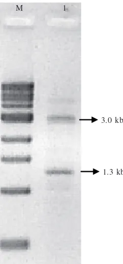
Inverse PCR generated two DNA bands, i.e.: 1.3 kb and 3.0 kb (Fig 1). It is surprising that the same nucleic acid sequence in Xag chromosome could result in more one band in PCR product. It is possible that the primer could be complemented with two sites on the chromosome until it formed two amplicons. For this study, we only analyzed 1.3 kb fragment because the DNA sequence has aligned nucleotide with previous DNA sequence (Pratiwi 2004).
Inverse PCR product was purified and inserted to pGEM-T Easy (3.015 kb), giving pFpGEM-T3551 (Fig 2). Plasmid verification was done with restriction analysis which showed that the insert was 1.3 kb long and the nucleotide did not have EcoRI, PstI and SacI sites (data not shown).
The DNA sequence was analyzed employing BLAST Program (NCBI). BLASTN analysis revealed that 1.3 kb nucleotides was homologous with Xanthomonas axonopodis pv. citri str. 306. Alignment analysis of 1.3 kb nucleotides showed a 99% identity with Xanthomonas axonopodis citri str. 306, and E-value of zero. Nucleotides of 1.3 kb encoding Inner Membrane Proteins (IMPs) with an identity of 90% and Cystein Proteases (CPs) with an identity of 99% in Xanthomonas axonopodis pv. citri str. 306. Open reading frame (ORF) analysis revealed the presence of two genes in different operons. IMPs and CPs consisted of 182 and 153 amino acids, respectively (data not shown). Genes of imps are involved in pathogenicity (von Heijne 1992). The genes of imps were found at the start codon (ATG), but the stop codon (TGA) was as found in the Pratiwi (2004) sequence after assembly. Frequency for TGA in Xanthomonas axonopodis citri str. 306 is about 70.89% (http://rice.tigr.org/tigr_scripts/CMR2/codon_tables). Putative rbs (Ribosome Binding Site) and the promoter of imps genes (Tang 1991; Katzen et al. 1996; Baldini et al. 1999) have been found (Fig 3). The transposon inserted at the C-terminal region of the genes (Fig 3 and 4).
The quality of isolated total RNA was confirmed by denaturing agarose gel electrophoresis. Two intact ribosomal bands characteristic of undegraded RNA were observed. The sample RNAs also had a normal spectra (A260/A280 = 2.0) and could be used for cDNA synthesis and RT-PCR. Two intact ribosomal bands showed that from upper to lower, 23S rRNA, 16S rRNA. Usually, sizes of ribosomal RNAs in bacteria are 1.5 kb and 2.9 kb for 16S rRNA and 23S rRNA respectively (data not shown).
Fig 2 Plasmid pFT3551.
Fig 1 Agarose gel electrophoresis profile of IPCR product. M: 1 kb DNA ladder (New England Biolabs,Beverly, USA). 1: IPCR.
Fig 3 DNA sequence of imps genes, amino acids, and position of transposon insertion (black arrow). -35, -10: promoter, rbs: ribosome binding site; start: start codon; stop: stop codon.
acagtggcggtcgtcgaataaccgg
-35
gtcgcgcacttcaccttcgcttcaaattcgcctcaaacctgcgac
-10
cgaaagtgactgttgacacgactcttagagaccataagaatcaac
R K * terminator terminator
Stop
tccactgaattggtactttccagtcagggccgttgatcagcaatg
pFT3551 as the positive control. Distilled water, Y16S and M16S are the negative control and have no amplicon. Yield of amplicon of YR32 was much more than of M715. The gene for inner membrane proteins has been successfully transcribed in YR32, but not in M715 (Fig 6).
In this research, we used of universal primers for 16S rDNA. The aim was to examine the quality of first strand cDNA synthesized. No PCR product after amplification by primers shows that the cDNA formed originated from transcript mRNA of inner membrane protein genes in Xag. pFT3551 (4.3 kb) which is a DNA plasmid that possesses inner membrane proteins and cystein proteases genes from Xag. pFT3551 was amplified by inner membrane protein primers and has the same product length (about 375 bp). This is demonstrates that the oligonucleotides have the correct sequence for DNA coding for bacterial of inner membrane proteins.
DISCUSSION
Pustule disease is one of the five main diseases in soybeans. Pustule disease attacks soybean’s leaves and the symptom of pustule disease is chlorosis with a yellow spot in the centre. Pustule disease in soybeans causes a decrease in productivity in Indonesia.
In our laboratory, the mechanism of pathogenicity has been studied since 1995. Rukayadi (1998) have been constructed the mutant M715. M715 has a phenotype of De Lorenzo et al. (1990) described that mini Tn5-Kmr has
stop transcription points at two DNA flanks of the resistance antibiotic gene. The signal of the mRNA transcript in M715 was weaker than inYR32. The level of transcription in M715 was lower than in YR32.
0.6 1.5
2.9
kb M 1 2 3 4 1 2 3 4 kb
a b
Fig 5 RNA electrophoresis and RNA-blot analysis of YR32 and M715. a. RNA electrophoregram of YR32 and M715, b. RNA-blot of YR32 and M715 with inner membrane proteins probe. Lane 1 and 2: YR32, lane 3 and 4: M715; lane M: 1 kb DNA ladder (New England Biolabs, Beverly, USA).
miniTn5-Kmr-Tpr
Fig 4 Physical map of the genes and position of transposon insertion ORF 1: cystein proteases. ORF2: inner membrane proteins, ORF3: TonB dependent-receptor.
ORF3 ORF2
BstZ17i Xcml
Nsil ORF1
Alul Hincll
Msel Kpnl Sall Notl
2198 bp
375 bp M YR32 M715 ddH2O Y16S M16S pFT3551
Fig 6 RT-PCR of inner membrane proteins from total RNA sample. M: Nugen Marker; YR32: Xag YR32 (RT of YR32, PCR with imps primer); M715: Mutant M715 (RT of M715, PCR with imps primer); ddH2O: negative control (Distillated water, PCR with imps primer); Y16S:
negative control (RT of YR32, PCR with 16S rDNA primer); M16S: negative control (RT of M715, PCR with 16S rDNA primer); pFT3551: positive control (pFT3551, PCR with imps primer).
kb
1.25
0.7
non-pathogenicity in soybean and no hypersensitive reaction in tomato. Mutant M715 was constructed by transposon mini Tn5-Kmr-Tpr. Our research revealed that mini Tn5-Kmr -Tpr was inserted in the inner membrane proteins genes. Imps genes were involved in the protein transportation apparatus especially of virulence, toxins, and pathogenicity factor determinants transported from the cell to the environment (von Heijne 1992). In E. coli, Imps function in colicin transportation (Marchler-Bauer and Bryant 2004). Quinaud et al. (2005) and Edqvist et al. (2003) revealed that in Pseudomonas aeruginosa and Yersinia, Imps was a part of the Type III secretion system (T3S) that plays key roles in pathogenicity and are employed to inject toxin (effectors) directly into the cytoplasm of target cells.
Analysis of mRNA showed that inner membrane proteins was successfully transcribed in Xag YR32 but not in M715. The amplicon formed in M715 was weaker than in YR32. This phenomenon strongly indicated that some factor interfered in M715 expression and we consider that this is because M715 was interrupted by a transposon. The mRNA of imps in M715 was not stable and had shorter half-life. Additionally the size of translated protein differed from that of the wild type. Transposon mutagenesis caused a change of nucleotide sequence in the position of transposon insertion. In our research, we used the composite transposon mini Tn5-Kmr-Tpr ,derived from Tn5, which has terminator transcription regions at two flanking antibiotic resistance genes (de Lorenzo et al. 1990).
Bioinformatics analysis revealed that the transposon was inserted in the C-terminus of the imps gene. This conclusion was strongly supported by northern blotting analysis. Northern blotting indicated the inner membrane protein gene system was monocistronic, because size of its the transcript was the same as for the genes (about 600 bp). This phenomenon supported bioinformatics analysis which showed that in the DNA sequence of imps genes was a terminator stop transcription. In addition to the size of the transcript mRNA in M715 was shorter than in YR32 (Fig 5). If the transposon inserted close to the end of C-terminus in M715, then the result would be that RNA polymerase could be stopped in stop transcription of the transposon at the C-terminal end of the genes.
The C-terminal region of proteins has a functional role in whole protein structure. Tateno et al. (2006) described the C-terminus deletions and site-directed mutagenesis in membrane calcium channel would result in misfolding of the C-terminus and/or inaccessibility to trafficking/sorting machineries. Takazaki et al. (2006) explained that mutation at the C-terminus of a human anion transporter affected the rate of conformational change of this protein. They concluded that the C-terminal region has a functional role in the conformational change capacity that is necessary for anion transport. Han et al. (2006) reported the C-terminal region of a potassium channel plays a critical role in the localization and gating of the channel.
The mutagenesis transposon in imps has a similar result. Mutation at the C-terminus of Imps affected protein functionality. A mutant Imps was recognized by the signal recognition particle (SRP) at the hydrophobic N-terminal and transported to the bacterial inner membrane. When studying protein coded in the SecA-SecYEG complex, mutant Imps
have a changed configuration and this influences localization in the inner membrane. Changed localization and configuration of mutant Imps affects functionality of coded proteins. In conclusion, the phenotype of M715 was not pathogenic and gave no hypersensitive reaction in tomato plants (Rukayadi et al. 2000). Here we conclude that this phenotype is a result of damaged bacterial proteins Imps which have a role as effectors in protein translocation. We consider a defective (protein) transportation system is the reason for the loss of pathogenicity by Xag.
ACKNOWLEDGEMENT
This report is part of AF dissertation and primarily funded by the Research Center for Microbial Diversity, Bogor Agricultural University. We thank Yul Kurniarun and Research and Development PT. Charoen Phokphand Indonesia for the laboratory facilities used in RNA analysis.
REFERENCE
Akhdiya A. 2000. Gene cloning that involved pathogenicity in
Xanthomonasaxonopodis pv. glycines [Thesis]. Bogor: Bogor
Agricultural University.
Altschul SF, Gish W, Miller W, Myers EW, Lipman DJ. 1997. Basic local alignment search tool. J Mol Biol 215:403-410.
Baldini RL, Tahara ST, Rosato YB. 1999. A rolling circle miniplasmid
of Xanthomonas campestris pv. glycines: The nucleotide sequence
and its use as a cloning vector. Plasmid 42:126-133.
de Lorenzo DV, Herrero M, Jakubzi U, Timmis KN. 1990. Mini-Tn5 transposon derivatives for insertion mutagenesis, promoter probing, and chromosomal insertion of cloned DNA in Gram-Negative Eubacteria. J Bacteriol 72:6568-6572.
Edqvist PJ et al. 2003. YscP and YscU Regulate Substrate Specificity of the Yersinia Type III Secretion System. J Bacteriol 185:2259-2266.
Han W, Nattel S, Noguchi T, Shrier A. 2006. C-terminal domain of KV4.2 and associated KCHIP2 interactions regulate functional expression and gating of KV4.2. J Biol Chem (in press). Hwang PL. Harsono HD, Shaw PD. 1992. Use of detached soybean
cotyledons for testing pathogenicity of Xanthomonas campestris
pv. glycines. Plant Dis 76:182-183.
Katzen F, Becker A, Zorreguita A, Puhler A, Lelpi L. 1996. Promoter analysis of the Xanthomonas campestris pv. campestris gum operon directing biosynthesis of the xanthan polysaccharide. J
Bacteriol 178:4313-4318.
Lazo GR, Roffey R, Gabriel DW. 1987. Conservation of plasmid DNA sequences and pathovar identification of strains of
Xanthomonas campestris. Phytopathology 77:448-453.
Marchler-Bauer A, Bryant SH. 2004. CD-Search: protein domain annotations on the fly. Nucleic Acids Res 32:327-331. Mesak FM, Suwanto A, Tjahjono B, Guhardja E. 1994. Modifikasi
bioesei kotiledon kedelai untuk uji patogenisitas Xanthomonas
campestris pv. glycines. J Il Pert Indon 4:77-82.
Pratiwi E. 2004. Analysis of DNA sequence involved in pathogenicity and design of specific PCR primer of Xanthomonas axonopodis
pv. glycines [Dissertation]. Bogor: Bogor Agricultural University.
Quinaud M et al. 2005. The PscE-PscF-PscG Complex Controls Type III Secretion Needle Biogenesis in Pseudomonas
aeruginosa. J Biol Chem 280:36293-36300.
Rukayadi Y. 1998. Construction of partial genetic map and epiphytic survival of non-pathogenic mutant of Xanthomonas axonopodis
(campestris) pv. glycines YR32 [Dissertation]. Bogor: Bogor
Agricultural University.
Rukayadi Y, Suwanto A, Tjahjono B, Harling R. 2000. Survival and ephiphytic fitness of a nonpathogenic mutant of Xanthomonas
campestris pv. glycines. ApplEnviron Microbiol 66:1183-1189.
Takazaki S et al. 2006. The functional role of arginine 901 at the C-terminus of the human anion transporter band 3 protein. J
Biochem 139:903-912.
Tang J. 1991. Genetic and molecular analysis of a cluster of rpf genes involved in positive regulation of synthesis of extracellular enzymes and polysaccharide in Xanthomonas campestris pv.
campestris.Mol Genet 226:409-441.
Tateno T, Nakamura N, Hirata Y, Hirose S. 2006. Role of C-terminal of Kir7.1 potassium channel in cell-surface expression. Cell Biol Int 30:270-277.
von Heijne G. 1992. Membrane protein structure prediction. Hydrophobicity analysis and the positive-inside rule. J Mol Biol 225:487-494.
Wahyudi AT, Takeyama H, Matsunaga T. 2001. Isolation of
Magnetospirillum magneticum AMB-1 mutants defective in
bacterial magnetic particle synthesis by transposon mutagenesis.
Appl Biochem Biotechnol 91:147-154.