Cytology technique: development of a simple spot method for cultured
cell suspension
Jeanne A. Pawitan,1 Lia Damayanti,1 Arleni,2, 3 Ni M. Swantari4
1 Department of Histology, Faculty of Medicine, University of Indonesia, Jakarta, Indonesia 2 Depatment of Biology, Faculty of Medicine, University of Indonesia, Jakarta, Indonesia
3 Immunology and Endocrinology Integrated Laboratory, Faculty of Medicine, University of Indonesia, Jakarta, Indonesia 4 Division of Plastic Surgery, Department of Surgery, Faculty of Medicine, University of Indonesia, Jakarta, Indonesia
Abstrak
Tujuan Mengembangkan cara spot sederhana untuk melekatkan suspensi sel hasil kultur pada kaca objek.
Metode Kami membandingkan tiga cara, masing-masing pada kaca objek biasa dan khusus (Shandon-Polysin). Ketiga cara tersebut adalah membuat sediaan spot kecil secara langsung, atau dengan menambahkan 3 atau 10 µl fetal bovine serum (FBS) per 20 µl sampel. Dengan demikian, secara keseluruhan ada 6 cara. Hasilnya dinilai secara kualitatif dalam hal warna latar belakang, warna dan keutuhan batas spot, dan bagian yang terlipat dan terlepas. Selanjutnya, untuk tiap kaca objek, jumlah sel utuh yang melekat dihitung, dan persentase sel utuh yang melekat per jumlah sel yang dibuat spot juga dihitung. Perbedaan sel utuh yang melekat di antara keenam cara di atas dianalisis dengan ANOVA menggunakan program SPSS 13.0 for Windows.
Hasil Tidak ada perbedaan bermakna di antara keenam cara di atas dalam hal persentase sel utuh yang melekat (P= 0,804), walaupun cara yang menggunakan kaca objek khusus tanpa penambahan FBS (kadar FBS inal 5%) menghasilkan persentase sel utuh melekat yang paling tinggi, dengan latar belakang bersih tanpa lipatan.
Kesimpulan Kami telah mengembangkan cara spot sederhana untuk membuat sediaan sitologi suspensi sel hasil kultur, dan hasil terbaik didapat dengan menggunakan kaca objek khusus dengan suspensi sel berkadar FBS 5%. (Med J Indones 2010; 19:26-31)
Abstract
Aim To develop a simple spot method to attach cultured cells in suspension on to a glass slide.
Methods We compared three approaches using both conventional and special glass slide (Shandon-Polysin)., either without additional fetal bovine serum (FBS), or with addition of 3 or 10 µl of FBS to a 20 µl sample (altogether there were six approaches). The slides were examined qualitatively for the background color, boundary color and intactness, and whether there were folded and detached parts. Further, for each slide, the attached intact cells were counted, and the percentage of attached intact cells per number of spotted cells was calculated. The difference in attach intact cells between different approaches was analyzed by ANOVA using SPSS 13.0 for windows.
Results There were no signiicant difference in the percentage of attached intact cells between the six approaches (P=
0.804), though the approach using special glass slide without additional FBS (FBS inal concentration 5%) yield the highest percentage of attached intact cells, showed clean background without folded parts.
Conclusions We have developed a simple spot method for cultured cell suspension, and the best approach to make spot specimen is using special glass slide with 5% FBS in the cell suspension. (Med J Indones 2010; 19:26-31)
Key words:spot specimen, special glass slide, fetal bovine serum
Correspondence email to: [email protected]
Visualization of stained cultured cells is usually done by staining of either cultured cells on chamber slides,1,3 on coverslips, glass slide in culture dishes, or
wells of tissue culture plates.4-7 These methods require
an additional culture that need at least one day for culturing and additional culture medium. Moreover, when various kinds of staining are needed, the number of culture dishes, wells or chamber slide rises according to the number of staining method used, and thus will increase the cost.
Another method is by making cytology specimens from cultured cell suspension using a cytospin apparatus.8
This method needs a cytospin apparatus that is not always available in a basic laboratory. Further, the use of disposable cytospin chambers raises the cost, while using reusable chambers often leads to contamination, if the chambers are not washed thoroughly.9
Vol. 19, No. 1, February 2010 Simple spot method 27
Methods
This research is part of a research on adipose tissue derived stem cells, which has got an approval from the ethical committee of the Faculty of Medicine University of Indonesia. This research was done in the Immunology and Endocrinology Integrated Laboratory, and the Department of Histology, Faculty of Medicine, University of Indonesia, from October 2009 to January 2010.
sample and sample processing
The samples were adipose tissue derived trypsinized cultured cell suspensions. The trypsin in the cell suspensions was neutralized by equal volume of
culture medium containing 10% fetal bovine serum
(FBS), Penicillin - Streptomycin and Amphotericin B in Dulbeco Minimal Eagle Medium (DMEM).
Procedure of spot method
The cells in cell suspensions were counted in Neubauer chamber, on an inverted microscope. Then 1-2 µl of cells in aliquot samples were spotted on to clean glass slide, using a micro pipette and a yellow tip. If air bubbles developed, they were broken by a sewing needle. We tried various approaches i.e.: Approach-1: spotting the cell suspension directly on conventional glass slide; approach-2: spotting the cell suspension directly on special glass slide; approach-3: before spotting on to conventional glass slide, 20 µl of cell suspension was mixed with 3 µl FBS; approach-4: before spotting on to special glass slide, 20 µl of cell suspension was mixed with 3 µl FBS;
approach-5: before spotting on to conventional glass slide,
20 µl of cell suspension was mixed with 10 µl FBS; and approach-6 before spotting on to special glass slide, 20 µl of cell suspension was mixed with 10 µl FBS
The spotted cell suspensions were air dried and ixed using 100% ethyl alcohol (proanalysis, Merck) for 5 minutes, stained by hematoxylin and eosin (HE)
staining, cleared in xylene (proanalysis, Merck), mounted in mounting medium (Entellan) and covered by a coverslip (22 x 22mm, Assistent).
he staining procedure
Before staining, the dried cell suspension were
rehydrated using ethyl alcohol 70%, 50% and distilled water for 5 minutes each subsequently. Then the slides
were immersed in hematoxylin solution for 1 minute, washed in running tap water, followed by 3 dips in 0.1N HCL, several dips in distilled water, and immersed in eosin solution for 3 minutes. After that, the slides were washed in distilled water, followed by dehydration in
50%, 70%, 95%, and 100% ethyl alcohol for 5 minutes
each subsequently, dipped several times in xylene, and
inally a drop of mounting medium was added on to the
glass slide to cover the spot and covered by a coverslip.
Every approach was done in 5 replicates. The number
of attached intact cells on each glass slide was counted under an ordinary light microscope, with 400 times of
magniication. Cell counting was done on the whole
spotted specimen.
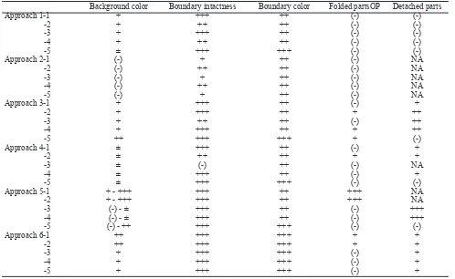
Further the background was examined for its color, and the boundary was examined for the color and intactness. Our preliminary attempts to make spot specimens showed that the spotted cell suspension formed a thin layer when it was dried. In some of them, part of the thin layer was detached or folded. Therefore, in addition to the above examinations, the whole specimen was examined whether there were folded or detached parts, and they were qualitatively evaluated.
Background color Boundary intactness Boundary color Folded partsOP Detached parts
Approach 1-1 + +++ ++ (-) (-)
-2 + ++ ++ (-) (-)
-3 + +++ ++ (-) (-)
-4 + ++ ++ (-) (-)
-5 ± +++ +++ (-) (-)
Approach 2-1 (-) + ++ (-) NA -2 (-) ++ ++ (-) NA -3 (-) + ++ (-) NA -4 (-) ++ ++ (-) NA -5 (-) + ++ (-) NA Approach 3-1 + +++ ++ (-) + -2 + +++ ++ + ++
-3 + ++ ++ (-) ++
-4 + +++ ++ + ++
-5 ++ +++ +++ + (-)
Approach 4-1 ± +++ ++ (-) + -2 ± ++ ++ + +
-3 ± (-) ++ (-) NA -4 ± +++ ++ (-) +
-5 ± +++ +++ (-) (-)
Approach 5-1 + - +++ +++ ++ +++ NA -2 + - +++ +++ ++ +++ NA -3 (-) - ± +++ ++ (-) +++
-4 (-) - ± +++ ++ (-) +++
-5 (-) - ++ +++ +++ (-) (-)
Approach 6-1 ++ +++ +++ + + -2 ++ +++ +++ + +
-3 + +++ +++ (-) +
-4 + +++ +++ (-) +
-5 + +++ +++ (-) +
NA= not applicable, background and boundary color: (-)= very clear, ±= clear with some pale dots, += pale, ++= lightly colored, +++= colored, back
-ground boundary: (-)= not seen, += 0-25% intact, ++= 26-75% intact, +++= >76% intact, folded parts: (-)= not seen, += 0-25% folded, ++= 26-50% folded, +++= >50% folded, detached parts: (-)= not seen, += 0-25% detached, ++= 26-50% detached, +++= >50% detached
Table 1. Backgrounds and images of the six approaches
Figure 1. Background and boundary color assessment
Vol. 19, No. 1, February 2010 Simple spot method 29
Background evaluation (color and boundary), folded and detached parts for every slide was qualitatively evaluated, noted, and presented descriptively. Further, the number of cells in every cell suspension was noted. The number of cells spotted was calculated from the amount spotted and the number of cells in the suspension, and then noted. The number of attached intact cells on each glass slide was noted, and the percentage of attached intact cells from the amount of spotted cells was calculated. compared by ANOVA. Before ANOVA procedure was done, homogeneity and normality of the data were tested using Levene test and 1 sample Kolmogorov Smirnov test, respectively. Finally, the procedure with the highest percentage and most clear background, least folded and detached part was regarded as the best method.
Results
In approach 1 and 2, the cell suspensions were spotted directly on to glass slides, without additional FBS. The
FBS concentration in the two approaches was 5% (10%
FBS in the medium was diluted by equal amount of
trypsin). The inal FBS concentration in both approach 3 and 4, and both approach 5 and 6 could be calculated and yielded around 20% and 38% FBS respectively.
The background image of the six approaches can be
the percentage of attached intact cells between the six approaches (P= 0.804).
Table 1 shows that additional FBS caused colored background. As clear background is preferred, additional FBS did not give good result in term of background color. In contrast, we preferred darker and intact boundaries when counting the cells, as sharp boundary helped to guide the examiner to what extent the slide should be moved. However, sometimes the boundaries were severely damaged (Table 1). Therefore counting took more time, as we had to move further than the boundary, and additional FBS did not give concluding result in term of boundary intactness. Moreover, table 1 shows that additional FBS tended to increase folded and detached part.
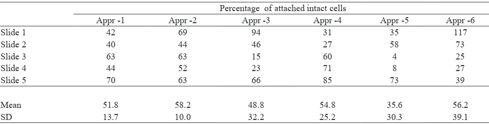
Table 2 shows that approaches using special glass
slides i.e. approach-2 without additional FBS (inal FBS concentration 5%) followed by approach-6 with
the highest concentration of FBS showed the highest
yield of attached intact cells, that were 58.2% and 56.2% respectively.
disCussion
Addition of FBS (5-10%) in cell suspension to be
cytocentrifuged is recommended in making cytological specimens.10 In order to preserve the cells and to get
more intact cells, we used additional FBS (approach 3-6) to neutralize the trypsin.
In term of the background color, clear background is preferred. However, this study showed that additional Percentage of attached intact cells
FBS caused colored background. This result is in line with the recommendation to reduce protein content as excess protein may interfere with staining reaction.9
Further, as additional FBS tended to increase folded and detached part, but did not give concluding result in term of boundary intactness, additional FBS did not give any additional value to the quality of the specimens in term of background color, cell layer and boundary intactness, and prevention of cell layer folding.
In term of the amount of attached intact cells, additional FBS might count for the increase in intact cells, as FBS can neutralize the damaging effect of trypsin. Moreover, additional FBS might increase attached cells as it may serve as adhesive between the cells and glass slide, such as the use of albumin as glass slide coating.9 Although there was no signiicant difference between
the 6 approaches, approach-2 using special glass slides
without additional FBS (inal FBS concentration 5%) showed the highest yield of attached intact cells (58.2%) (Table 2). This result showed that 5% FBS is enough to
neutralize the damaging effect of trypsin, and to serve as adhesive.
In contrast, approach-5 that contained the highest FBS
concentration using conventional glass slide yielded the lowest number of attached intact cells. This result may be due to the increase in detached part from the conventional glass slide due to excess FBS that negate the increase in the number of attached intact cells. However, approach-6 that also contained the highest
FBS concentration as in approach-5 yielded the second
highest number of attached intact cells. The reason is supposed to be due to the use of special glass slide in approach-6, as the special glass slide is claimed to be superior compared to conventional glass slide in term of cell attachment, which might play a role in the prevention of the detachment of the cell layer.
The limitation of this study was that counting was done by a single examiner, and the blinding method only
worked for the 3 kinds of FBS inal concentration, as
the conventional and special glass slide was readily distinguishable.
This spot method is very simple and was proven to endure routine HE staining that uses dips in acid solution. To know whether the specimen can endure immunocytochemical staining that involves severe treatments without detachment of the cell layer, further study should be done.
If this simple method can endure immunocytochemical staining, it will be very valuable in stem cell research,
as it can be used for the identiication of the various
kinds of stem cells and the calculation of the proportion of a certain kind of stem cell using 100-200 cells only. In conclusion, we have developed a simple spot method for cultured cell suspension, and the best approach to make spot specimen is using special glass slide with
5% FBS in the cell suspension.
Acknowledgment
The authors would like to thank Mrs. Trimiatun for her technical assistance in preparing the samples, and Dr. Gentur Sudjatmiko spBP for facilitating the collection of adipose tissue specimens that were used to prepare the samples. This study was funded by DRPM UI, al. Multilineage cells from human adipose tissue: implications for cell-based therapies. Tissue Eng.(2001; 7: 211–26. 3. Kern S, Eichler H, Stoeve J, Klüter H, Bieback K.
Comparative Analysis of Mesenchymal Stem Cells from Bone Marrow, Umbilical Cord Blood, or Adipose Tissue. Stem Cells. 2006;24;1294-1301. Available from: http:// www.StemCells.com.
4. Jurgens WJFM, Oedayrajsingh-Varma MJ, Helder MN, Doulabi BZ, Schouten TE, Kuik DJ, et al. Effect of tissue-harvesting site on yield of stem cells derived from adipose tissue: implications for cell-based therapies. Cell Tissue Res. 2008; 332:415–26.
5. Mitchel JB, Mc Intosh K, Zvonic S, Garret S, Floyd ZE, Kloster A, et al. Immunophenotype of human adipose-derived cells: temporal changes in stromal-associated and stem cell–associated markers. Stem Cells. 2006;24:376–85 6. Oedayrajsingh-Varma MJ , van Ham SM, Knippenberg
M, Helder MN, Klein-Nulend J, Schouten TE, et al. Adipose tissue-derived mesenchymal stem cell yield and growth characteristics are affected by the tissue-harvesting procedure. Cytotherapy. 2006; 8:166–77.
7. Kretlow JD, Jin YQ, Liu W, Zhang WJ, Hong TH, Zhou G, et al. Donor age and cell passage affects differentiation potential ofmurine bone marrow-derived stem cells. BMC Cell Biology. 2008;9:60. Available from: http://www. biomedcentral.com/1471-2121/9/60. (13 pages).
Vol. 19, No. 1, February 2010 Simple spot method 31
cell subpopulations identiied in fresh human adipose tissue stromal vascular fraction and in the derived mesenchymal stem cells. Journal of Translational Medicine. 2007;5:55. Available from: http://www.translational-medicine.com/ content/5/1/55. 10 pages.
9. Bales CE. Laboratory techniques. In: Koss LG. Koss’ diagnostic cytology and its histopathologic bases. 5th
edition. Vol.II. Philadelphia: Lippincott William and Wilkins; 2006. p. 1569-1634.