How to cite:
Mansyur M, Yudaningtyas E, Prawiro SR, Widjajanto E (2018) The Effect of Low Power Ultrasonic Wave Exposure to Suppress Methicillin-Resistant Staphylococcus aureus (MRSA) In Vitro. J. Trop. Life. Science 8 (2): 144 – 150.
*Corresponding author: Mas Mansyur
Biomedical Department and Biomolecular Research, Faculty of Medicine, University of Wijaya Kusuma
Surabaya, Indonesia.
The Effect of Low Power Ultrasonic Wave Exposure to
Suppress Methicillin-Resistant
Staphylococcus aureus
(MRSA)
In Vitro
Mas Mansyur 1*, Erni Yudaningtyas 2, Sumarno Reto Prawiro 3, 5, Edi Widjajanto 4, 5
1 Biomedical Department and Biomolecular Research, Faculty of Medicine, University of Wijaya Kusuma, Surabaya, Indonesia. 2 Department of Electrical Engineering, Faculty of Engineering, Brawijaya University, Malang, Indonesia
3 Department of Microbiology, Faculty of Medicine, Brawijaya University, Malang, Indonesia 4 Department of Clinical Pathology, Faculty of Medicine, Brawijaya University, Malang, Indonesia
5 dr. Saiful Anwar Public Hospital, Malang, Indonesia
ABSTRACT
The incidence of methicillin-resistant Staphylococcus aureus (MRSA) infection keeps increasing in every part of the
world. Currently, the infection prevalence of MRSA has reached 70% in Asia. In Indonesia in 2006 the prevalence was 23.5%; the infection prevalence of MRSA in RS Atmajaya Jakarta reached 47%, in RSUP Dr. Moh. Husin Palembang reached 46%, and RSUD Abdul Moeloek Lampung in 2013 reached 38.4%. MRSA is multiresistant to antibiotics and is hard to kill compared to most other negative gram bacteria. The purpose of this research is to find the lethal power and exposure of ultrasonic waves to kill MRSA, monitoring its effects via changes in shape, size, structure and Gram staining as indicators. The observations were done macroscopically by culturing the MRSA in a petri dish filled with Chromagar MRSA medium, while the morphological observations of MRSA were done by SEM, changes in the structure using TEM, and changes in the color of MRSA cells using Gram staining. Ultrasonic wave exposure, at a lethal power = 8.432 watt, killed a significant percentage of MRSA over the control (p = 0.000). The death indicators of the MRSA due to exposure to ultrasonic waves of various power were: changes in shape of MRSA affected by ultrasonic power (p = 0.005), changes in size is not affected by ultrasonic power (p= 0.470), the stain of MRSA cell staining from purple to pink affected by ultrasonic power (p = 0.000), all compared with the control. MRSA died due to necrosis, with physical evidence of the MRSA death such as mechanical stress marked by swollen MRSA cell, shift cell wall, crack and tears, cavitation marked by pieces of MRSA cell in the field of view due to explosions inside the cell, change to an irregular cell shape, and changes in color from black to transparent.
Keywords: Cavitation, mechanical stress, MRSA, SEM, TEM, ultrasonic
INTRODUCTION
Methicillin-resistant Staphylococcus aureus(MRSA)
has developed from selection pressures caused by inap-propriate antibiotic therapy exposure [1]. MRSA is mul-tiresistant to many antibiotics used to control negative Gram bacteria [2]. Thus, MRSA is one of the most prominent pathogens causing health problems in the human community as well as livestock [3, 4, 5, 6, 7]. MRSA must be eradicated by effective methods that will not cause further resistance to develop.
waves with low frequency and are used to break cell
membranes and make the cells lyse [10 – 16]. Cavitation
produced by ultrasonic waves can break bacterial cell walls, as well as structural and functional components via intracellular cavitation [17, 18]. Ultrasonic energy absorption by enzymatic proteins can cause changes in enzymatic activity. The hypothesis of frequency reso-nance explains two biological mechanisms that might change protein function as the result of ultrasonic en-ergy absorption [19].
When bacteria are in intensive fields of ultrasonic waves with high frequency, then the bacteria will suffer great vibratory disruptions and a large voltage, in which sealing and stretching occur because of differences of pressure between the inside and outside of the cell wall. When the strain in the cell wall is large enough to exceed the limit of elasticity then the cell wall will be torn and the bacteria will die. Another mechanism of interaction of ultrasonic waves with bacteria is the effect of cavita-tion. The effect of cavitation can break molecular bonds.
The broken H2O molecule forms H+, OH-, and HO2
radicals as well as a great deal of H2O2, which will
oxi-dize organic molecules throughout the bacterial cell causing cell death [20].
Various experiments have studied controlling
Esch-erichia coli bacteria using ultrasonic waves. Dehghani and Hadi (2005) used 42 kHz frequency, 70-watt power waves and were able to kill 99.80% of the bacteria [21]. Herceg et al. (2013) used 20 kHz frequency, 600-watt power waves, which combined with high temperatures, were able to kill E. coli 3014, S. aureus 3048, Salmonella
sp. 3046, Listeria monosytogeneses ATCC 23074, and
Bacillus cereus 30 [2]. Kumar et al. (2014) tested various parameters to decrease bacterial populations in sludge, i.e., frequency (35 kHz and 130 kHz), power 250 watts and time period (5, 10, 20 and 30 minutes) [22]. As the frequency and time period increased, the bacterial pop-ulation was decreased. It was also observed that a 130 KHz frequency was more effective than 35 kHz. Li et al. (2016) used ultrasonic waves with a frequency of 20 kHz
with a power and irradiation time of 60 W·m-2 and 0 to
20 minutes, respectively. The rates of killing of E. coli is
bigger than S. aureus [23].
The ultrasonic method has advantages over other methods to control MRSA, such as 1. It does not use antibiotics so that it does not cause antibiotic resistance; 2. It kills MRSA physically and chemically simultane-ously and there is no chance to cause antibiotic re-sistance; 3. Uses relatively less power (maximum 8.4
watts) than similar methods in other research; 4. Physi-cal proof of the death of MRSA and the causes can be clearly observed; 5. It has the potential to be developed as a wound debridement because it can selectively dis-solve fibrin without harmful macroscopic changes in granulation tissue [24]. Many studies have used ultra-sonic waves to kill bacteria, however, the mechanism of the death and study of the physical evidence of the dead bacteria have never been done. This research aims to find the lethal power of the ultrasonic wave exposure to kill MRSA in vitro while studying the mechanism of death using the indicators changes in shape, size, struc-ture and Gram staining of MRSA cells.
MATERIALS AND METHODS
MRSA isolates were obtained from the Microbiol-ogy section of RS Dr. Soetomo, grown in Luria Bertani Broth Miller M1245-500G then diluted by graphic wa-ter. A total of 100 ml MRSA suspension exposed by an ultrasonic wave with frequency 26 [25] and a power of 2, 3, 4, 5, or 6 watts for 2 minutes. Then, the bacteria were cultured in a petri dish filled with Chromagar MRSA MR 500 POOO204, observing the grown MRSA population after 24 hours, using a Quebec colony coun-ter, to create a regression equation to obtain a lethal power curve (100% cell death).
The macroscopic observation was done by the TPC method; microscopic observation using SEM was per-formed to understand changes in morphology of MRSA cells, changes in the structure of MRSA cells using TEM, and the changes in cell staining using Gram staining. This research was approved by the ethics committee of Medical Faculty of Wijaya Kusuma Surabaya University. No.485/SLE/FK/UWKS/IX/2014.
The death indicators of MRSA were changes in: shape, size, structure and cell staining, assessed via elec-tron microscopy and Gram staining. The death percent-age of MRSA (the amount of dead MRSA in a treatment divided by dead MRSA in the control multiplied by 100%) of the various treatments was statistically ana-lyzed using ANOVA, while the more specific effects were tested via regression. Before performing the ANOVA test, the data were first tested for normality and homogeneity. The level of confidence used in all tests
was α = 0.05. The statistical analysis process used
soft-ware program MINITAB 16.
RESULTS AND DISCUSSION
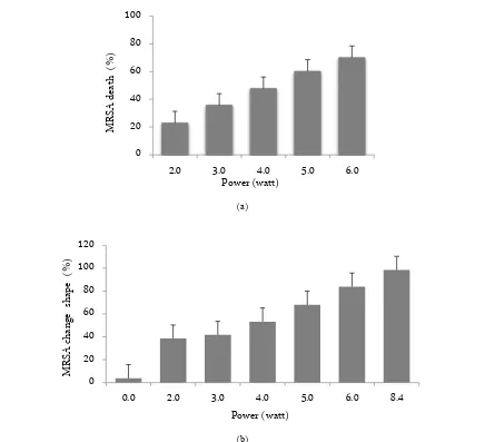
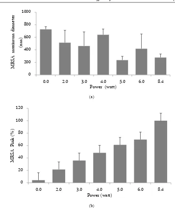
the death percentage of MRSA (p = 0.000), while the lethal power as found via regression was 8.432 watts. The death indicators of MRSA affected by ultrasonic wave are changes in shape (p = 0.005), size (p = 0.70); qualitatively there were changes in the structure of the cell and cellular uptake of Gram staining (p = 0.000), all compared with the control (Figure 1). The treatment of ultrasonic wave causing changes in the morphology and size of MRSA cell. Moreover, the ultrasonic wavelength also increases the MRSA death (Figure 2). The MRSA was stained with the Gram staining, the live cell color is purple but the death cell is a pink color.
This study we employed the square ultrasonic wave signal, which is induced more damage to the microbe compare to other signal forms [26]. This research is us-ing direct contact method, which means the ultrasonic
transducer is inserted into the bottom of the vessel which filled with MRSA suspension. The power of ul-trasonic wave exposure (P) had a very significant effect on the death percentage of MRSA (K), with p = 0.000, and the effect as stated by regression equation: K(P) = - 0.188 + 11.881P and lethal power 8.432 watts. These re-sults are supported by the indicators of death, as noted by MRSA cell observation with SEM and TEM. This le-thal power is relatively small compared to the similar research using ultrasonic wave. The death of the cells happened due to necrosis.
The power of ultrasonic waves could disturb the li-pid membrane, which affects bacteria growth or inhibits the growth process altogether [27], while the shift of me-chanical power of the ultrasonic wave could cause the separation of multi-molecular complex and cause the
(a)
(b)
Figure 1. Data distribution of the effect of power ultrasonic wave exposure to the MRSA death (a). Data distribution of the effect of power ultrasonic wave exposure to the changes of MRSA shape. Shown that one of indicators of MRSA death is the change of MRSA shape (b).
0 20 40 60 80 100
2.0 3.0 4.0 5.0 6.0
MRSA
de
ath
(
%)
Power (watt)
0 20 40 60 80 100 120
0.0 2.0 3.0 4.0 5.0 6.0 8.4
MRSA
cha
n
ge
shap
e
(
%)
loss of function or decreasing activity [19]. When the
(a)
(b)
Figure 2. Data distribution of the effect of power ultrasonic wave exposure to the diameter of MRSA, statistically the power of ultrasonic does not affect the of MRSA (p = 0.470) but physically different (a). Data distribution of the effect of power ultrasonic wave exposure to the change color of MRSA that have died (pink MRSA) (b). From the data shown that the changes of MRSA are an indicator of MRSA death.
Figure 3. MRSA control (a, b, and c): Because of the chemical effect of H2O2 of MRSA cell wall which exposed by ultrasonic with 4 watt and 6 watts power is broken in the cell wall so the cytoplasm and organelles are out from the cell. Cell occurred mechanical stress was caused by ultrasonic exposed with 8.4 watts power, Pieces of cells occur because of the intracellular cavitation caused by ultrasonic exposure with 8.4 watts power (e). (Observed by TEM with magnification 20,000×, scale 200 nm)
0 200 400 600 800 1000
0.0 2.0 3.0 4.0 5.0 6.0 8.4
MRS
A
m
ax
imum
di
ame
te
r
(n
m
)
Power (watt)
0 20 40 60 80 100 120
0.0 2.0 3.0 4.0 5.0 6.0 8.4
MRSA
Pi
n
k
(%)
stretch of the cell wall is beyond the elasticity limit, then the cell will be torn and the bacteria will die [20]. From the data analysis, it is shown that, when the MRSA was in the continuous ultrasonic field, periodic changes in pressure inflicts mechanical stress, which is marked by enlargement or shrinkage of the MRSA cell. If the elas-ticity limit of the MRSA cell wall is exceeded, the cell was torn, disrupting cell function and causing the MRSA cells to die (Figure 2).
Cavitation is regarded as the main mechanism that increases membrane permeability [28, 29, 30, 31]. Cavi-tation happens next to the surface; broken bubbles cause liquid with high velocity inflict shearing off next to the surface [32]. The extracellular cavitation causes a solid turbulation, while intracellular cavitation causes an ex-plosion from the inside of the cell. When the suspension of MRSA was in a continuous ultrasonic wave field, then intracellular or extracellular cavitation occurred. When intracellular cavitation happens in the MRSA cells, then microbubbles will arise with increasing magnitude, which causes the density to decline so that the MRSA cells will keep moving to the surface of the suspension and make hydrostatic pressure decline. If the pressure inside the MRSA cell is higher than outside the cell, this will inflict an explosion that is marked by broken cell walls (Figure 3).
Free radicals are formed by continuous irradiation into MRSA suspension [20]. These free radicals with join with a hydrogen atom in the water so that will
pro-duce hydrogen peroxide (H2O2) [33]. Ultrasonic waves
in liquid media caused mechanical effects (microstream-ing, high shear force, shockwave) and sonochemical re-actions (free radical, hydrogen peroxide), which finally caused interference or cell disruption of bacteria [34, 35, 36]. There are multiple effects of ultrasonic wave inhibi-tion in microbe cells, including the formainhibi-tion of pores, thinning cell wall, interference with the cell membrane, release of cytoplasmic contents and damage to the DNA structure [37, 38]. If the suspension of MRSA exposed by ultrasonic wave continuously, hydrogen peroxide will react with MRSA cell walls so that the cell wall experi-ences thinning, which causes the cell to swell and in par-ticular parts forming holes. Through these holes, the cy-toplasm will leak out of the cell; if this happens contin-uously, then the cell will not be able to retain its form and will die. Combinations of various frequencies of ul-trasonic waves with the various power of ulul-trasonic waves in a simultaneous experiment are needed to fur-ther characterize the optimum frequency and power to kill MRSA in vitro.
CONCLUSION
The power of wave influences very significantly to the death percentage of MRSA, with lethal power at 8.432 watts. Changes in shape, size, structure and cell staining due to mechanical stress, cavitation and chemi-cal effect were observed in ultrasound-treated cells. Dead MRSA cell happened due to necrosis, will change in shape from round to not round, the mean of the maxi-mum diameter can be smaller or larger from the mean of maximum diameter of control MRSA.
ACKNOWLEDGMENT
The author thanks, Drs. Tri Anggono Prijo.; Dr. drh. Dadik Rahardjo, M.Kes.; Rini Purbowati, S.Si., M.Si., and all technicians in Laboratory of Electron Mi-croscope (SEM), Malang State University.; all techni-cians in Laboratory of Electron Microscope (TEM), Uni-versity of Gajah Mada, Yogyakarta,; all technicians in Laboratory of Microbiology and Histology, University of Wijaya Kusuma, Surabaya, for their technical support to this experiment.
REFERENCES
1. Nurkusuma D (2009) Faktor yang berpengaruh terhadap metichillin-resistant Staphylococcus aureus (MRSA) pada kasus infeksi luka pasca operasi di Ruang Perawatan Bedah Rumah Sakit Dokter Kariadi Semarang. Master Thesis. Uni-versitas Diponegoro.
2. Herceg Z, Ksenija M, Šalamon BS et al. (2013) Effect of high intensity ultrasound treatment on the growth of food spoilage bacteria. Original scientific paper. University of Za-greb, Faculty of Food Technology and Biotechnology. 3. Song JH, Hsueh PR, Chung DR et al. (2011) Spread of
methicillin-resistant Staphylococcus aureus between the community and the hospitals in Asian countries: An AN-SORP study. Journal of Antimicrobial Chemotherapy 66 (5): 1061 – 1069. doi: 10.1093/jac/dkr024.
4. Stefani S, Chung DR, Lindsay JA et al. (2012) Meticillin-resistant Staphylococcus aureus (MRSA): global epidemiol-ogy and harmonization of typing methods. International Journal of Antimicrobial Agents 39 (4): 273 – 282. doi: 10.1016/j.ijantimicag.2011.09.030.
5. Schaumburg F, Kock R, Mellmann A et al. (2012) Popula-tion dynamics among methicillin-resistant Staphylococcus aureus isolates in Germany during a 6-year period. Journal of Clinical Microbiology 50 (10): 3186 – 3192. doi: 10.1128/JCM.01174-12.
aureus in the community in Portugal: evidence for the blur-ring of community-hospital boundaries. European Journal of Clinical Microbiology and Infectious Diseases 32 (10): 1269 – 1283. doi: 10.1007/s10096-013-1872-2.
7. Xiao M, Wang H, Zhao Y et al (2013) National surveillance of methicillin-resistant Staphylococcus aureus in China highlights a still-evolving epidemiology with 15 novel emerging multilocus sequence types. Journal of Clinical Mi-crobiology 51 (11): 3638 – 3644. doi: 10.1128/JCM.01375-13.
8. Hess CL, Howard MA, Attinger CE (2003) A review of me-chanical adjuncts in wounds healing: Hydrotherapy, ultra-sound, negative pressure therapy, hyperbaric oxygen, and electrostimulation. Annals of Plastic Surgery 51 (2): 210 – 217. doi: 10.1097/01.SAP.0000058513.10033.6B.
9. Breuing KH, Bayer L, Neuwalder J, Orgill DP (2005) Early experience using low-frequency ultrasound in chronic wounds. Annals of Plastic Surgery 55 (2): 183 – 187. doi: 10.1097/01.sap.0000168695.20350.07.
10. Scherba G, Weigel RM, O'Brien WD (1991) Quantitative assessment of the germicidal efficacy of ultrasonic energy. Applied and Environmental Microbiology 57 (7): 2079 – 2084.
11. Lillard HS (1994) Decontamination of poultry skin by soni-cation. Food Technology 48 (12): 72 – 73.
12. Raso J, Pagan R, Condon S, Sala FJ (1998) Influence of tem-perature and pressure on the lethality of ultrasound. Applied and Environmental Microbiology 64 (2): 465 – 471. 13. Vollmer AC, Kwakye S, Halpern M, Everbach EC (1998)
Bacterial stress responses to 1-Megahertz pulsed ultrasound in the presence of microbubbles. Applied and Environmen-tal Microbiology 64 (10): 3927 – 3931.
14. Belgrader P, Hansford D, Kovacs GT et al. (1999) A min-isonicator to rapidly disrupt bacterial spores for DNA anal-ysis. Analytical Chemistry 71 (19): 4232 – 4236. doi: 10.1021/ac990347o.
15. Singer A J, Coby CT, Singer Jr AHHCT, Tortora GT (1999). The effects of low-frequency ultrasound on Staphylococcus epidermidis. Current Microbiology 38 (3): 194 – 196. 16. Cochran SA, Prausnitz MR (2001) Sonoluminescence as an
indicator of cell membrane disruption by acoustic cavitation. Ultrasound in Medicine and Biology 27 (6): 841 – 850. doi: 10.1016/S0301-5629(01)00382-9.
17. Chandrapala J, Oliver C, Kentish S, Ashokkumar M (2012) Ultrasonics in food processing - food quality assurance and food safety. Trends in Food Science and Technology 26 (2): 88 – 98. doi: 10.1016/j.tifs.2012.01.010.
18. Chandrapala J, Oliver C, Kentish S, Ashokkumar M (2012) Ultrasonics in food processing. Ultrasonics Sonochemistry 19 (5): 975 – 983. doi: 10.1016/j.ultsonch.2012.01.010.
19. Johns LD (2002) Nonthermal effects of therapeutic ultra-sound: The frequency resonance hypothesis. Journal of Ath-letic Training 37 (3): 293 – 299.
20. Ackerman E (1988) Biophysical science. New Jersey, Eng-lewood Cliffs.
21. Dehghani HM (2005) The effectiveness of ultrasound on the destruction of E. coli. American Journal of Environmental Sciences 1 (3): 187 – 189. doi: 10.3844/ajessp.2005.187.189. 22. Kumar R, Yadav N, Rawat L, Goyal MK (2014) Effect of two waves of ultrasonic on waste water treatment. Journal of Chemical Engineering and Process Technology 5 (3): 193 – 198. doi:10.4172/2157-7048.1000193.
23. Li J, Ahn J, Liu D et al. (2016) Evaluation of ultrasound-induced damage to Escherichia coli and Staphylococcus au-reus by flow cytometry and transmission electron micros-copy. Applied and Environmental Microbiology 82 (6): 1828
– 1837. doi: 10.1128/AEM.03080-15
24. Stanisic MM, Provo BJ, Larson DL, Kloth LC (2005) Wound debridement with 25 kHz ultrasound. Advances in Skin and Wound Care 18 (9): 484 – 490. doi: 10.1097/00129334-200511000-00012.
25. Mansyur M, Yudaningtias E, Prawiro SR, Widjajanto E (2017) Low-Frequency ultrasonic wave to kill methicillin-re-sistant Staphylococcus aureus in vitro due to mechanical stress, cavitation, and chemistry effect. Turkish Journal of Biology, in press.
26. Hudori (2002) Studi daya reduksi desinfektans gelombang ultrasonik terhadap bakteri E. coli dengan variasi bentuk ge-lombang. Logika 7 (8): 32 – 38.
27. Cho JH, Kim EK, So JS (1995) Improved transformation of Pseudomonas putida Kt2440 by electroporation. Biotechnol-ogy Techniques 9 (1): 41–44. doi: 10.1007/BF00152998. 28. Tachibana K, Uchida T, Ogawa K et al. (1999) Induction of
cell membrane porosity by ultrasound. Lancet 353: 1409. doi: 10.1016/S0140-6736(99)01244-1.
29. Deng CX, Sieling F, Pan H, Cui J (2004) Ultrasound-in-duced cell membrane porosity. Ultrasound in Medicine and Biology 30 (4): 519 – 526. doi: 10.1016/S0140-6736(99)01244-1.
30. Newman CMH, Bettinger T (2007) Gene therapy progress and prospects: ultrasound for gene transfer. Gene Therapy 14 (6): 465 – 475. doi:10.1038/sj.gt.3302925.
31. Frenkel V (2008) Ultrasound-mediated delivery of drugs and genes to solid tumors. Advanced Drug Delivery Reviews 60 (10): 1193 – 1208. doi: 10.1016/j.addr.2008.03.007. 32. Pitt WG, Ross SA (2003) Ultrasound increase the rate of
bacterial cell growth. Biotechnology Progress 19 (3): 1038 – 1044. doi: 10.1021/bp0340685.
densed tannin). Chemosphere 56 (10): 981 – 987. doi: 10.1016/j.chemosphere.2004.05.022.
34. Rastogi NK (2011) Opportunities and challenges in the ap-plication of ultrasound in food processing. Critical Reviews in Food Science and Nutrition 51 (8): 705 – 722. doi: 10.1080/10408391003770583.
35. Ashokkumar M (2011) The characterization of acoustic cav-itation bubbles—an overview. Ultrasonics Sonochemistry 18 (4): 864 – 872. doi: 10.1016/j.ultsonch.2010.11.016. 36. Golmohamadi A, Moller G, Powers J, Nindo C (2013)
Ef-fect of ultrasound frequency on antioxidant activity, total
phenolic and anthocyanin content of red raspberry puree. Ultrasonics Sonochemistry 20 (5): 1316 – 1323. doi: 10.1016/j.ultsonch.2013.01.020.
37. Carel JA, Garcia-Perez JV, Benedito J, Mulet A (2012) Food process innovation through new technologies: Use of ultra-sound. Journal of Food Engineering 110 (2): 200 – 207. doi: 10.1016/j.jfoodeng.2011.05.038.
38. de São José JFB, de Andrade NJ, Ramos AM et al. (2014) Decontamination by ultrasound application in fresh fruits and vegetables. Food Control 45: 36 – 50. doi: 10.1016/j.foodcont.2014.04.015.