ORIGINAL ARTICLE
Conditioned medium from normoxia
(WJMSCs-norCM) and hypoxia-treated
WJMSCs (WJMSCs-hypoCM) in inhibiting
cancer cell proliferation
Wahyu Widowati
a,*, Laura Wijaya
b, Harry Murti
b,
Halida Widyastuti
b, Dwi Agustina
b, Dian Ratih Laksmitawati
c,
Nurul Fauziah
d, Sutiman B. Sumitro
e, M. Aris Widodo
f,
Indra Bachtiar
b,**
aMedical Research Center, Faculty of Medicine, Maranatha Christian University, Bandung, West Java,
Indonesia
bStem Cell and Cancer Institute, Jakarta, Indonesia
cFaculty of Pharmacy, Pancasila University, Jakarta, Indonesia
dBiomolecular and Biomedical Research Center, Aretha Medika Utama, Bandung, West Java, Indonesia eDepartment of Biology, Faculty of Science, Brawijaya University, Malang, East Java, Indonesia fPharmacology Laboratory, Faculty of Medicine, Brawijaya University, Malang, East Java, Indonesia
Received 5 June 2014; received in revised form 10 August 2014; accepted 12 August 2014
Available online 2 October 2014
KEYWORDS
anticancer;
conditioned medium; hypoxic;
normoxic; Wharton’s jelly
mesenchymal stem cells
Abstract Mesenchymal stem cells (MSCs) have unique properties, including high proliferation rates, self-renewal, multilineage differentiation ability, wide multipotency, hypoimmunogeni-city, noninduction of teratomas, and anticancer properties. MSCs can be isolated from embry-onic and extraembryembry-onic tissues as well as adult organs. Human Wharton’s jelly stem cell-conditioned medium possesses anticancer properties and inhibits the growth of solid tumors. Lower oxygen concentration or hypoxic condition can increase the proliferation of MSCs, but there are no differences in surface markers. We determined the osteocyte, chondrocyte, and adipocyte differentiation of normoxic WJMSCs (nor-WJMSCs) and hypoxic 2.5%, hypoxic 5% (hypo-WJMSCs); from a different passage (P4 and P8), we determined the inhibitory effect of WJMSCs-norCM and WJMSCs-hypoCM on the proliferation of human cancer cells including cervical (HeLa), liver (HepG2), prostate (pc3), ovarian (skov3), and oral squamous (hsc3) cancer cell lines compared to normal cells including mouse fibroblast (NIH3T3), human
* Corresponding author. Medical Research Center, Faculty of Medicine, Maranatha Christian University, Jl. Prof. drg. Surya Sumantri no 65 Bandun 40164, West Java, Indonesia.
** Corresponding author. Stem Cell and Cancer Institute, JL.A. Yani no 2 Pulo Mas, Jakarta 13210, Indonesia.
E-mail addresses:wahyu_w60@yahoo.com(W. Widowati),nantenine@yahoo.com(I. Bachtiar).
http://dx.doi.org/10.1016/j.bgm.2014.08.008
2214-0247/Copyrightª2014, Taiwan Genomic Medicine and Biomarker Society. Published by Elsevier Taiwan LLC. All rights reserved. Available online atwww.sciencedirect.com
ScienceDirect
fibroblast, and human mesenchymal stem cells (hMSCs). Surfacer marker expression of nor-WJMSCs-and hypo-WJMSCs from P4 and P8 were>95% for CD90, CD73 and CD105 and<2% for CD14, CD19, CD34, CD45, and HLDA-II. Nor-WJMSCs and hypo-WJMSCs from P4 and P8 un-derwent differentiation to osteocyte, chondrocyte, and adipocyte. WJMSCs-norCM and WJMSCs-hypoCM could inhibit proliferation of various cancer cell lines with minimum inhibitory concentration (IC50) 51.690e81.440% and cause low inhibition of the normal cells with IC50 136.290e185.339%. WJMSCs-norCM and WJMSCs-hypoCM were not cytotoxic toward normal cells. Nor-WJMSCs and hypo-WJMSCs from P4 and P8 showed no significant differences in MSC surface marker expression or differentiation. WJMSCs-norCM and WJMSCs-hypoCM could inhibit proliferation in various cancer cell lines, and were safe for normal cells.
Copyrightª2014, Taiwan Genomic Medicine and Biomarker Society. Published by Elsevier Taiwan LLC. All rights reserved.
Introduction
Umbilical cord matrix contains an inexhaustible, noncon-troversial source of stem cells.1e4. Postnatal stem cells are
offered for use less often because of possible moral/ethical conflict.4 The umbilical cord mesenchymal stem cells
(UCMSCs) derived from human umbilical cord Wharton’s jelly (WJMSCs) exhibit low immunogenicity and low immu-nity after cytotherapy.5The UCMSCs are more useful and simpler in donor accessibility isolation, expansion, prolif-erative capacity, and banking capability; can be used in clinical and experimental therapy6,7; have a higher prolif-eration rate and self-renewal capacity than adult tissue-derived MSCs2,8; and a short doubling time.9,10 MSCs possess strong immunosuppressive properties and can be used for autologous and allogeneic therapy.11,12
Research has shown that MSCs, including UCMSCs, bone marrow MSCs, and adipose tissue MSCs, have anticancer activity and have been shown to inhibit the proliferation of cancer cells in bothin vitroandin vivoassays.5
Hypoxic conditions (1e3% O2) are more beneficial for
MSCs because low oxygen tension is more suitable for MSC physiology in the bone marrow (2e7% O2). Previous
research has shown that MSCs cultured in hypoxic condi-tions (2e5% O2) could maintain their viability.13The hypoxic
microenvironment can lead to a substantial increase in the proliferation rate and population doubling time, but no differences in surface markers of MSCs has been shown. The hypoxic 2.5% O2yield has the highest proliferation and the
lowest population doubling and population doubling time.10 The hypoxic condition induces molecular responses including angiogenesis, metabolic change, and metastasis; it also induces the secretion of growth factor and cytokine in MSCs, and elevated the secretion of transforming growth factor-b1 (TGF-b1). In addition, the hypoxic condition can enhance cancer cell growth through the MSCs effect by secretion and expression of TGF-b1.14
We conducted the continuing research to elucidate the osteocyte, chondrocyte, and adipocyte differentiation of normoxia-treated WJMSCs (nor-WJMSCs) and hypoxia-treated WJMSCs (hypo-WJMSCs) from early and late pas-sage (P4 and P8), to evaluate the WJMSCs-nor conditioned medium (norCM) and WJMSCs-hypoCM toward cancer cell lines including HeLa, HepG2, pc3, skov3, and hsc3 compared to human fibroblast, NIH3T3, and human mesenchymal stem cells (hMSCs).
Materials and methods
Isolation and cultivation of WJMSCs
Isolation and cultivation of MSCs from umbilical cord was as described by Widowati et al,10fresh human umbilical cords (UC;nZ5) were obtained from women aged 25e40 years
after full-term births (normal vaginal delivery), all donors signed a written informed consent, and guidelines were approved by the Institutional Ethics Committee at the Stem Cell and Cancer Institute, Jakarta, Indonesia and from the Institutional Ethics Committee collaboration between Maranatha Christian University, Bandung, Indonesia and Immanuel Hospital Bandung, Bandung, Indonesia.
Isolated WJMSCs from UC were rinsed in normal saline (0.9% w/v sodium chloride) and cut into very small explants (1e2 mm), then plated on tissue culture plastic plates. The
explants were cultured in minimum essential medium-a
with 2 mM GlutaMAX (Invitrogen, Carlsbad, CA, USA), sup-plemented with 20% fetal bovine serum (FBS; Invitrogen) and penicilline streptomycine amphotericin B (100 U/mL, 100 mg/mL, and 0.25 mg/mL; Invitrogen). Cultures were incubated in a humidified atmosphere with 5% CO2at 37C,
replacing medium every 5 days for 21 days. The cells were harvested and replated at a density 8103cells/cm2when
cells reached 80e90% confluence. WJMSCs were cultured in
95% air (21% O2), and 5% CO2 for normoxic and hypoxic
conditions (5% O2and 2.5% O2).10,15
Cell surface phenotype and multipotent differentiation
53321-100), CD73 (BD550257), CD 90 (abcam 226), CD 34 (BD 348053), CD45 (BD 555482), CD14 (abcam 28061-100), CD 19 (abcam 1168-500), and HLA-DR (abcam 23901); FITC-conjugated: mIgG1 (BD349041 and BD 349043), and mIgG2 (BD349053) then added to each FACS tube: isotype mIgG2a-PE, CD105-mIgG2a-PE, HLA class II-PE; isotype mIgG1-mIgG2a-PE, CD73-mIgG2a-PE, CD19-PE; isotype mIgG1-FITC, CD 34-FITC, CD45-FITC, CD14-FITC, after incubation at 4C for 15 minutes. The cells were analyzed by flow cytometry with a FACS CaliburTM 3 argon laser 488 nm (Becton Dickinson Biosciences, Franklin Lakes, NJ, USA) using CellQuest Pro Acquisition on the BD FACStationTM Software. The experiments and measurement of surface marker were performed in triplicate.10
For osteogenic differentiation, WJMSCs (P4 and P8) were seeded at density 1104cells/cm2in culture dishes using
StemPro Osteogenesis Differentiation Kit (Gibco A10072-01, Invitrogen) for 3 weeks. Calcium deposits were visualized using Alizarin red S (Biochemicals and Life Science Research Products, Amresco 9436). For chondrogenic differentiation of WJMSCs were seeded at density 1 104 cells/cm2 in culture dishes using StemPro Chondrogenesis Differentiation Kit (Gibco A10071-01, Invitrogen) for 2 weeks. Chondrocytes were visualized using Alcian blue (Amresco, 0298). Adipo-genic differentiation of WJMSCs was done using StemPro Adipogenesis Differentiation Kit (Gibco A10070-01, Invi-trogen, Carlsbad, CA, USA) for 2 weeks. We used Oil Red O (Sigma 00625, St Louis, USA) to confirm lipid droplets.16e18
Preparation of conditioned media from normoxia-or hypoxia-treated WJMSCs
WJMSCs of P4 were used for the experiments. The WJMSCs were seeded at a density of 8103 cells/cm2under
nor-moxia (20% O2and 5% CO2) and hypoxia (5% O2and 20% CO2)
for 72 hours; when cultures reached 80e90% confluence,
cells were harvested. The medium was collected and centrifuged at 3000gfor 4 minutes at room temperature, and the supernatant was filtered by a 0.22-mm MillexeGV
Filter Unit with Durapore (SLGV 033 RS, Millipore Corpora-tion, Billerica, MA, USA) and used as WJMSCs-CM.18,19
Anticancer assay
The cancer cell lines of cervical (HeLa- ATCC CCL-2), liver (HepG2- ATCC HB-8065), prostate (PC3-ATCC CRL-1435), ovarian (SKOV3-ATCC HTB-77), oral squamous (HSC3; ATCC, Manassas, VA, USA), mouse fibroblast (NIH3T3-ATCC CRL-1658), human fibroblast (primary cells), and hMSCs (primary cells from Wharton’s jelly) were obtained from Stem Cell and Cancer Institute, Jakarta, Indonesia. The cells were grown and maintained in Dulbecco modified Eagle’s medium sup-plemented with 10% FBS (Invitrogen), 100 U/mL penicillin (Invitrogen), and 100mg/mL streptomycin (Invitrogen), and incubated at 37C in a humidified atmosphere and 5% CO
2.20,21
The cell viability assay uses an optimized reagent con-taining resazurin converted to fluorescent resorufin by viable cells that absorbs the light at 490 nm. Briefly, cells were seeded at density 5103in 96-well plates for 24 hours of incubation20,21; cells were supplemented by WJMSCs-norCM
and WJMSCs-hypoCM in various concentrations (100%, 75%, 50%, and 0%) then incubated for 72 hours. The anticancer
activity of WJMSCs-CM was noted for cancer cell lines including HeLa, HepG2, PC3, SKOV3, HSC3. We used NIH3T3, human fibroblast, and hMSCs as controls to determine the cytotoxic effect of WJMSCs-CM in normal cells. MTS [3- (4,5-dimethylthiazol-2-yl)-5-(3-carboxyme-thoxyphenyl)-2-(4-sulfophenyl)-2H-tetrazolium] assay (Promega, Madison, WI, USA) was used to determine cell viability. MTS was added at 10mL to each well. The plate was incubated at 5% CO2,
37C for 4 hours. The absorbance of the cells was measured
at 490 nm using a microplate enzyme-linked immunosorbent assay reader (Multiskan Go, Thermo Scientific, USA). The data were presented as number of viable cells and the per-centage of viability.20,21
Statistical analysis
To verify the statistical significance of all parameters, the data were calculated and expressed in means and standard deviation (M SD). To compare treatments, analysis of variance (ANOVA) was used, andp<0.05 were considered
as statistically significant, along with Tukey honestly sig-nificant difference post hoc test and 95% confidence in-terval. The median inhibitory concentration (IC50) of
cytotoxic effect was analyzed using probit analysis. Statis-tical analysis used the SPSS version 20.0 program (IBM SPSS Statistics for Windows, Version 20.0. Armonk, NY: IBM Corp, SPSS Inc., Chicago, IL, USA).
Results
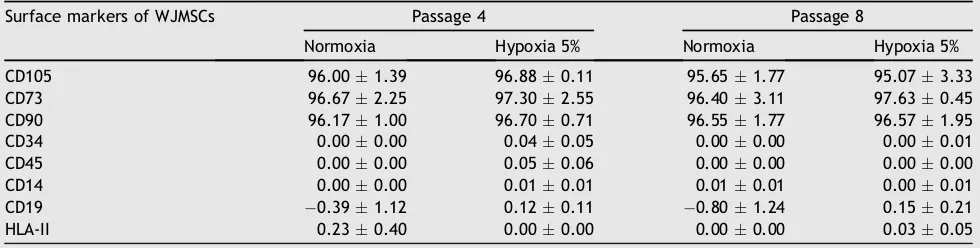
MSC markers by cell surface phenotype
Flow cytometry analysis showed that for cultured cells under normoxia and hypoxia (5% O2) for P4 and P8, WJ-MSCs
were positive for MSCs marker CD105, CD73, and CD90 and negative for CD34, CD45, CD14, CD19, and HLA-II. The ef-fect of oxygen level (normoxic and hypoxic 5% O2) and
passage (early passage P4 and late passage P8) on the surface marker of WJMSCs can be seen inTable 1. Surface marker expression of WJ-MSCs (P4 and P8) on hypoxia and normoxia were not significantly different (p>0.05).
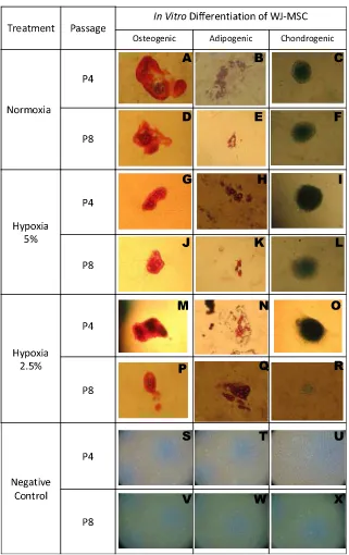
MSC differentiation
We examined the differentiation potentials (osteogenic, chondrogenic, and adipogenic differentiation) that were cultured in either normoxic (20% O2and 5% CO2) or hypoxic
(5% O2and 2.5%O2) conditions. After 3 weeks, WJMSCs were
cultured in osteogenesis differentiation medium, and the differentiated WJMSCs responses to either normoxia or hypoxia exhibited calcium deposits based on the staining of Alizarin red S. After 2 weeks, WJMSCs were cultured in chondrogenesis differentiation medium, and the differen-tiated WJMSCs in responses to either normoxia or hypoxia exhibited chondrocyte expression using Alcian blue. After 2 weeks, WJ-MSCs were cultured in adipogensis differentia-tion medium in response to either normoxia or hypoxia and exhibited lipid droplets using Oil Red O (Fig. 1).
Fig. 1 shows that WJ-MSCs incubated in normoxia (21%
O2) and hypoxia (5% O2 and 2.5% O2) differentiated to
Cytotoxic activity
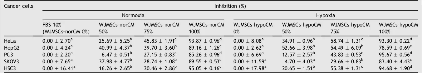
To determine the effect of both norCM and WJMSCs-hypoCM toward various cancer cell lines (HeLa, HepG2, PC3, SKOV3, and HSC3) and normal cells (NIH3T3, human fibro-blast, and hMSCs), the cell lines were cultured at density 5103in a 96-well plate. We determined the cell viability by MTS assay. WJMSCs-norCM and WJMSCs-norCM exhibited decreased viability in cancer cell lines in a concentration-dependent manner. The effect of WJMSCs-norCM and WJMSCs-hypoCM on the number of cancer cells can be seen
inTable 2. The number of cancer cells decreased along with
treatments, and the higher WJMSCs-CM concentration decreased the number of cancer cells. The effect of WJMSCs-norCM and WJMSCs-hypoCM toward inhibition of cancer cell viability can be seen inTable 3. As seen inTable 3, WJMSCs-norCM and WJMSCs-hypoCM could inhibit the proliferation of cancer cells including HeLa, HepG2, PC3, SKOV3, and HSC3 in a concentration-dependent manner.
The IC50 value of WJMSCs-norCM and WJMSCs-hypoCM
(concentration of anticancer candidate, which could inhibit 50% cell proliferation) was found to be 51.690e81.440%
(Table 4). Each sample (norCM and
WJMSCs-hypoCM) was done in triplicate and inhibition data were analyzed using probit analysis to obtain the IC50. As shown in
Table 4, WJMSCs-norCM and WJMSCs-hypoCM exhibited a
cytotoxic effect toward HeLa, HepG2, PC3, SKOV3, and HSC3 cells, and the highest anticancer activity was WJMSCs-hypoCM in HepG2 with IC5051.690% and in HeLa with IC50
61.425%. The highest anticancer activity of WJMSCs-norCM with IC50 was 64.424% in HepG2. WJMSCs-hypoCM had
higher anticancer activity to inhibit HeLa, HepG2, pc3, and hsc3 cell lines compared to WJMSCs-norCM.
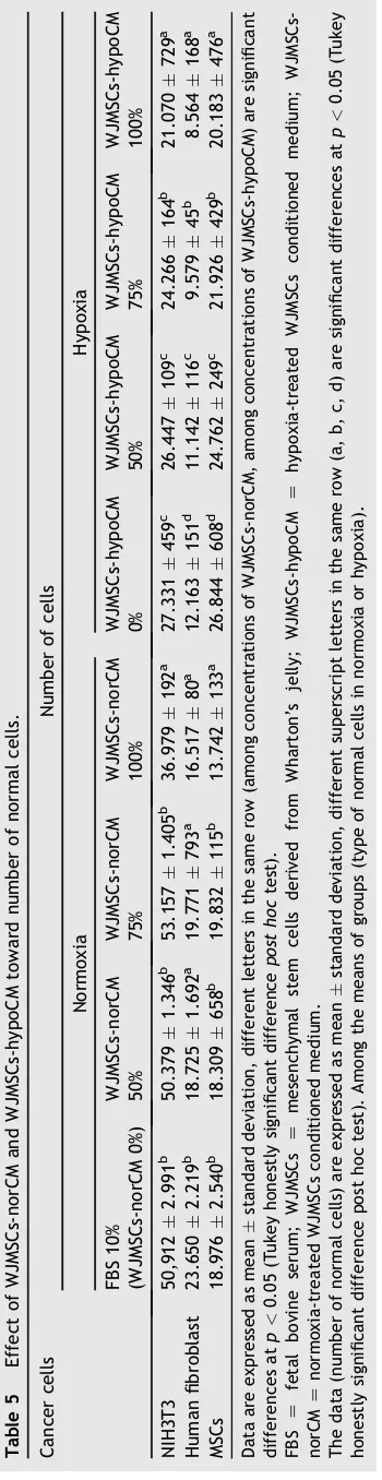
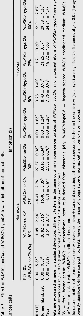
The selective cytotoxic effect of WJMSCs-norCM and WJMSCs-hypoCM was carried out in NIH3T3, human fibro-blast, and hMSCs. The effect of WJMSCs-norCM and WJMSCs-hypoCM on the number of normal cells can be seen
in Table 5. The effect of hypoCM and
WJMSCs-norCM on inhibition of normal cells can be seen in
Table 6. The IC50 value of norCM and
WJMSCs-hypoCM was found to be 136.290e185.339% (Table 7). As
shown inTables 5e7, WJMSCs-norCM and WJMSCs-hypoCM demonstrated a low cytotoxic effect, inhibited low cell proliferation in normal cells, including NIH3T3, human
fibroblast cells, and hMSCs. norCM and
WJMSCs-hypoCM were not toxic toward normal cells with
IC50>100% concentration of CM.
Discussion
WJ-MSCs was positive for CD105, CD73, and CD90 and negative for CD34, CD45, CD14, CD19, and HLA-II (Table 1). These data were consistent with previous research that hMSCs highly expressed CD105, CD73,1,10 and CD9022 and
had low expression of CD34, CD45, CD14, CD19, and
HLA-II.1,10,22The surface marker of WJMSCs of P4 and P8 both
normoxic and hypoxic 5% O2were not significantly different
(p>0.05); these data are consistent with previous research
that surface markers expression are positive for CD105, CD73, and CD90 (>95%) and negative for CD34, CD45, CD14,
CD19, and HLA-II (<2%).10,16
Pluripotency was confirmed by the ability of WJ-MSCs cells to differentiate into osteocytes, chondrocytes, and adipocytes.24As demonstrated inFig. 1, both nor-WJMSCs and hypo-WJMSCs differentiated to osteocytes, chon-drocytes, and adipocytes; these findings were consistent with previous research that MSCs possess an extensive po-tential to proliferate and differentiate. into osteocytes, adipocytes, and chondrocytes.22,24e26 MSCs can also be
isolated from umbilical cord Wharton’s jelly and markers expressed by flow cytometry; differentiate into osteoblast, adipocyte- and chondrocyte-like cells; and exhibit multi-potent differentiation multi-potential.27
After 2 weeks of exposure to adipogenesis induction medium, cells began to show a round shape and most of them contained cytoplasmatic vacuoles, intracellular accumulated lipids, and small oil droplets in the cytoplasm that were positive with Oil Red O staining (Fig. 1B, E, H, K, N, and Q). Control cells grew with proliferative medium were negative with Oil Red O staining (Fig. 1T and W).18,28,29 These data were validated with previous research, which found that reverse transcription-polymerase chain reaction analysis of adipogenic gene expression also revealed similar degrees of upregulation of lipoprotein lipase, adipocyte fatty acid-binding protein 2 (aP2), and peroxisome proliferator-activated receptorg2 (PPARg2).18,30
After 3 weeks of osteogenic induction, the cells pro-duced mineralized matrix by Alizarin red S staining (Fig. 1A, Table 1 Effect of oxygen level and type of passage toward surface markers for umbilical cord mesenchymal stem cells derived from human umbilical cord Wharton’s jelly.
Surface markers of WJMSCs Passage 4 Passage 8
Normoxia Hypoxia 5% Normoxia Hypoxia 5%
CD105 96.001.39 96.880.11 95.651.77 95.073.33
D, G, J, M, and P), cells displayed bone-like nodular ag-gregates of matrix mineralization,25 and were absent in
control cultures28(Fig. 1S and V). These data are consistent with those of a previous study finding that reverse transcriptase-polymerase chain reaction analysis of osteo-genic gene expression also revealed similar levels of upre-gulation of osteopontin and osteocalcin.18
The chondrogenic potential of the MSCs was confirmed with the presence of acidic proteoglycans; it was observed after 2 weeks of chondrogenic differentiation by Alcian blue staining,23,31 for chondrogenic extracellular matrix
containing hyaluronic acids31(Fig. 1C, F, I, L, O, and R), the negative controls of chondrogenic differentiation of WJMSCs were represented by MSCs of P4 and P8 not cultured into differentiation media31 (Fig. 1U and X). Ac-cording toFig. 1, WJMSCs of early passage and late passage differentiated to osteocytes, chondrocytes, and adipo-cytes; these data were validated with the previous finding that MSCs could be expanded to 10 or 11 passages.32
Tables 2e4show that WJMSCs-norCM and WJMSCs-hypoCM
Table 2 Effect of WJMSCs-norCM and WJMSCs-hypoCM toward number of cancer cells.
Cancer cells Number of cells
Normoxia Hypoxia
FBS 10%
(WJMSCs-norCM 0%)
WJMSCs-norCM 50%
WJMSCs-norCM 75%
WJMSCs-norCM 100%
WJMSCs-hypoCM 0% WJMSCs-hypoCM 50%
WJMSCs-hypoCM 75%
WJMSCs-hypoCM 100%
HeLa 21,900591d 16,2733,340c 11,863419b 1,343211a 24,9132,012d 16,216240c 10,280326b 1,67055a HepG2 21,508912c 12,692940b 12,968775b 2,332270a 24,062630c 11,392959b 10,9521,466b 5,152167a PC3 20,701456d 19,3611,057c 15,081172b 3,051199a 22,7481,522d 19,888584c 12,778120b 984127a
SKOV3 18,1051,385d 11,229864b 12,902196c 1,89296a 17,9592,082c 17,115725c 12,632150b 2,982795a HSC3 18,5283,041c 15,515491b 12,885530b 91830c 17,6253,168d 13,985266c 7,865230b 938334a
Data are expressed as meanstandard deviation, different letters in the same row (among concentrations of WJMSCs-norCM and among concentrations of WJMSCs-hypoCM) are significant differences atp<0.05 (Tukey honestly significant differencepost hoctest).
FBS Z fetal bovine serum; WJMSCs Z mesenchymal stem cells derived from Wharton’s jelly; WJMSCs-hypoCM Z hypoxia-treated WJMSCs conditioned medium;
WJMSCs-norCMZnormoxia-treated WJMSCs conditioned medium.
The data (number of cancer cells) are expressed as meanstandard deviation, different superscript letters in the same row (a, b, c, d) are significant differences atp<0.05 (Tukey honestly significant difference post hoc test). Among the means of groups (type of cancer cell lines in normoxia or hypoxia).
Table 3 Effect of WJMSCs-norCM and WJMSCs-hypoCM toward inhibition of cancer cells.
Cancer cells Inhibition (%)
Normoxia Hypoxia
FBS 10%
(WJMSCs-norCM 0%)
WJMSCs-norCM 50%
WJMSCs-norCM 75%
WJMSCs-norCM 100%
WJMSCs-hypoCM 0%
WJMSCs-hypoCM 50%
WJMSCs-hypoCM 75%
WJMSCs-hypoCM 100%
HeLa 0.002.70a 25.695.25b 45.831.91c 93.870.96d 0.008.08a 34.910.96b 58.741.31c 93.300.22d
HepG2 0.004.24a 40.994.37b 39.703.60b 89.161.26c 0.002.62a 52.663.98b 54.496.09b 78.590.69c
PC3 0.002.20a 6.470.51b 27.150.83c 85.260.96d 0.006.69a 12.572.57b 43.830.53c 95.670.56d SKOV3 0.007.65a 37.984.77b 28.741.08b 89.550.53c 0.0011.59a 4.704.03a 29.660.83b 83.404.43c
HSC3 0.0016.41a 16.262.65b 30.462.86b 95.050.16c 0.0017.98a 20.651.51b 55.381.31c 94.681.90d
Data are expressed as meanstandard deviation, different letters in the same row (among concentrations of WJMSCs-norCM, among concentrations of WJMSCs-hypoCM) are significant differences atp<0.05 (Tukey honestly significant differencepost hoctest).
FBS Z fetal bovine serum; WJMSCs Z mesenchymal stem cells derived from Wharton’s jelly; WJMSCs-hypoCM Z hypoxia-treated WJMSCs conditioned medium;
WJMSCs-norCMZnormoxia-treated WJMSCs conditioned medium.
The data (inhibition of cancer cells) are expressed as meanstandard deviation, different superscript letters in the same row (a, b, c, d) are significant differences atp<0.05 (Tukey honestly significant difference post hoc test). Among the means of groups (type of cancer cell lines in normoxia or hypoxia).
medium
inhibits
cancer
cell
proliferation
various activities. These data were validated with previous studies that hMSCs can be used for neoplastic transformation and can be developed for novel anticancer therapeutics33;
human Wharton’s jelly stem cells inhibited certain solid tumors.4,34e36 UCMSCs significantly inhibit proliferation of
cancer cell lines byin vivoandin vitroassay.4,37 Unengi-neered human and rat UC-MSCs significantly attenuate pro-liferation of breast cancer cell linesin vitroandin vivo,4rat
mammary tumor cells,37human lung cancer cells,38mouse Lewis lung carcinoma cells,39 and mouse pancreatic
carci-noma cells.5,40 Human umbilical cord mesenchymal stem cells (hUCMSCs) are able to inhibit breast cancer cell prolif-eration (MDA-MB-231) in a severe combined immunodefi-ciency (SCID) mouse model through secretion of dickkopf and suppression of the Wnt pathway.35 hWJMSCs-conditioned
medium (hWJSC-CM; 50%) or hWJSCs-cell lysate (hWJSC-CL) 15mg/mL for 48e72 hours inhibit cancer cell proliferation in
breast adenocarcinoma (MDA-MB-231), ovarian carcinoma (TOV-112D), and osteosarcoma (MG-63) cells. The cancer cell lines exhibited cell shrinkage, blebbing, and vacuolations compared to controls.41 The inhibition was 20e26% and
31e46% for hWJSC-CM and hWJSC-CL, respectively, for all
three cancer cell lines. Cell cycle assays show increases in sub-G1 and G2/M phases for all three cancer cell lines sug-gestive of apoptosis and metaphase arrest.41 hWJSCs migrated to metastatic tumor sites in the lungs and reduced tumor burden after hWJSCs were administered intravenously 8 days after tumor transplantation in a rat model.4,37,42 Engineered hWJSCs-expressed human interferon-binhibited breast tumor growth in animal models.43 hWJSCs inhibit human mammary carcinoma proliferation.41
Conditioned medium and cell-free lysate of hWJSCs (hWJSC-CM and hWJSC-CL) inhibit the growth of a range of cancer cells, including breast cancer (MDA-MB-231) and ovarian cancer cells (TOV-112D), as well as osteosarcoma cells (MG-63).41 Exposure of the osteosarcoma cell lines SKES-1 and MG-63 to hWJSC-CL and hWJSC-CM results in cell death and significant growth inhibition in vitro. At the molecular level, there is a simultaneous upregulation of proapoptotic and autophagy-related genes, such as BAX, ATG-5, and BECLIN-1, and downregulation of prosurvival genes, such as BCL-2 and SURVIVIN. In vivo, there was a notable reduction in mammary tumor sizes and weights in immunodeficient mice at 6 weeks after the injections of Table 4 The IC50of WJMSCs-norCM and WJMSCs-hypoCM
in various cancer cell lines for 72 hours of incubation.
Cancer cell lines IC50(%)
WJMSCs-norCM WJMSCs-hypoCM
IC50 Z median inhibitory concentration; WJMSCs Z
mesen-chymal stem cells derived from Wharton’s jelly; WJMSCs-hypoCM Z hypoxia-treated WJMSCs conditioned medium;
WJMSCs-norCM Z normoxia-treated WJMSCs conditioned
hWJSC-CL and hWJSC-CM into these tumors. These findings suggest that hWJSC-CL and hWJSC-CM may interfere with the growth of mammary carcinoma and osteosarcoma cells via apoptosis and autophagy.41A similar cell death mech-anism is observed during co-culture of WJMSCs with the prostate cancer cell line (PC3). In the presence of WJMSCs, PC3 cells exhibit caspase 9/3, PARP, and BAX induction, c-Jun NH2-terminal kinase (JNK) activation, as well as a decrease in phosphatidylinositol 3-kinase (PI3K)/AKT (also known protein kinase B (PKB)) and extracellular signal-regulated kinase (ERK) phosphorylation. Simultaneously, there is a downregulation of prosurvival gene expressions, such as BCL-2, BCL-XL, SURVIVIN, Mcl-1, and cIAP-1.4,44e47
The tumoricidal activity of hWJSCs-CM is probably mediated by certain soluble factors secreted by these cells into their extracellular environment, such as interleukins, cell adhesion molecules, hyaluronic acid, growth factors, and glycosoaminoglycans.44,48,49Indeed, proteomic analysis of hWJSC-CM shows significantly high levels of interleukins (IL-1a, IL-6, IL-7, and IL-8), stem cell factor, human growth factor, and intercellular adhesion molecule-1.44Moreover,
the extracellular matrix of WJMSCs also contains dickkopf-1, a protein known to suppress the Wnt signaling pathway.35,48 Likewise, bone marrow MSCs conditioned medium suppresses the proliferation of two hepatoma cell linesin vitroand induces tumor regression in a hepatoma SCID mouse xenograft model by means of Wnt signaling pathway regulations.45,47,48Engineered bone marrow MSCs
are found to secrete IL-12, which inhibits the growth of melanoma and cervical cancer cells through the induction of a tumor-specific T cell responsein vivo.45Moreover, bone marrow MSCs also express several suicide genes, which halt the proliferation of prostate cancer cells in an athymic murine model.45In addition to the upregulation of several proapoptotic and tumor suppressor genes in hWJSCs, tran-scriptomic studies have also found an increased expression of several cytokines in these cells, such as IL-12a, which are thought to induce apoptosis and thereby mediate the anticancer effects of hWJSCs, CM, and
hWJSC-CL.50,51
The IL-12 gene promoted the activation of the cellular immune response via expression of a Th1-type cytokine profile and was associated with the inhibition of tumor growth.3,52 IL-12 treatment represents a novel approach
for gene therapy against cervical cancer.51IL-8 of hWJSCs killed the cancer cells.41 Hyaluronan oligosaccharides
T
Table 7 The median inhibitory concentration IC50 of
WJMSCs-norCM and WJMSCs-hypoCM in various normal cells for 72 hours of incubation.
Normal cells IC50(%)
WJMSCs-norCM WJMSCs-hypoCM
NIH3T3 136.29 159.33
Human fibroblast 148.47 152.48
MSCs 140.44 185.34
IC50Zmedian inhibitory concentration; MSCsZmesenchymal
stem cells; WJMSCsZmesenchymal stem cells derived from
Wharton’s jelly; WJMSCs-hypoCMZ hypoxia-treated WJMSCs
conditioned medium; WJMSCs-norCM Z normoxia-treated
inhibited the growth of osteosarcoma cell lines (MG-63 and LM-8)53and glycosoaminoglyans inhibited the cell prolifer-ation of osteoblasts and osteosarcoma cells.54
UC-MSCs expressed the multiple tumor suppressor gene.5 hUCMSC are able to inhibit human breast cancer cells by attenuating primarily the AKT and mitogen-activated pro-tein kinase pathways and stimulating the intrinsic apoptosis pathway.5 hUCMSC attenuated the growth of cancer cells
and mainly by attenuation of Erk-1/2 and PI3K/AKT signaling and intrinsic apoptosis.5
Nor-WJMSCs and hypo-WJMSCs from P4 and P8 showed no significant differences in MSCs surface marker expres-sion and MSCs differentiation. norCM and WJMSCs-hypoCM could inhibit cells proliferation in various cancer cell lines, and were not toxic for normal cells.
Conflicts of interest
All contributing authors declare no conflicts of interest.
Acknowledgments
The authors gratefully acknowledge the financial support from the Ministry of Research and Technology (Research Grant No KP-2013-0715 and KP-2014-0713). This research was also supported by the Stem Cell and Cancer Institute, Jakarta, Indonesia.
References
1.Weiss ML, Medicetty S, Bledsoe AR, et al. Human umbilical cord matrix stem cells: preliminary characterization and ef-fect of transplantation in a rodent model of Parkinson’s dis-ease.Stem Cells. 2006;24:781e792.
2.Weiss ML, Troyer DL. Stem cells in the umbilical cord.Stem Cell Rev. 2006;2:155e162.
3.Weiss ML, Anderson C, Medicetty S, et al. Immune properties of human umbilical cord Wharton’s jelly-derived cells.Stem Cells Express. 2008:2865e2874.
4.Ayuzawa R, Doi C, Rachakatla RS, et al. Naive human umbil-ical cord matrix derived stem cells significantly attenuate growth of human breast cancer cells in vitro and in vivo.
Cancer Lett. 2009;280:31e37.
5.Tamura M, Kawabata A, Ohta N, et al. Wharton’s jelly stem cells as agents for cancer therapy.The Open Tissue Eng Regen Med J. 2011;4:39e47.
6.Prasanna SJ, Gopalakrishnan D, Shankar SR, et al. Proin-flammatory cytokines, IFNgamma and TNFalpha, influence immune properties of human bone marrow and Wharton jelly mesenchymal stem cells differentially. PLoS One. 2010;5: e9016.
7.Puranik SB, Nagesh A, Guttedar RS. Isolation of mesenchymal-like cells from Wharton’s jelly of umbilical cord.IJPCBS. 2012; 2:218e224.
8.Can A, Karahuseyinoglu S. Concise review: human umbilical cord stroma with regard to the source of fetus-derived stem cells.Stem Cells. 2007;25:2886e2895.
9.Bongso A, Fong C-F, Gauthaman K. Taking stem cells to the clinic: major challenges.J Cell Biochem. 2008;105:1352e1360. 10.Widowati W, Wijaya L, Bachtiar I, et al. Effect of oxygen tension on proliferation and characteristics of Wharton’s jelly-derived mesenchymal stem cells. Biomarkers Genomic Med.2014;6:43e48.
11. Jones BJ, McTaggart SJ. Immunosuppression by mesenchymal stromal cells: from culture to clinic.Exp Hematol. 2008;36: 733e741.
12. Menon LG, Shi VJ, Carroll RS.Mesenchymal Stromal Cells as a Drug Delivery System. Boston, MA 02115, USA: Stem Book; 2009:1e14. The Stem Cell Research Community.
13. Grayson WL, Zhao F, Bunnell B, et al. Hypoxia enhances pro-liferation and tissue formation of human mesenchymal stem cells.Biochem Biophys Res Commun. 2007;358:948e953. 14. Hung SP, Yang MH, Tseng KF, et al. Hypoxia-Induced secretion
of TGF-b1 in mesenchymal stem cell promotes breast cancer cell progression.Cell Transplantation. 2013;22:1869e1882. 15. Nekanti U, Dastidar S, Venugopal P, et al. Increased
prolif-eration and analysis of differential gene expression in human Wharton’s jelly-derived mesenchymal stromal cells under hypoxia.Int J Biol Sci. 2010;6:499e512.
16. Dominici M, Le Blanc K, Mueller I, et al. Minimal criteria for defining multipotent mesenchymal stromal cells. Cytother-apy. 2006;8:315e317.
17. Zheng L, Zhang D, Chen X, et al. Antitumor activities of human placenta-derived mesenchymal stem cells expressing endostatin on ovarian cancer.Plos One. 2012;7:e39119. 18. Jun EK, Zhang Q, Yoon BS, et al. Hypoxic conditioned medium
from human amniotic fluid-derived mesenchymal stem cells accelerates skin wound healing through TGF-b/SMAD2 and PI3K/Akt pathways.Int J Mol Sci. 2014;15:605e628. 19. Nakahara M, Okumura N, Kay EP, et al. Corneal endothelial
expansion promoted by human bone marrow mesenchymal stem cell-derived conditioned medium. Plos One. 2013;8: e69009.
20. Widowati W, Mozef T, Risdian C, et al. Anticancer and free radical scavenging potency of Catharanthus roseus, Den-drophthoe petandra, Piper betleandCurcuma mangga ex-tracts in breast cancer cell lines.Oxid Antioxid Med Sci. 2013; 2:137e142.
21. Widowati W, Wijaya L, Wargasetia TL, et al. Antioxidant, anticancer, and apoptosis-inducing effects ofPiperextracts in HeLa cells.J Exp Integr Med. 2013;3:225e230.
22. Shen Z-Y, Zhang J, Song H-L, et al. Bone-marrow mesen-chymal stem cells reduce rat intestinal ischemia-reperfusion injury, ZO-1 downregulation and tight junction disruption via a TNF-a-regulated mechanism. World J Gastroenterol. 2013;19:3583e3595.
23. Cardoso TC, Ferrari HF, Garcia AF, et al. Isolation and char-acterization of Wharton’s jelly-derived multipotent mesen-chymal stromal cells obtained from bovine umbilical cord and maintained in a defined serum-free three-dimensional sys-tem.BMC Biotechnology. 2012;12(8):1e12.
24. Jaiswal RK, Jaiswal N, Bruder SP, et al. Differentiation to the osteogenic or adult human mesenchymal stem cell adipogenic lineage is regulated by mitogen-activated protein kinase.
J. Biol. Chem. 2000;275:9645e9652.
25. Oswald J, Boxberger S, Jørgensen B, et al. Mesenchymal stem cells can be differentiated into endothelial cells in vitro.
Stem Cells. 2004;22:377e384.
26. Heino TJ, Hentunen TA. Differentiation of osteoblasts and osteocytes from mesenchymal stem cells.Curr Stem Cell Res Ther. 2008;3:131e145.
27. Zhang YN, Lie PC, Wei X. Differentiation of mesenchymal stromal cells derived from umbilical cord Wharton’s jelly into hepatocyte-like cells.Cytotherapy. 2009;11:548e558. 28. Conconi MT, Burra P, Di Liddo R, et al. CD105(þ) cells from
Wharton’s jelly show in vitro andin vivo myogenic differ-entiative potential.Int J Mol Med. 2006;18:1089e1096. 29. Amable PR, Teixeira MVT, Carias RBV, et al. Protein synthesis
30. Wang D, Chen K, Du WT, et al. CD14þmonocytes promote the immunosuppressive effect of human umbilical cord matrix stem cells.Exp Cell Res. 2010;316:2414e2423.
31. Corotchi MC, Popa MA, Remes A, et al. Isolation method and xeno-free culture conditions influence multipotent differen-tiation capacity of human Wharton’s jelly-derived mesen-chymal stem cells.Stem Cell Res Ther. 2013;4:1e18. 32. Nakamizo A, Marini F, Amano T, et al. Human bone
mar-rowederived mesenchymal stem cells in the treatment of
gliomas.Cancer Res. 2005;65:3307e3318.
33. Serakinci N, Guldberg P, Burns JS, et al. Adult human mesenchymal stem cell as a target for neoplastic trans-formation.Oncogene. 2004;23:5095e5098.
34. Rachakatla RS, Marini F, Weiss ML, et al. Development of human umbilical cord matrix stem cell-based gene therapy for exper-imental lung tumors.Cancer Gene Ther.2007;14:828e835. 35. Sun L, Wang D, Liang J, et al. Umbilical cord mesenchymal
stem cell transplantation in severe and refractory systemic lupus erythematosus.Arthritis Rheum. 2010;62:2467e2475. 36. Chao K-C, Tang H-Y, Chen M-Y. Human umbilical cord
mesenchymal stem cells suppress breast cancer tumouri-genesis through direct cellecell contact and internalization.
J Cell Mol Med. 2012;16:1803e1815.
37. Ganta C, Chiyo D, Ayuzawa R, et al. Rat umbilical cord stem cells completely abolish rat mammary carcinomas with no evidence of metastasis or recurrence 100 days post-tumor cell inoculation.Cancer Res. 2009;69:1815e1820.
38. Matsuzuka T, Rachakatla RS, Doi C, et al. Human umbilical cord matrix-derived stem cells expressing interferon-beta gene significantly attenuate bronchioloalveolar carcinoma xenografts in SCID mice.Lung Cancer. 2010;70:28e36. 39. Doi C, Egashira N, Kawabata A, et al. Angiotensin II type 2
receptor signaling significantly attenuates growth of murine pancreatic carcinoma grafts in syngeneic mice.BMC Cancer.
2010;10(67):1e13.
40. Doi C, Maurya DK, Pyle MM, et al. Cytotherapy with naive rat umbilical cord matrix stem cells significantly attenuates growth of murine pancreatic cancer cells and increases sur-vival in syngeneic mice.Cytotherapy. 2010;12:408e417. 41. Gauthaman K, Fong CY, Cheyyatraivendran S, et al. Human
umbilical cord wharton’s jelly stem cell (hwjsc) extracts inhibit cancer cell growthin vitro.J Cell Biochem. 2012;113: 2027e2039.
42. Maurya DK, Doi C, Kawabata A, et al. Therapy with unengi-neered naive rat umbilical cord matrix stem cells markedly inhibits growth of murine lung adenocarcinoma.BMC Cancer. 2010;10:1e10.
43.Chamberlain G, Fox J, Ashton B, et al. Concise review: mesenchymal stem cells: their phenotype, differentiation capacity, immunological features, and potential for homing.
Stem Cells. 2007;25:2739e2749.
44.Fong CY, Gauthaman K, Suganya C, et al. Human umbilical cord Wharton’s Jelly stem cells and its conditioned medium support hematopoietic stem cell expansion ex vivo. J Cell Biochem. 2012;113:658e668.
45.Seo SH, Kim KS, Park SH, et al. The effects of mesenchymal stem cells injected via different routes on modified IL-12-mediated antitumor activity.Gene Ther. 2011;18:488e495. 46.Cavarretta IT, Altanerova V, Matuskova M, et al. Adipose
tissue-derived mesenchymal stem cells expressing prodrug converting enzyme inhibit human prostate tumor growth.Mol Ther. 2011;18:223e231.
47.Han I, Yun M, Kim E-O, et al. Umbilical cord tissue-derived mesenchymal stem cells induce apoptosis in PC-3 prostate cancer cells through activation of JNK and downregulation of PI3K/AKT signaling.Stem Cell Res Ther. 2014;5:1e9. 48.Qiao L, Xu Z, Zhao T, et al. Suppression of tumorigenesis by
human mesenchymal stem cells in a hepatoma model. Cell Res. 2008;8:500e507.
49.Angelucci S, Marchisio M, Di Giuseppe F, et al. Proteome analysis of human Wharton’s jelly cells during in vitro expansion.Proteome Sci. 2010;8:18e25.
50.Kobayashi M, Fitz L, Ryan M, et al. Identification and purifi-cation of natural killer cell stimulatory factor (NKSF), a cytokine with multiple biologic effects on human lympho-cytes.J Exp Med. 1989;170:827e845.
51.Wolf SF, Temple PA, Kobayashi M, et al. Cloning of cDNA for natural killer cell stimulatory factor, a heterodimeric cyto-kine with multiple biologic effects on T and natural killer cells.J Immunol. 1991;146:3074e3081.
52.Paz FG, Marina VM, Ortega AM, et al. The relationship be-tween the antitumor effect of the IL-12 gene therapy and the expression of Th1 cytokines in an HPV16-positive murine tumor model.Mediators Inflamm. 2014;2014:1e10.
53.Hosono K, Nishida Y, Knudson W, et al. Hyaluronan oligosac-charides inhibit tumorigenicity of osteosarcoma cell lines MG-63 and LM-8 in vitro and in vivo via perturbation of hyaluronan-rich pericellular matrix of the cells.Am J Pathol.
2007;171(1):274e286.