EISSN: 2086-4094 DOI: 10.4308/hjb.21.2.87
_________________
∗Corresponding author. Phone: +62-21-7560563 ext 102,
Fax: +62-21-7566922, E-mail: [email protected]
Characterization of Trypsin-Like Protease
of
Lactobacillus plantarum
FNCC 0270
Trismilah Margono1,2∗, Wahono Sumaryono2, Amarila Malik3, Mohamad Sadikin1
1Biomedical Sciences of Faculty of Medicine, University of Indonesia, Jalan Salemba Raya No. 6, Jakarta 10430, Indonesia
2Agency for the Assessment and Application of Technology, LAPTIAB, Bld. 610-612, PUSPIPTEK, Tangerang 15314, Indonesia
3Faculty of Pharmacy, University of Indonesia, Depok Campus, Depok 16424, Indonesia
Received October 16, 2013/Accepted May 9, 2014
Trypsin is an enzyme that has a unique mechanism of cutting peptide bonds specifically at the carboxyl side of
lysine or arginine amino acids, with another amino acid. This study aims to analyze a trypsin-like protease (TLP) found in Lactobacillus plantarum FNCC 0270, by performing partial proteomic tests, i.e. MALDI-TOF/TOF, and standard bioinformatics tools. SDS-PAGE analysis showed 4 protein bands. Two bands of the (P1 and P2) showed molecular weights equivalent to 47.35 and 38.42 kD, each generating 8 and 11 peptide fragments respectively. According to information in www.ncbi.nlm.nih.gov/genbank/structures, the structure of serine protease HtrA (subs. plantarum L. plantarum ST–III) consists of three domains. Using Clone Manager® software by aligning two sequences we obtained eleven. The Lactobacillus produces of the trypsin-like serine protease has 40-90% similarity. Using the Clustal W2 software we passed the 11 sequences through multiple alignments, and found that the isolate L. plantarum is closely related to L. buchneri, L. brevis, and L. malefermentans on the phylogenetic tree. Alignment analysis results showed that all 8 peptide fragments of band 1 and 11 peptide fragments of band 2, of the SDS-PAGE, were located in the active domain region of the fourth trypsin-like serine protease producing Lactobacilli.
Keywords: trypsin-like protease, Lactobacillus, Lactobacillus plantarum FNCC 0270, peptides
___________________________________________________________________________ INTRODUCTION
According to Jellouli et al. 2009, trypsin is one member of a large family of serine proteases which
specifically hydrolyse proteins and peptides at the
carboxyl group of arginine and lysine residues. The trypsin enzyme serves to convert trypsinogen into active trypsin, and to hydrolize the protein produced in the pancreas in the digestion process.
To date, much research has been conducted on
the isolation of trypsin from various species of fish including pomfret fish, bigeye snapper, red snapper, chinook salmon, monterey sardines, mandarin fish,
and skipjack (Khantaphan & Benjakul 2010). Trypsin can also be isolated from pork and beef. However, isolation from these sources may be problematic because there is a fear the spread of bovine spongiform enchephalopathy (mad cow disease). Lactic acid bacteria (LAB), on the other hand, are harmless, and received GRAS status (Generally Recognized As Safe) from the FDA, indicating that it is safe for general use in food products. LAB produce a variety of exopolysaccharide (EPS) that
have been widely applied in the food industry and also have potential uses in the pharmaceutical and healthcare industries (Malik et al. 2012). However, information about the commercial potential and
scientific characteristics of the trypsin produced by
LAB is still very limited.
The National Center for Biotechnology Information (NCBI) makes particular note of the fact that trypsin is obtained from Lactobacillus is a trypsin-like proteases (TLP) instead of trypsin. NCBI also points out that, in order to obtain a trypsin-like proteases from some strains of Lactobacillus, it is necessary
to use a specific method for bioinformatics analysis
of the genome that produces trypsin-like proteases: the method of conceptual translation.
In this study, we obtained TLP proteins using
experimental methods i.e. extraction and purification
of the TLP protein, and then analyzed them. Analysis of the TLP protein was performed using proteomics, and by searching for trypsin-like serine proteases of Lactobacillus with bioinformatics information on the NCBI website.
The LAB we used as the source for TLP was a
flexible and versatile species that is encountered in a
variety of environmental niches and can be used as a probiotic supplement in dairy products and other foods.
This research is expected to find the characteristics
of the TLP protein L. plantarum FNCC 0270 obtained with experimental methods.
MATERIALS AND METHODS
Microbe. The Lactic Acid bacteria used to obtain TLP was from a collection of L. plantarum FNCC 0270 in the Laboratory for Technology Development of Agro-Biomedical Industries (LAPTIAB)-BPP Technology.
Extraction and Purification of TLP. To obtain TLP we followed a process of several stages: (Pato
et al. 2005; Wulansari et al. 2012; Suri et al. 2013) namely, the “rejuvenated ” of L. plantarum FNCC 0270 isolates in a media broth [Deman ROGOSA and Sharp broth (MRSB ) 5.2%] which was then incubated at 37 oC, pH 8, and agitated at 50 rpm for
24 h, furthermore named culture. The inoculum was 10% (v/v) of the culture in MRSB medium, which was then incubated at 37 oC, pH 8, and agitated
at 50 rpm for 18 h. The stater medium consisted: 3.64% baker’s yeast, 1.21% glucose, 0.13% skim milk, and then inoculated was 5% (v/v) of the inoculum medium, which was then incubated at 37
oC, pH 8, and agitated at 150 rpm for 6 h. Stater
was separated from the liquid via centrifugation at a speed of 6000 rpm, at 4 oC for 15 min. The resulting
separated stater was aseptically inoculated into 3.5 L of production medium composed of 3.64% baker’s yeast, 1.21% glucose, and 0.13% skim milk, at 37 oC, pH 8, 77 rpm agitation, aeration of 0.5 vvm,
and fermentation for 24 h. TLP enzyme purification
was conducted (Kishimura et al. 2006; Khanthapan
& Benjakul 2010) to obtain trypsin from fish. In that study, the steps included; ultrafiltration,
NH2SO4 precipitation, dialysis, ion - exchange
chromatography and affinity chromatography. Measurement of Protein Content. Protein content was measured by the Bradford method (1976). A total of 30 mL enzyme was reacted with 1.5 mL of Bradford reagent and then incubated 20 min. Absorbance was measured at 595 nm wavelength. Measurement used of the blank 30 mL of distilled water were reacted with 1.5 mL of Bradford reagent. Using a standard protein Bovine Serum Albumin (BSA).
Electrophoresis. Electrophoresis was performed using sodium dodecyl sulfate polyacrylamide gel (SDS-PAGE) 12% at pH 7.5. Protein markers
were prepared by dissolving 7.5 mL with 2.5 mL protein marker sample buffer. Electrophoresis was conducted at 125 v, 100 A for 1.5 h. The electrophoresis gel consisted of Tris-HCl buffer pH 8.8 and 6.8 respectively, each for the separating gel and stacking gel, along with 10% SDS, acrylamid, 10% APS, TEMED and distilled water. SDS-PAGE was stained with commasie brilliant blue (CBB). The protein marker used is of Low Molecular Weight (Amersham Pharmacia Biotech; Upsala Sweden) and contains phosphorylase b (97 kDa), albumin (66 kDa), ovalbumin (45 kDa), carbonic anhydrase (30 kDa), trypsin inhibitor (21.1 kDa), and lactalbumin (14.4 kDa).
Equipments and Softwares. We use an Ultra Performance Liquid Chromatography-Mass Spectrometer (UPLC-MS) using ACQUITY UPLC BEH C18 column, 1.7 µL, 2.1 x 50 mm, gradient elution method with eluent: (airbidest + 0.1% formic acid and acetonitrile + 01% formic acid), at a rate of
flow of 0.3 mL/min, column temperature 40 oC, 10 μL
injection. We also used a MALDI-TOF/TOF mass spectrometer with Proteomics Analyzer 41800 [AB Scex], Proteomics International Pty Ltd. (Western Australia 6009). Spectra were analyzed for protein
identification using Mascot sequence matching
software [Matrix Science] by Ludwig NR database. Software used in this study for bioinformatics analysis included: Clone Manager, Clustal W2, BLAST, PyMOL, SWISS, Expasy-protparam, Expasy-TMHMM, Expasy-Protscale, Signal P, and Target P.
RESULTS
Electrophoresis results of our multi-phase treatment to TLP can be seen in Figure 1. Data interpretation of the TLP on wells G was obtained via regression equation Y = -1162 X + 4984, and the estimated
MW (kD) of the first band (P1) was calculated as to 47.353 kD, of the second band (P2) as 38.42 kD, of the third band (P3) as 21.398 kD and of the fourth band (P4) as 12.957 kD.
Mass spectra values of the TLP for P1 was 1802 m/z, for P2 was 1579 m/z, for P3 was 1209 m/z, and for P4 was 985 m/z. We calculated the estimated molecular weight of each fragment, obtaining the following measures: P1 was 46.23 kD; P2 was 37, 67 kD; P3 was 20.93 kD; and P4 was 13.89 kD.
The results of SDS-PAGE electrophoresis staining with CBB, showed that bands P1 and P2 are have MW values of 47.353 and 38.42 kD. Eight (8) fragments
obtained from the first band (P1) and eleven (11)
1. The first band P1 have four fragments of the amino acid greater ten were pep1_1, pep1_2, pep1_3, pep1_5, and the second band P2 four fragments of the amino acid greater ten were pep2_1, pep2_3, pep2_4, pep2_6.
By searching through NCBI, 19 Lactobacillus
that produces a TLP were obtained using software Clone Manager®with program align two sequences, eleven Lactobacillus trypsin-like serine protease that has 40-90% similarity level were obtained as shown in Table 2.
Bioinformatics analysis of various trypsin-like serine protease from Lactobacillus were performed using software Clustal W2. Results of the analysis indicated the similarity between eleven (11)
Lactobacillus, reproducing of trypsin-like serine protease. Analysis of the phylogenetic tree with the software Clustal W2, eleven (11) Lactobacillus of trypsin-like serine protease producing, is shown in Figure 2. The result of analysis of the phylogenetic tree showed that L. plantarum protease has close relationship with L. buchneri, L. brevis, and L.
malefermentans. Conserved domain L. plantarum
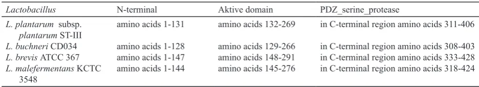
subsp. plantarum ST-III, GI: 308179226 from the NCBI site can be seen in Figure 3. Analysis of protein through the internet site of NCBI obtained structures of serine protease HtrA (L. plantarum
subs. plantarum ST-III) consists of three domains: 1.N-terminal: aa number from 1-27 and 28-131; 2. Active domain: aa number of 132-269 numbers; 3. PDZ_serine_protease: the C-terminal aa ranging from 311-406 then three (3) domains of L. buchneri,
L. brevis, and L. male-fermentans can be seen in
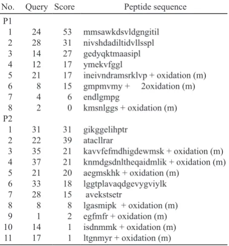
No. Query Score Peptide sequence
P1
ineivndramsrklvp + oxidation (m) gmpmvmy + 2oxidation (m) endlgmpg
kmsnlggs + oxidation (m)
gikggelihptr atacllrar
kavvfefmdhigdewmsk + oxidation (m) knmdgsdnltheqaidmlik + oxidation (m) aegmskhk + oxidation (m)
lggtplavaqdgevygviylk avekstsetr
lgasmipk + oxidation (m) egfmfr + oxidation (m) isdnmmk + oxidation (m) ltgnmyr + oxidation (m)
Description: oxidation(m) is a variable modification of Mascot
Search Results: PeptideView; https://sysbiomascot.wehi.edu.
au/mascot/cgi/peptide_view.pl?file=../d.
Table 2. The level of similarity (%) between Lactobacillus spp. producing trypsin-like serine protease by using Clone Manager®
software
C B M R Bch Cris Delb G S H P
L. casei ATCC-334, gi|116496224
L. brevis ATCC 367, gi|116496224
L. malefermentans KCTC 3548, gi|365901983
L. rhamnosus HN 001, gi|116628747
L. buchneri CD -034, gi|116496224
L. crispatus 214-1, gi|293380067
L. delbruecki ATCC BAA-365, gi|365901983
L. gasseri ATCC 33323,gi|116628747
L. suebicus KCTC 3549, gi|366051993
L. helveticus R0052, gi|116628747
L. plantarum subsp. plantarum ST-III, gi:308044717 54
Figure 1. Electrophoresis SDS_PAGE, CBB staining. M: marker,
A: crude enzymes, B: ultrafiltration (UF) 5 kD, C:
UF-PEG 20 KD, D: UF-membranes 30 kD (retentate), E:
Amino acid sequence (aa) the results of MALDI-TOF/TOF that the amount is greater than 10 for
the first band is 8 peptide (peptide 1_1; 1_2; 1_3;
1_5), of the second band 11 peptide (peptide 2_1; 2_3; 2_4; 2_6). Amino acid sequence of the region active domain (Trypsin_2) in the four Lactobacillus
were inserted into software Notepad for alignment analyzed by using Clone Manager®.
The results of analysis alignment the four
peptides from first band (P1) with L. plantarum
subsp. plantarum ST-III on active domain region (aa:132-269) indicates three peptides1_1; 1_2 and
1_5 had significant similarity at aa:132 + (19-70),
then the analysis of four peptides from second band (P2) indicates three peptide 2_1; 2_4 and 2_6 had
significant similarity at aa: 132 + (60-133) as shown
in Figure 4. The results from alignment of four
peptides from first band (P1) with L. buchneri CD034
on active domain region (aa: 129-266) indicates two
peptide 1_1 and 1_2 had significant similarity at aa:
129 + (29-75), then the analysis of four peptides from the second band (P2) indicates three peptide
2_1; 2_4 and 2_6 had significant similarity at aa:
132 + (59-136) as shown in Figure 5. The analysis
of the alignment, four peptides of the first band (P1) with L. brevis ATCC 367 on active domain region (aa: 148-291) indicates three peptides 1_1; 1_2; 1_3
had significant similarity at aa: 148 + (22-173), then
the analysis of four peptides from the second band (P2) indicates peptide 2_6 had significant similarity at a: 134 + (54-134) as shown in Figure 6. Analysis
results from alignment of four peptides from first L.casei
L.rhamnosus
L.brevis L.malefermentans L.plantarum L.buchneri L.suebicus L.crispatus L.helveticus L.delbrueckii L.gasseri
0.1
Figure 2. The phylogenetic tree based on the multiple sequence alignment of amino acids (aa), of eleven Lactobacilus
producing protease trypsin-like serine from the NCBI site using Clustal W2 program with the unweighted pair group method with arithmetic mean (UPGMA) and TreeViewX program.
Figure 3. Conserved domain from L. plantarum subsp. plantarum ST-III, GI : 308179226 result from NCBI www.ncbi.nlm.nih. gov/genbank/
Table 3. The three domains consisting of the N-terminal, active domain and PDZ_serine_protease from structures of trypsin-like serine protease from some Lactobacillus NCBI’s search results
Lactobacillus N-terminal Aktive domain PDZ_serine_protease
L. plantarum subsp.
plantarum ST-III
L. buchneri CD034
L. brevis ATCC 367
L. malefermentans KCTC 3548
amino acids 1-131
amino acids 1-128 amino acids 1-147 amino acids 1-144
amino acids 132-269
amino acids 129-266 amino acids 148-291 amino acids 145-276
in C-terminal region amino acids 311-406
in C-terminal region amino acids 308-403 in C-terminal region amino acids 333-428 in C-terminal region amino acids 318-424 Figure 4. Result of analysis alignment of peptide1_5 ( );
band (P1) with L. malefermentans KCTC 3548 at active domain region (aa: 145-276) peptide 1_1
and 1_2 show significant similarity at aa: 145 +
(33-106), then the alignment of analysis the four peptides from the second band (P2) had significant similarity at aa: 145 + (67-123) as can be seen in Figure 7.
Figure 5. Results of analysis alignment of peptide1_1 ( ); peptide 1_2 ( ); peptide1_5 ( ), and Peptide2_4 ( ); peptide2_6 ( ); peptide2_1 ( ) with L. buchneri CD034 in the active region domain (aa: 129-266).
Figure 6. Results of analysis alignment of peptide1_1 ( ); peptide 1_3 ( ); peptide1_2 ( ), and Peptide2_6 ( ) with L. brevis ATCC 367 in the active region domain (aa: 148-291).
Figure 7. Results of analysis alignment of peptide1_2 ( ); peptide 1_1 ( ); peptide2_6 ( ), and Peptide2_1 ( ) with L. malefermentans KCTC 3548 in the active region domain (aa: 145-276).
1 mankslikva vtalvaglig ggvaygginy fqnnniatss tsvptgsnks gststtnvkv 61 nvssqatkvf ennkaavvsv inlqkkssss swsgilggdd ssgsdsssss dsssskleey 121 segsgliykk sgdaayivtn nhvvsgssai rvimsdgtkl sakivgtdsv tdlavlkins 181 skvtktasfg nsdnikvget alaigspmgs nyattltqgi isakkrtvat tntsgqttgy 241 atviqtdtai nsgnsggplf niagqvigin smklasdnsg tsvegmgfai psnevvkiin 301 elvqkgevvr palgvatydl snisssdqks vlklptsvtk gvvimktysg spakaagltk 361 ydvitelggk kvtslatlrs alyahsvndt vtvkyyhngk lktanmklte ttktltkqsn
Figure 8. Amino acid sequence (aa) serine protease HtrA (L. plantarum subsp. plantarum ST-III) from NCBI.
gdaayivtn nhvvsgssai rvimsdgtkl sakivgtdsv tdlavlkins
181 skvtktasfg nsdnikvget alaigspmgs nyattltqgi isakkrtvat tntsgqttgy 241 atviqtdtai nsgnsggplf niagqvigi
Figure 9. Amino acid (aa) at region 132-269 /region_name=”Trypsin_2 /note=“Trypsin-like peptidase domain; pfam13365/db_ xref=”CDD from NCBI.
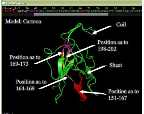
Figure 10. Three-dimensional models of Cartoon position partial sequences of amino acids (aa) trypsin-like protease (TLP) on P1 ( , , , ) are significant aa sequence similarity with trypsin 2 ( ) or trypsin-like peptidase domain of L. plantarum subsp.
plantarum ST-III (NCBI).
Model: Cartoon
Position aa to 169-173
Position aa to 164-169
Position aa to 198-202
Position aa to 151-167
Sheet Coil
Model: Cartoon
Position aa to 254-265
Position aa to 198-203
Position aa to 192-196
Sheet Coil
Figure 11. Three-dimensional models of Cartoon position partial sequences of amino acids (aa) trypsin-like protease (TLP) on P2 ( , , ) are significant aa sequence similarity with Trypsin 2 ( ) or trypsin-like peptidase domain of L. plantarum
Amino acid sequences from NCBI (http://www. ncbi.nlm.nih.gov/): YP_003923354.1 for serine protease HtrA [L. plantarum subsp. plantarum ST-III] and the area (region) aa 132-269/region_name = “Trypsin_2/note =” Trypsin-like peptidase domain; pfam13365/db_xref = “CDD: 205544” as shown in Figure 8 and 9 respectively.
Figure 10 and 11 shows the position of the amino
acid sequence (aa) TLP results are in significant
similarity with trypsin-like peptidase domains or trypsin 2 of L. plantarum subsp. plantarum ST-III (NCBI).
DISCUSSION
Molecular weight TLP is also estimated by UPLC-MS, using formic acid as the eluent. According to Lagrain et al. (2013) with formic acid as the eluent, the sensitivity of the MS detector clearly increased. This was also underlined by improved mass intensities of the detected proteins in the Base Peak Chromatogram (BPC). Results from electrophoresis and UPLC-MS of TLP L. plantarum FNCC 0270 there were four bands and equivalent forms with molecular weight ± 47, ± 38, ± 21, and ± 13 kD, respectively. In humans,
there are five forms of pancreatic trypsin namely
cationic trypsinogen (PRSS1), anionic trypsinogen (PRSS2), mesotrypsin (PRSS3), pancreasin, trypsin IV (Whitcomb & Lowe 2006). Typically, cationic trypsinogen represents about two-thirds of the total trypsinogen, while anionic trypsinogen about one-third. PRSS1 obtained by using software Prot Param BM 26.558; iso-electric point (PI) of 6.08 and 247 amino acids.
From NCBI search to get the serine protease
of L. plantarum ST-III subs. plantarum
(YP_003923354.1), L. buchneri (YP_006724721.1),
L. brevis (YP_794241.1), L. malefermentans
(ZP_09439806.1), all bioinformatics analysis of the genome that produces trypsin-like proteases, used approach to the conceptual translation method. They have a molecular weight, 42.97, 43.349, 46.124, 43.216 kD respectively (Makarova et al. 2006; Wang et al. 2011; Heinl et al. 2012). Trypsin from the Pyloric caeca red snapper brown striped had estimated molecular weight 23 kD. Usually trypsin
of fish has a molecular weight between 20-30 kD
(Khantaphant & Benjakul 2010).
Band of the P1 and P2 from SDS-PAGE electrophoresis staining with CBB, digested with chymotrypsin and peptides extracted according to standard techniques (Bringans et al. 2008). Then
the peptides were analyzed by Mascot sequence matching software and the results are shown in Table 1. Analysis of the phylogenetic tree showed that L. plantarum protease has close relationship with L. buchneri, L. brevis, and L. malefermentans. Therefore from the four Lactobacillus should be found conserved domain through website NCBI/ BLAST and obtained active domain (Trypsin_2) with amino acid (aa) different length (Marchler-Bauer et al. 2011). The fragments of TLP by L. plantarum FNCC 0270 (pep1_1, pep1_2, pep1_3, pep1_5 and pep2_1, pep2_3, pep2_4, pep2_6) with alignment analysis, respectively indicates the similarity although only small at amino acid (aa) the active domain region of trypsin2 from L. plantarum subs. plantarum ST-III, L. buchneri, L brevis, and L. malefermentans using Clone Manager software (Figure 4-7). Based on these conditions it is assumed that the TLP by L. plantarum FNCC 0270 protein isolated in this study, is a member of serine protease group of L. plantarum.
Sequence aa of HtrA serine [L. plantarum subsp.
plantarum ST-III] and trypsin–like peptidase domain or Trypsin 2, with some software can describe the characteristics of these proteins. Three-dimensional images of a trypsin–like peptidase domain or trypsin 2 of L. plantarum subsp. plantarum ST-III (NCBI) obtained by the software SWISS Model and PyMOL with inserted aa sequences Trypsin 2 at program PyMOL (Arnold et al. 2006; Benkert et al. 2011).
L. plantarum subsp. plantarum ST-III obtained nine (9) form sheet (strand) and four (4) form coil. This is in accordance with the three-dimensional image as shown in Figure 10 and 11.
The Signal P software can predict the end of protein and cut off the protein (Petersen et al. 2011). Region of the end protein C that carries the code location HtrA serine protein [L. plantarum
subsp. plantarum ST-III] predicted cleavage site between at position 26 and 27 value 0.141: AYG-GI; measure D, position 1-26, value 0.457, cutoff 0.450, SP = Yes. The region of the end protein C that carries the code location domain trypsin-like peptidase or trypsin 2 from L. plantarum subsp.
plantarum ST-III predicted at positions 19, value 0.147, measure D, position 1-31, value 0.224, cutoff 0.450 and SP = No.
Prot Scale software (protein_expasy) at analyze proteins with aa scale (scale Hphob. HPLC) of the serine protease HtrA [L. plantarum subsp. plantarum
ST-III] had scores between -1.5 s/d +4. Scores (-) is hydrophilic on the surface of the protein and scores (+) is hydrophobic in the protein, where as the trypsin-like peptidase domain or trypsin 2 from
L. plantarum subsp. plantarum ST-III had scores between -1 - +3.25.
Software Prot Param (protein_expasy) to find
information about protein. The serine protease HtrA [L. plantarum subsp. plantarum ST-III] obtained MW: 43101.4; PI: 9.56; aa number: 420; Formula C1865H3079N515O635S7 and amount Atomic number: 6101, the half-life (in vivo) estimated >20 h for the yeast and >10 h for Escherichia coli and 30 h for mammalian reticulocytes (in vitro). Trypsin-like peptidase domain or trypsin 2 of L. plantarum subsp.
plantarum ST-III acquired MW:13881.5; PI: 9.35; aa number: 138; Formula C598H994N168O205S2 and amount Atomic number: 1967, the estimated half-life equal to the serine protease HtrA.
Isolated TLP of L. plantarum FNCC 0270 was little but unique. Full genome sequencing of
Lactobacillus and then gene annotation and de novo
assembled to track the presence of genes encoding trypsin-like proteases.
ACKNOWLEDGEMENT
Our thanks to the Ministry of Research and Technology (No.:105/M/KP/VIII/2009), BPPT, which has provided funding to complete this study, as well as all those who participated in this study.
REFERENCES
Arnold K, Bordoli L, Kopp J, Schwede T. 2006. A web-based environment for protein structure homology modeling.
Bioinformatics 22:195-201. http://dx.doi.org/10.1093/ bioinformatics/bti770
Benkert P, Biasini M, Schwede T. 2011. Toward the estimation of the absolute quality of individual protein stucture models.
Bioinformatics 27:343-350. http://dx.doi.org/10.1093/ bioinformatics/btq662
Bradford M. 1976. A rapid and sensitive method for quantitation of microgram quantities of protein utilizing the principle of protein-dye bind. Anal Biochem 72:248-254. http://dx.doi. org/10.1016/0003-2697(76)90527-3
Bringans S, Eriksen S, Kendrick T, Gopalakrishnakone P, Livk A, Lock R, Lipscombe R. 2008. Proteomic analysis of the venom of Heterometruslongimanus (Asian black scorpion).
Proteomics 8:1081-1096. http://dx.doi.org/10.1002/ pmic.200700948
Heinl S, Wibberg D, Eikmeyer F, Szczepanowski R, Blom J, Linke B, Goesmann A, Grabherr R, Schwab H, Puhler A, Schluter A. 2012. Insights into the completely annotated genome of Lactobacillus buchneri CD 034, a strain isolated from stable grass silage. Biotechnology 161:153-166. Jean-Michel C, Notredame C. 2007. Bioinformatics for
Dummies. 2nd ed. Wiley Publishing, Inc. 111 River street , Hoboken, NJ 07030-5774, p 411-414.
Jellouli K, Bougatef A, Daassi D, Balti R, Barkia A, Nasri M. 2009. New alkaline trypsin from the intestine of
Grey triggerfish (Balistescapricus) with high activity
at low temperature: purification and characterization. Food Chem 116:644-650. http://dx.doi.org/10.1016/j. foodchem.2009.02.087
Kishimura H, Tokuda Y, Klomklao S, Benjakul S, Ando S. 2006. Enzymatic characteristics of trypsin from pyloricceca of spotted mackerel (Scomber Australisicus). Food Biochem 30:466-477. http://dx.doi.org/10.1111/j.1745-4514.2006.00076.x
Khantaphan S, Benjakul S. 2010. Purification and characterization
of trypsin from the pyloric caeca of brownstripe red snapper (Lutjanus vita). Food Chem 120:658-664. http://dx.doi. org/10.1016/j.foodchem.2009.09.098
Lagrain B, Brunnbauer M, Rombouts I, Koehler P. 2013.
Identification of intact high molecular weight glutenin
sub units from the wheat proteome using combined liquid chromatography-electrospray ionization mass spectrometry.
PLoS ONE 8: e58682. http://dx.doi.org/10.1371/journal. pone.0058682
Malik A, Hermawati AK, Hestiningtyas M, Soemiati A, Radji M. 2012. Isolation and screening of lactic acid bacteria molecular gene carrie glukansukrase of food and beverages containing sugar. Makara (Science) 14:57-62.
Marchler-Bauer A, Lu S, Anderson JB, Chitsaz F, Derbyshire MK, DeWeese-Scott C, Fong JH, Geer LY, Geer RC, Gonzales NR, Gwadz M, Hurwitz DI, Jackson JD, Ke Z, Lanczycki CJ, Lu F, Marchler GH, Mullokandov M, Omelchenko MV, Robertson CL, Song JS, Thanki N, Yamashita RA, Zhang D, Zhang N, Zheng C, Bryant SH. 2011. CDD: a conserved domain database for the functional annotation of proteins. Nucleic Acids Res 39 (Database issue): D225-9. http://dx.doi.org/10.1093/nar/gkq1189 Pato U, Ali M, Parlindungan AK. 2005. Taurocholate
deconjugation and cholesterol binding by indigenous dadih lactic acid bacteri. Hayati 12:103-107.
Petersen IN, Brunak S, Heijne G, Nielsen H. 2011. Discriminating signal peptides from trans membrane regions. Nat Methods 8:785-786. http://dx.doi.org/10.1038/ nmeth.1701
Suri WL, Syukura S, Jamsarib. 2013. Optimization of protease activity from lactic acid bacteria (Lab) Pediococcus pentosaceus isolated from soursop fermentation (Annona muricata L.). Jurnal Kimia Unand 2:18-25.
Wang Y, Chen C, Ai L, Zhou F, Zhou Z, Wang L, Zhang H, Chen W, Guo B. 2011. Complete genome sequence of the probiotic Lactobacillus plantarum ST-III. Bacteriology
193:313-314. http://dx.doi.org/10.1128/JB.01159-10 Whitcomb DC, Lowe ME. 2006. Human pancreatic digestive
enzymes. Dig Dis Sci 52:1-17. http://dx.doi.org/10.1007/ s10620-006-9589-z
Wulansari D, Wahyuntari B, Trismilah, Nurhasanah A. 2012. The effect of growth medium pH towards trypsin_like activity produced by lactic acid bacteria. Microbiol Indones