Available Online at www.jbpr.in
Journal of Biomedical and Pharmaceutical Research, Volume 3, Issue 4, 2014, 63-69
P
a
g
e
6
3
P
a
g
e
6
3
P
a
g
e
6
3
P
a
g
e
6
3
RESEARCH ARTICLE
BRINE SHRIMP LETHALITY TEST AND ANTI-PROLIFERATION TEST AGAINST HUMAN
CANCER-ORIGIN CELL LINES USING ETHANOLIC AND WATER EXTRACTS OF
SELAGINELLADOEDERLEINII
HIERON
J.T. Priscilla1, S. Geethaa1, S. Sreeramanan 2, M.T. Ong1*
1
Institute for Research in Molecular Medicine (INFORMM), Universiti Sains Malaysia, 11800 USM, Pulau Pinang, Malaysia.
2
School of Biological Sciences, Universiti Sains Malaysia, 11800 USM, Pulau Pinang, Malaysia.
Received 15 July 2014; Accepted 26 August 2014
INTRODUCTION:
Medicinal plants form a rich source of novel drugs that forms the ingredients in traditional systems of medicine, modern medicines, nutraceuticals, food supplements, folk medicines, pharmaceutical intermediates, bioactive principles and lead compounds in synthetic drugs [1]. In Malaysia, the practice of traditional medicine is common among various ethnic groups such as the Malays, Chinese, Indians and aborigines with the knowledge being passed down through the generations. The practice relies on the use of medicinal plants, properties of which are mainly known via traditional knowhow and/or empirical observations. According to the World Health Organization (WHO), 80 % of people still rely on plant-based traditional medicines for primary health care globally. It is therefore crucial to scientifically evaluate and validate the properties of medicinal plants as reported in traditional use.
Selaginella doederleinii HIERON has been reported to be
a medicinal plant, but has not been widely used, either traditionally or in modern medicine. Its role in medicinal
use is yet to be determined. The genus Selaginella is composed of about 700 species and belongs to the kingdom of Plantae, the family of Selaginellaceae. They belong to a group of lycopods, whose members go by common names of ground pines and club mosses.
Selaginella doederleinii HIERON is a well-known perennial
pteridophyte plant growing in South and Southwestern China at low altitude [2], and it has been used in traditional Chinese medicine [3].
Selaginella can be found in pharmacopoeia Asia, Africa,
Latin America, but is not found in Europe and North America [4]. This is due to the habitat conditions of the plant. Humid conditions with higher rain fall and level of a relatively higher slope supports the life of this plant. The local name of Selaginella in Malaysia is cakar ayam, name given by referring to the leaf shape that resembles the scales on a chicken leg on either side of the stem [5].
Selaginella has many other local names, such as rumput
solo, cemara kipas gunung (Java), paku rane (Sunda) and
shi shang bai (石上柏 ) or juan bai (卷柏) (Chinese).
ABSTRACT
Most of the properties of herbal products claimed in traditional use are based on traditional knowhow and/or empirical observations. Support from scientific evaluation and validation is thus crucial. Selaginella doederleinii
HIERON is a member from the family of Selaginellaceae, a perennial, fern-like plant, and spike moss. It is known in traditional medicine to be useful to treat many diseases. However, proper scientific experimental results have not been reported thus far. The present study was conducted with the objectives of evaluating the phytochemical compositions, antimicrobial activities, and toxicity of the plant extracts. Qualitative phytochemical analysis on ethanolic (SDE) and water extracts (SDW) revealed the presence of saponins, tannins and cardiac glycosides in the extracts. The 50% lethal concentration (LC50) in brine shrimp lethality test (BSLT) using SDE and SDW after 24 hours
of exposure was found to be >1000µg/ml. Strong antimicrobial activity was observed against Escherichia coli,
Pseudomonas aeruginosa, Staphylococcus aureus, Bacillus subtilus. Cancer-origin cell lines MDA-MB231 and
HepG2 were found to be the most susceptible, among other tested cell lines, with the treatments of SDE (LC50
=306 µg/ml) and SDW (LC50= 329 µg/ml) respectively. Amelioration with further subfractionation would increase
the specific antiproliferation activities of these subfractions.
P
a
g
e
6
4
P
a
g
e
6
4
P
a
g
e
6
4
P
a
g
e
6
4
P
a
g
e
6
4
P
a
g
e
6
4
P
a
g
e
6
4
P
a
g
e
6
4
P
a
g
e
6
4
P
a
g
e
6
4
P
a
g
e
6
4
P
a
g
e
6
4
P
a
g
e
6
4
P
a
g
e
6
4
P
a
g
e
6
4
P
a
g
e
6
4
P
a
g
e
6
4
P
a
g
e
6
4
P
a
g
e
6
4
P
a
g
e
6
4
P
a
g
e
6
4
The most commonly used part of Selaginella is the leaves, reason for the plant to be termed as leaf herb. In traditional medicine it can be used singularly or in combination with other plants, fresh or dried, consumed immediately or cooked before consumption [5]. These plants are sweet and have a warm effect [6].Traditional treatment is carried out by boiling the plant parts as a drink for the patient. Approximately 15-30 grams (for normal people) or 50-100 grams (for medically diagnosed cancer patients) of Selaginella doederleinii extract is boiled for 3-4 hours before consumption. The present study was aimed to evaluate the anti-microbial activities and to identify phytochemical compositions of Selaginella
doederleinii. Preliminary cytotoxic effect on biological
system as well as anti-proliferation properties of the plant extracts against cancer-origin cell lines were evaluated using brine shrimp lethality test and MTT assay respectively.
MATERIALS AND METHODS:
Plant materials:
The Selaginella doederleinii leaves and stems were
collected from Air Itam Dam in Penang, Malaysia. Identification and classification of the plant material were carried out by a botanist from the School of Biological Sciences at Universiti Sains Malaysia. The specimen voucher (HerbariumUSM 11260) was deposited in the herbarium for future reference.
Extract Preparation:
The dried leaves and stems were ground to fine powder, further extracted with ethanol and water following the solvent to dry weight ratio 10ml: 1 g [7] with several daily shakings at room temperature. The extracts were filtered through filter paper (WhatmanTMNo.1) and further concentrated in a rotary evaporator (RII0 Buchi, Switzerland) at 40°C. The resinous residue from plant material was dried in oven at 37°C for 4 days and subjected to lyophilisation (Labconco, Freezone 4.5 liter, Kansas City, MO). The working concentration of ethanolic extract (SDE) was kept at 100 mg/ml in DMSO and aqueous extract (SDW) in water respectively. These were stored at -20˚C until tested.
Phytochemical Tests:
Qualitative phytochemical tests were carried out on the crude extracts and on the powdered specimens of
Selaginella doederleinii Hieron extracts using standard
procedures to identify the constituents as described previously [8-13].Tests were carried out in three replicates. The concentration of Selaginella doederleinii Hieron extract used in each phytochemical test was 10 mg/mL. a) Alkaloids: 5mL of the extract was added to 2mL of 1% aqueous hydrochloric acid (HCl). To this acidic medium,
1ml of Dragendroff’s reagent was added. An orange or red precipitate produced immediately indicates the presence of alkaloids.
b) Tannins: About 10mg of the dried sample was boiled in 2mL of water in a test tube and then filtered. A few drops of 0.1 % of ferric chloride solution was added to the filtrate and observed for brownish green or a blue-black coloration.
c) Saponins: About 10mg of the dried sample was shaken with water in a test tube and warmed in a water bath. Frothing, which persist on warming was taking as a preliminary evidence for the presence of saponins. Few drops of olive oil was added and vigorously shaken. Formation of soluble emulsion in the extract indicates the presence of saponins.
d) Terpenoids/Steroids (Salkowski test): Five milliliter of aqueous extract mixed with 2mL of chloroform and 3mL of concentrated sulphuric acid was carefully added to form a layer. A reddish brown coloration on the interface confirmed the presence of terpenoids.
e) Cardiac Glycosides (Keller-Killani test): Five milliliter of aqueous extract was treated with 2mL of glacial acetic acid which containing one drop of ferric chloride solution. This was underlayed with 1mL of concentrated sulphuric acid. A brown ring on the interface indicates a deoxysugar characteristic of cardenolides. A violet ring may appear below the brown ring, while in the acetic acid layer, a greenish ring may form just gradually throughout the thin layer.
f) Reducing sugars in glycosides (Fehling’s test): The residue was re-dissolved in water in the water bath .Two milliliter of the extract in a test tube was added with 1mL each of Fehling’s solution A and B. The mixture was shaken and heated in a water bath for 10minutes. A brick-red precipitate indicates a reducing sugar test [8; 14-
15]
.
Brine Shrimp Lethality Test (BSLT):
The procedure for BSLT was modified from the assay described by Mc Laughlin and groupers [16]. Brine shrimp
(Artemia salina) eggs (SandersTM Great Salt Lake, Brine
Shrimp Company L.C., U.S.A.) were hatched in artificial sea water prepared from commercial sea salt (38 g sea salt/liter deionized water) with constant light source and oxygen supply after 24 hours of incubation. Selaginella
doederleinii Hieron ethanolic (SDE) and aqueous (SDW)
P
a
g
e
6
5
P
a
g
e
6
5
P
a
g
e
6
5
P
a
g
e
6
5
P
a
g
e
6
5
P
a
g
e
6
5
P
a
g
e
6
5
P
a
g
e
6
5
P
a
g
e
6
5
P
a
g
e
6
5
P
a
g
e
6
5
P
a
g
e
6
5
P
a
g
e
6
5
P
a
g
e
6
5
P
a
g
e
6
5
P
a
g
e
6
5
P
a
g
e
6
5
P
a
g
e
6
5
P
a
g
e
6
5
P
a
g
e
6
5
P
a
g
e
6
5
the number of living nauplii was counted after the incubation period. Lethal concentration for 50% mortality (LC50) for brine shrimp with 95% confidence level was
determined by Probit analysis [17] on a Finney computer program BioStat™ 2009 (Analyst Soft Inc., Vancouver, Canada).Percentage of mortality was corrected for the natural mortality observed in the negative controls using Abbott’s formula, P= (pi-C)/ (1-C), where pi denotes the
observed mortality rate and C means the natural mortality [17].
Antimicrobial Susceptibility Test:
Microbial strains:
Shigella flexneri (ATCC® No. 12022TM), Escherichia coli
(ATCC® No. 25922TM), Staphylococcus aureus (ATCC® No. 25923TM), Bacillus subtilus (lab isolate), Bacillus cereus
(ATCC® No. 10876TM), Pseudomonas aeruginosa (ATCC® No. 27853TM), Aspergillus niger (ATCC® No. 9142TM) and
Candida albicans (ATCC® No. 10231TM) were used for the
evaluation. The bacterial strains were grown in nutrient broth at 37°C and maintained in nutrient agar slant at 4°C. However, the fungal strains were grown and maintained in Potato Dextrose Agar (PDA) at 28°C for
A.niger and at 37°C for C. albicans.
Disc Diffusion Method:
The Selaginella doederleinii Hieron lyophilized extracts
were tested for antimicrobial activity by the disc diffusion method according to the NCCLS (2001). Overnight suspension cultures of microbial strains were adjusted according to 0.5 McFarland standards (105 CFU/ml). One hundred microliter of adjusted culture was spread on the Mueller Hilton Agar (MHA) media for bacteria and (PDA) for fungi. Sterile discs were prepared and placed on the culture spread agar media. Each sterile disc was
impregnated with 20μl of the extract (50mg/ml of crude
extract), Chloramphenicol (30μg/ml, as positive control for bacterial strains), Amphotericin B (0.2mg/ml, as positive control for fungal strains), Phosphate Buffer Saline (PBS) and Tween 20% (as negative controls). The inoculated plates were incubated at 37°C for 24 h for bacterial strains and 48 h for fungal strains [18] to examine the zones of inhibition.
Minimum Inhibitory Concentration (MIC) tests:
a) MIC for antibacterial test:
The extract was assayed for antibacterial using agar dilution method in 24-well microtiter plate. The inoculums of microorganisms were prepared from 18-hour nutrient broth cultures. The suspension cultures of tested microbial strains were adjusted according to 0.5 McFarland standards (105 CFU/ml). A series of two-fold dilutions of each extract (500mg/ml), ranging from 20 to
0.625mg/ml, were prepared in MHA and later inoculated with 2µl suspension of the test organisms. MHA with only Chloramphenicol (30µg/ml) was used as positive growth control and MHA with 4% of DMSO was used as negative growth control. Inoculated plates were incubated at 37˚C. The minimum inhibitory concentration (MIC) was determined as the lowest concentration of each extract that completely inhibited growth of microorganism up to
24hours, whereas the minimum bactericidal
concentration (MBC) was the lowest concentration at which no growth was observed after incubation.
b) MIC for antifungal test:
A serial two fold dilutions of SDE and SDW extracts was carried out in PDA media from 20mg/ml to 0.625mg/ml. The mixture of PDA media and extract was transferred into 24-well plates aseptically. Upon solidification of the agar medium, 2µl of standardized culture of Aspergillus
niger andCandida albicans suspension was added to each
plate. The plates were incubated at 28°C ± 5˚C for 48 hours. The PDA media without extract served as negative control and PDA media with Amphotericin B (200µg/ml) served as positive control. The MIC is regarded as the lowest concentration of the extract that did not show any visible growth after 7 days of incubation (compared with control) [19]. The presence of one or two colonies was disregarded [20].
Cytotoxicity:
Cell culture:
Hs27 (ATCC® No.CRL-1634TM), HepG2 (ATCC® No.HB-8065TM), MCF-7 (ATCC® No.HTB-22TM) and MDA-MB 231 (ATCC® No. HTB-26TM) cells were cultured in Dulbecco’s Modified Eagle’s Medium (DMEM)(Gibco®Invitrogen, Carlsbad, CA, USA) supplemented with fetal calf serum (10% v/v), penicillin (100units/ml), streptomycin (100 μg/ml) and amphotericin B (0.025μg/ml). Medium for Hs27 was supplemented with additional 4.5g/L of glucose. Cultures were maintained at 37°C in a water-saturated atmosphere containing 5% CO2. Cell counts
were performed using a Neubauer haemocytometer under light microscope and living cells were identified by Trypan blue staining method. Approximately 2500 cells were seeded in each well of a 96-well culture plate and were starved in DMEM under cell culture incubation conditions for 48 hours prior to cell-based assay.
Measurement of cell growth inhibition:
P
a
g
e
6
6
P
a
g
e
6
6
P
a
g
e
6
6
P
a
g
e
6
6
P
a
g
e
6
6
P
a
g
e
6
6
P
a
g
e
6
6
P
a
g
e
6
6
P
a
g
e
6
6
P
a
g
e
6
6
P
a
g
e
6
6
P
a
g
e
6
6
P
a
g
e
6
6
P
a
g
e
6
6
P
a
g
e
6
6
P
a
g
e
6
6
P
a
g
e
6
6
P
a
g
e
6
6
P
a
g
e
6
6
P
a
g
e
6
6
P
a
g
e
6
6
(Mosmann ,1983) [21]. Two thousand five hundred cells were seeded into each well of 96-well plates and cultured in the CO2 incubator at 37°C. After 48 hours, the cells
were treated with various serial-diluted concentrations of
Selaginella doederleinii extract respectively and returned
into the CO2 incubator at 37°C. The absorbance at 570 nm
was read on a spectrophotometric plate reader (Multiskan spectrum, Thermo Electron Co.,Waltham, Massachusetts, USA). The proportion of surviving cells was calculated as [(OD of drug-treated sample – OD of blank)/ (OD of control – OD of blank)] × 100%. Dose-response curves were constructed using Probit analysis (Finney, 1962) to obtain the LC50 values. All experimental
data were derived from at least 3 independent experiments [22].
Wound Healing Test:
Hs27 (ATCC® No. CRL-1634TM) cells were seeded at 1x10⁵ cells/well into 24-well culture plates and cultured in DMEM containing 10% of FBS. These were then incubated for 24 hours. The confluent monolayer cells were wounded using micropipette tip, and cellular debris
present was removed by washing twice with PBS. The wounded monolayers were incubated for 48 hours in complete media containing various concentrations of crude extract and the potential bio-active fractions. Complete media served as positive control whereas water and medium containing DMSO were used as negative controls. Photographs of the wounds were taken at various incubation time periods (0 hour, 2 hours, 4 hours, 8 hours, 12 hours and 24 hours).
Statistical analysis:
Data are presented as mean ± S.E.M. of triplicate determinations, except when results of plots are shown, in which case a representative experiment is depicted in each figure. Statistical significance was indicated when p < 0.05.
RESULTS:
Phytochemical analysis was conducted to qualitatively identify the class of compounds found in SDE and SDW. The screening revealed the presence of alkaloids, tannins, saponins and cardiac glycosides in these fractions [Table 1].
Table 1: Phytochemical analysis of Selaginella doederleinii Hieron extract.
Class of chemical compounds Ethanolic
extract(SDE)
Aqueous
Extract(SDW)
Tannins + +
Alkaloids + +
Saponins Terpenoids
Cardiac glycosides Reducing sugar
+ - + -
+ - - -
*Note: +presence, - absence of the secondary metabolites in the extract tested
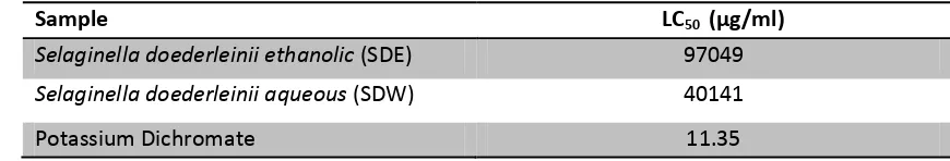
Brine shrimp lethality test (BSLT) [23] was conducted to assess the cytotoxicity level of the extracts. The values of lethal concentration for 50% mortality (LC50) after 24
hours of exposure for SDE and SDW fractions were found
to be less than 1000µg/ml as shown in [Table 2]. Potassium dichromate, as positive control, showed an LC50 of 11.35µg/ml.
Table 2: Brine Shrimp toxicity expressed as LC50value via Probit analysis.
Sample LC50 (µg/ml)
Selaginella doederleinii ethanolic (SDE) 97049
Selaginella doederleinii aqueous (SDW) 40141
Potassium Dichromate 11.35
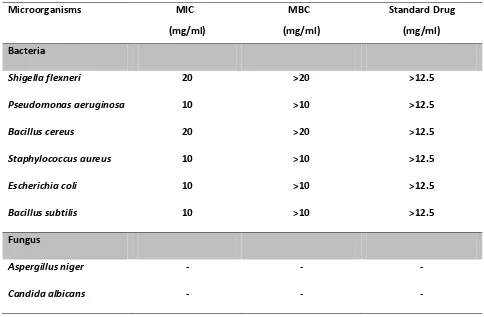
Disc diffusion assays showed that no detectable zone of inhibition, representing antibacterial or antifungal
P
a
g
e
6
7
P
a
g
e
6
7
P
a
g
e
6
7
P
a
g
e
6
7
P
a
g
e
6
7
P
a
g
e
6
7
P
a
g
e
6
7
P
a
g
e
6
7
P
a
g
e
6
7
P
a
g
e
6
7
P
a
g
e
6
7
P
a
g
e
6
7
P
a
g
e
6
7
P
a
g
e
6
7
P
a
g
e
6
7
P
a
g
e
6
7
P
a
g
e
6
7
P
a
g
e
6
7
P
a
g
e
6
7
P
a
g
e
6
7
P
a
g
e
6
7
agar dilution method did show observable growth inhibition in the tested microorganisms with SDE. Standard antibiotic Chloramphenicol was used as positive
control. Antifungal activity was not observed with SDE against the two tested fungal strains [Table 3].
Table 3: The MIC and MBC values are mean of three replicates for each tested microorganism.
Microorganisms MIC MBC Standard Drug
(mg/ml) (mg/ml) (mg/ml)
Bacteria
Shigella flexneri 20 >20 >12.5
Pseudomonas aeruginosa 10 >10 >12.5
Bacillus cereus 20 >20 >12.5
Staphylococcus aureus 10 >10 >12.5
Escherichia coli 10 >10 >12.5
Bacillus subtilis 10 >10 >12.5
Fungus
Aspergillus niger - - -
Candida albicans - - -
Table 4: Probit analysis results showing the LC50 at various time points post-treatment with SDE and SDW with 95 % confidence level.
*N/A denotes the value was too high and therefore not included.
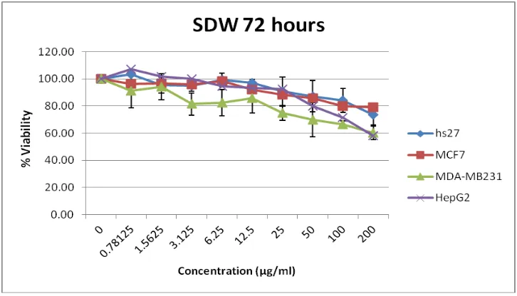
HepG2 cells showed susceptibility towards SDW at 72 post-treatment hours [Figure 1]. At this time point the susceptibility was not observed in both breast cancer-origin cell lines or in Hs27 (non-cancer-cancer-origin cell line) within the tested concentration range (0-200µg/ml). The LC50 value was found to be 329µg/ml. At 48 hours
post-treatment with SDW, no significant antiproliferation
activity was observed in MCF-7 and Hs27 cells, whereas MDA-MB231 was found to be susceptible to the treatment with an LC50 value of 539µg/ml [Table 4].
It has been found that SDE and SDW exhibited promotional effects of wound healing within the concentrations range from 1 µg/ml to 10µg/ml.
SDE SDW
24 hours 48 hours 72 hours 24 hours 48 hours 72 hours
Hs27 LC50 N/A 1470µg/ml 900µg/ml N/A 9146µg/ml 3814µg/ml
MCF7 LC50 N/A N/A N/A N/A N/A 6409µg/ml
MDA_MB231 LC50 2002µg/ml 306µg/ml 776µg/ml 1348µg/ml 539µg/ml 710µg/ml
P
a
g
e
6
8
P
a
g
e
6
8
P
a
g
e
6
8
P
a
g
e
6
8
P
a
g
e
6
8
P
a
g
e
6
8
P
a
g
e
6
8
P
a
g
e
6
8
P
a
g
e
6
8
P
a
g
e
6
8
P
a
g
e
6
8
P
a
g
e
6
8
P
a
g
e
6
8
P
a
g
e
6
8
P
a
g
e
6
8
P
a
g
e
6
8
P
a
g
e
6
8
P
a
g
e
6
8
P
a
g
e
6
8
P
a
g
e
6
8
P
a
g
e
6
8
Figure 1: The graph shows the viability at 72 hours post-treatment with SDW shows the growth of cancer origin specially MDA-MB231 and HepG2 were reduced compared to that of non-cancer origin cells (Hs27).
DISCUSSION:
There has been a remarkable growth of the usage of herbal medicine in many parts of the world. Herbal medicine is popularly used because of its origin that is the nature, fewer side effects, more efficient and less expensive [24]. Many have raised the issue on the efficacy and safety of the herbal medicine usage. For these,
S.doederleinii ethanolic and water extracts were further
evaluated.
Phytochemical screening is an important step in the study of medicinal plant and reveals the relationship of the phytochemical groups to the therapeutic uses. Accordingly, alkaloids, tannins, saponins and cardiac glycosides are considered as key ingredients in traditional Chinese medicines, and are responsible for most of the observed biological effects observed [25]. Crude extracts and pure substances with LC50 value lower than
1000μg/ml are considered bioactive in toxicity level evaluation of plant extracts using BSLT [16]. These results indicate the non-toxicity of the fractions. Thus, these findings corroborate the folk medicinal use of the plant in the treatment for both human and animals.
The difference in the phytochemical properties in the extract and its extractant reveals the differences observed in the antimicrobial results. In disc diffusion assay, the investigated plant did not show any antimicrobial activity but the result was otherwise in agar dilution method. It is possible that the active compounds were trapped in the discs and were not soluble, hence not diffusible, in the media to exert inhibitory activities against the tested microorganisms [16]. Thus, it can be concluded that SDE posses antimicrobial properties against the tested bacteria B.subtilis, S. aureus, E. coli, P.
aeruginosa at 10mg/ml, and this property was exerted
against S. flexneri , B. cereus at 20mg/ml.
In studies examining the anti-proliferative activity of herbal extracts, MDA-MB231 cells were found to be more susceptible at 72 hours post-treatment with SDE, with a value of LC50 = 306µg/ml, compared to that of MCF-7 cell
line [Table 4]. These might suggest that the active compound(s) in SDE might act through the pathway(s) involving caspase 3, and estrogen and EGF receptors which might not be crucial for the antiproliferation property observed [26]. HepG2, human liver cancer-origin cells, showed susceptibility towards SDW at 72 hours post-treatment where by 50% reduction was seen in cell viability. The cell viability assay carried out using MTT, showed no significant signs in MCF-7 and Hs27 cell lines and less susceptibility in MDA-MB231 for the treatment of SDW extract. Thus, it can be concluded that the active compound(s) in SDW are likely to cause the anti-proliferative action in HepG2. These active compound(s) found tallies the results observed in the phytochemical screening tested in the study.
P
a
g
e
6
9
P
a
g
e
6
9
P
a
g
e
6
9
P
a
g
e
6
9
P
a
g
e
6
9
P
a
g
e
6
9
P
a
g
e
6
9
P
a
g
e
6
9
P
a
g
e
6
9
P
a
g
e
6
9
P
a
g
e
6
9
P
a
g
e
6
9
P
a
g
e
6
9
P
a
g
e
6
9
P
a
g
e
6
9
P
a
g
e
6
9
P
a
g
e
6
9
P
a
g
e
6
9
P
a
g
e
6
9
P
a
g
e
6
9
P
a
g
e
6
9
CONCLUSION:
In conclusion, SDE and SDW possess phytochemicals such as alkaloids, tannins, cardiac glycosides and saponins which could potentially be the leading cause of anti-proliferative action observed in MDA-MB231 and HepG2. The mechanisms of the selective cytotoxicity against cancer-origin cell lines need to be further studied in view of the development of anti-cancer treatments.
ACKNOWLEDGEMENT:
J.T Priscilla and S. Geethaa are recipients of “MyBrain15” Scholarship from the Malaysia Ministry of Education (MOE), Malaysia.
REFERENCES:
1. Ncube NS, Afolayanc AJ, Okoh AI. Assessment techniques of antimicrobial properties of natural compounds of plant origin: current methods and future trends. Afr. J. Biotechnol. 2008;7:1797-1806. 2. Lin RC, Skaltsounis AL, Seguin E, Tillequin F, Koch M.
Phenolic Constituents of Selaginella doederleinii. Planta Med. 1994;60:168-170.
3. Chao LR, Seguin E, Tillequin F, & Koch, M. New Alkaloid Glycosides from Selaginella-Doederleinii. J. Nat. Prod. 1987; 50:422-426.
4. Duke J, Bogenschutz-Godwin MJ, du Cellier J & P-AK., D. Handbook of medicinal herbs,CRC Press Boca Raton; Flourida, 2002.
5. Dalimartha S. Atlas of Indonesian Medicinal Plants. Trubus Agriwidya. Yogyakarta.[Indonesia]; 1999. 6. Dan Bensky, Steven Clavey & Stõger, E. Chinese
herbal medicine;Materia Medica, 3rd Edition Eastland Press.Seattle, WA; 2004.
7. Green RJ. Antioxidant Activity of Peanut Plant Tissues.
Masters Thesis, North Carolina State University, USA. (2004).
8. Adegoke AA, Iberi PA, Akinpelu DA, Aiyegoro OA, Mboto CI. Studies on phytochemical screening and antimicrobial potentials of Phyllanthus amarus against multiple antibiotic resistant bacteria. IJARNP. 2010;3:6-12.
9. Aiyelaagbe OO, Osamudiamen PM. Phytochemical Screening for Active Compounds in Mangifera indica
Leaves from Ibadan, Oyo State. Plant Sciences Research. 2009;2: 11-13.
10. Kumar A, Ilavarasan R, Jayachandran T, Decaraman, M, Aravindhan P, Padmanabhan N, Krishnan MRV. Phytochemicals investigation on a tropical plant,
Syzygium cumini from Kattuppalayam, Erode district,
Tamil Nadu, South India. PJN. 2009;8:83-85.
11. Majaw S, Moirangthem J. Qualitativeand Quantitative Analysis of Clerodendron colebrookianum Walp.
Leaves and Zingiber cassumunar Roxb. Rhizomes. Ethnobot. Leaflets. 2009; 13: 578- 589.
12. Raaman N. Phytochemical technique. New India Publishing Agency, New Delhi;2006
13. Edeoga HO, Okwu DE, Mbaebie BO. Phytochemical constituents of some Nigerian medicinal plants. Afr. J. Biotechnol. 2005;4:685-688.
14. Sofowora A. Medicinal Plants and Traditional Medicine in Africa. 2nd Edition Spectrum Books Limited, Ibadan, Nigeria. pp. 1-153.1993
15. Parekh J, Chanda S. Antibacterial and phytochemical studies on twelve species of Indian medicinal plants. Afr. J. Biomed. Res. 2007;10:175-181.
16. Mclaughlin JL, Rogers LL, Anderson JE. The use of biological assays to evaluate botanicals. Ther. Innov. Regul. Sci. 1998;32(51).
17. Finney DJ. Probit analysis. University Press,
Cambridge 1962.
18. Talib WH, Mahasneh AM. Antimicrobial cytotoxicity and phytochemical screening of Jordanian plants used in traditional medicine. Molecules. 2010; 15(3): 1811-1824.
19. Turkoglu A, Duru EM, Mercan N. Antioxidant and Antimicrobial Activity of Russula delica From: An Edidle Wild Mushroom. EJAC. 2007; 2:54-67.
20. Hammer KA, Carson CF, Riley TV. Antimicrobial activity of essential oils and other plant extracts. J appl microbiol. 1999;86:985.
21. Mosmann T. Rapid colorimetric assay for cellular growth and survival: Application to proliferation and cytotoxicity assays. J.Immunol.Methods. 1983; 65:55-63.
22. Lee YK, Lay LK, Mahsufi MS, Guan TS, Elumalai S, Thong OM. Anti-proliferation effect of Hevea brasiliensis latex B-serum on human breast epithelial cells. Pak J Pharm Sci. 2012; 25:645-50.
23. Meyer BN, Ferrigni NR, Putnam JE, Jacobsen LB, Nichols DJ, McLaughlin J. Brine shrimp: a convenient general bioassay for active plant constituents. Planta Med. 1982;45:31-40.
24. Eloff JN, Ntloedibe DT, Van Brummelen R. A simplified but effective method for the quality control of medicinal plants by planar chromatography. Afr. J. Tradit. Complement. Altern. Med. 2011;8(5S).
25. Liu J, Henkel T. Traditional Chinese medicine (TCM): are polyphenols and saponins the key ingredients triggering biological activities? Curr. Med. Chem. 2002;9:1483–1485.