Indonesian Journal of Biotechnology
VOLUME 22(1), 2017, 31–38 | RESEARCH ARTICLE
Mid-gesta onal exposure to histone deacetylase inhibitor
suberoylanilide hydroxamic acid influence cor cal interneuron and
astrocyte in mouse brain
Nunung Yuniar 1,2,∗, Berry Juliandi1,3, Tsukasa Sanosaka1, and Kinichi Nakashima1
1Laboratory of Molecular Neuroscience, Graduate School of Biological Sciences, Nara Ins tute of Science and Technology, 8916-5
Takayamacho, Ikoma, Nara, 630-0192 Japan
2Laboratory of Pharmacology and Toxicology, Department of Pharmacology and Clinical Pharmacy, Faculty of Pharmacy, Universitas
Gadjah Mada, Sekip Utara, Sleman, Yogyakarta 55281, Indonesia
3Division of Animal Biosystema cs and Ecology, Department of Biology, Bogor Agricultural University, Jalan Raya Darmaga, Bogor 16680,
Indonesia
∗Corresponding author:nunung@mail.ugm.ac.id
ABSTRACTSuberoylanilide hydroxamic acid (SAHA) has been reported preclinically to diffuse across the placenta and to be found in fetal plasma, sugges ng that it can influence the fetus if taken by a pregnant cancer pa ent. In utero exposure of SAHA to mouse embryos during mid-gesta on was found to perturb cor cogenesis. However, the influence of in utero administra on of SAHA to mouse embryos during mid-gesta on on astrocyte, glial cell, and inhibitory neurons (interneurons) is yet to be reported. Pregnant dams were divided into control and SAHA groups and given methyl cellulose (as control) and SAHA orally once a day for 3 days during mid-gesta on, star ng from embryonic day (E)12 un l E14. Astrocyte, interneuron, and behavior analyses were performed on the pups from postnatal day 7 un l adulthood (3 months old). Brains were har-vested and immunohistochemistry, Western Blot, and RT-PCR were performed on their cortex area. Transient exposure of SAHA to mouse embryos resulted in a decrease and increase in cor cal astrocyte and interneuron, respec vely. Meanwhile, adult SAHA mice displayed significantly increased anxiety, decreased memory, altered long-term cogni ve func ons, and re-duced social interac ons. Our study suggests that exposure to SAHA during prominent neurogenic periods might imbalance the normal excitatory:inhibitory neuron ra o required for the precise regula on of physiological func ons in the brain.
KEYWORDSastrocyte; cortex; interneurons; neuronal cells; SAHA
1. Introduc on
Stem cells drive the formation of tissues and organs through the development and remain in adult niche set-tings for tissue repair and maintenance. In early develop-ment, embryonic stem cells (ESCs) present in the inner cell mass of the blastocyst sense gradients of differentia-tion factors that instruct the formadifferentia-tion of mesoderm, endo-derm, and ectoderm. Specification of the central nervous system (CNS) begins with neural tube formation from ectoderm. ESCs develop into neural stem cells (NSCs). NSCs can self-renew and are multipotent, can give rise to its progenitors and in addition to neurons instead of glial cells (astrocytes, oligodendrocytes) in the CNS. Thus, ow-ing to the self-renew ability and potential to generate vari-ous neural cell types, NSCs has profound implications for brain form and function as well as the repair of damaged tissue (e.g. spinal cord injury), neurodegeneration (Parkin-son disease), and brain tumors (Ramasamy et al. 2013).
During corticogenesis where neurogenesis precedes gliogenesis, NSC undergoes fate restriction due to their potency change. In the neurogenic phase, NSCs produce different types of neurons populated in the different layer cortex. Neurogenesis occurred in an inside-out fashion where deep-layer (DL) neuron are arising and migrate first, while upper layer (UL) neuron are born and migrate later. DL neurogenesis occurs during mid-gestation, whereas UL neurogenesis occurs around late gestation followed by gliogenesis. The corticogenesis is complete on postnatal day 7 (Hevner et al. 2003).
treat-ment of mouse ESCs culture system that mimics cortico-genesis in vitro (Namihira et al. 2008;Okano and Tem-ple 2009;Juliandi et al. 2010a). In addition, we also re-ported that in utero exposure of SAHA to mouse embryos during mid-gestation, the time when prominent neurogen-esis occurred, resulted in reduced NSC marker expression and perturbed corticogenesis by increased and decreased number of UL and DL cortical excitatory neurons, respec-tively. Furthermore, a determinant of UL neuronal lineage
Satb2was also up-regulated, whereas those of DL ones,
Fezf2 and Ctip2, were down-regulated by SAHA treat-ment. UL neurogenesis was enhanced due to the increase of proliferating Tbr2+ intermediate progenitor cells that are committed to differentiation into UL neuron following SAHA administration. An identical effect was observed in vitro upon SAHA application to differentiating embryonic NSCs derived from mouse cortices. Collectively, these results suggest that proper regulation of HDACs is criti-cal for precise embryonic corticogenesis (Yuniarti et al. 2013).
Suberoylanilide hydroxamic acid (SAHA) is one of the epi-drugs developed for cancer treatment that works epigenetically by inhibiting HDACs (O’Connor et al. 2006;Marks 2007). SAHA has been reported to diffuse across placenta and found in fetal plasma in preclinical studies, suggesting that it can influence fetuses if taken by pregnant cancer patients. The defect affected by fetal ex-posure to SAHA apparently persisted. In adult hippocam-pus of mice that treated by SAHA during mid-gestation, the number of NSC was still lower (Yuniarti et al. 2013). Given the fact that NSCs lining the embryonic neural tube give rise to the entire repertoire of neurons, astrocytes, and oligodendrocytes of the adult CNS, the decreased of NSCs in embryonic and adult SAHA-treated brains would influ-ence the subsequent generation of glial cells, astrocytes and oligodendrocytes, that generally follows neurogene-sis in the developing mammalian brain. However, there is no report about the influence of in utero administration of SAHA to mouse embryos during mid-gestation on astro-cyte, one of the glial cells that support life of a nerve cell, and on inhibitory neurons (interneurons) considering that to support physiological function of brain particularly in cortical area required normal excitatory:inhibitory neuron ratio of 5:1.
2. Materials and methods
2.1. Drug prepara onSuberoylanilide hydroxamic acid (SAHA; Cayman Chem-ical, Ann Arbor, MI) was dissolved in dimethyl sulfox-ide (DMSO; Nacalai Tesque, Kyoto, Japan) and then dose formulations were prepared as suspensions in 0.5% (w/v) methyl cellulose (Wako, Osaka, Japan). The formulations were freshly prepared and briefly mixed before use.
2.2. Animals treatment
All mice used in this study were handled according to the animal experimentation guidelines of Nara Institute of Sci-ence and Technology that comply with National Institutes of Health Guide for the Care and Use of Laboratory Ani-mals and animal treatment guidelines for preclinical study of Integrated Testing and Research Laboratory Universi-tas Gadjah Mada. All efforts were made to minimize the number of animals used and their suffering. Mice were individually housed on 12-h light/dark cycle at a constant temperature of 23–24°C, 50–70% humidity with free ac-cess to pellet diet and water. Time-pregnant C57BL/6J mice were orally administered with SAHA (50 mg/kg) or equal volume of 10% (v/v) DMSO in methyl cellulose as Control, once a day for 3 consecutive days starting from embryonic day (E) 12.5 until E14.5, a period of promi-nent cortical neurogenesis. The pregnant mice in Control and SAHA groups were allowed to give birth and their offsprings were sacrificed after 7 days old (for astrocyte analysis). For interneuron analysis and behavior test, the same groups of 3 months old mice were used. Another set of pregnant mice in Control and SAHA groups were sacri-ficed on E15.5 for proliferating cell analysis. Brains were harvested and immunohistochemistry, Western Blot, and RT-PCR were performed to their cortex area.
2.3. Tissue prepara on
Pregnant dams were sacrificed by cervical dislocation. Embryos were removed by cesarean section and immedi-ately embryonic brains were collected in ice-cold phos-phate buffered saline (PBS). Postnatal brains were col-lected after pups were deeply anesthetized by hypother-mia before transcardially perfused with PBS and then 4% paraformaldehyde (PFA) in PBS. To harvest adult brains, mice were deeply anesthetized with i.p. injection of somnophentyl and perfused via the ascending aorta with PBS and PFA in PBS afterward. The dissected brains were fixed in 4% PFA in PBS, followed by incubation in a series of sucrose gradient in PBS before being embedded in Opti-mal Cutting Temperature compound (Sakura Finetek, Tor-rence, CA) and stored at -80°C until use. Cryostat sections of embryonic brains (in coronal plane with 20 μm thick-ness), postnatal and adult brains (in coronal plane with 40 μm thickness) were serially cut on a Leica CM 1900 (Leica Microsystems, Wetzlar, German) and mounted on MAS-coated glass slides (Matsunami Glass), while post-natal and adult brains were floated with PBS in 6-well or 12-well chamber slides (Nunc, Greiner). Before used for immunohistochemistry, the postnatal and adult brain sec-tions were washed 3 times with PBS and then mounted on MAS-coated glass slides.
2.4. Immunohistochemistry
Next, sections were incubated overnight at 4°C with ap-propriate primary antibodies. The primary antibodies used were rabbit anti-Parvalbumin (1:500; Abcam) and mouse anti-pH3 (1:1000; Cell Signaling, Danvers, MA), and rabbit anti-cleaved Notch (1:100; Cell Signaling). Anti-gen retrieval was conducted by AntiAnti-gen Retrieval solu-tion (Dako, Glostrup, Denmark) for 15 min at 90°C in water bath for detection of some antigens when using Notch and Parvalbumin antibodies. After 3 washes with PBS, the sections were incubated for 1–2 h at RT with the appropriate secondary antibodies. The secondary anti-bodies used were Cy3-conjugated donkey anti-mouse and FITC-conjugated donkey anti-rabbit (all 1:500; Jackson ImmunoResearch Laboratories, West Grove, PA). After 3 washes with PBS, nuclei were stained for 15 min at RT with Hoechst 33258 (Nacalai Tesque). Sections were washed with PBS, cover slips were placed on the sections with Immu-Mount (Thermo Scientific, Pittsburgh, PA), and viewed and photographed using fluorescence micro-scope Axiovert 200M or Zeiss LSM 710 (Carl Zeiss, Got-tingen, Germany) equipped with a camera and appropriate epifluorescence filters.
2.5. Quan ta ve real- me PCR (qRT-PCR)
Total RNAs from embryonic and postnatal cortices were isolated using Sepasol-RNA I Super G (Nacalai Tesque), and treated with DNase I (Promega, Madison, WI). cDNAs were synthesized from 1 μg total RNA with SuperScript VILO cDNA Synthesis Kit (Invitrogen) as recommended by the manufacturer. qRT-PCR was per-formed by MX3000p (Agilent Technologies, Santa Clara, CA) using KAPA SYBR FAST qPCR Master Mix Univer-sal kit with ROX as a reference dye (KAPA Biosystems, Boston, MA). The expression of target genes was normal-ized to that of glyceraldehyde 3-phosphate dehydrogenase (GAPDH). The gene-specific primers were as follows (5′-3′): Hes5: Hes5-S, AAGAGCCTGCACCAGGACTA;
Coronal sections from at least 3 different areas of medial ganglion eminence (MGE) and lateral ganglion eminence (LGE) were carefully matched between groups based on Hoechst staining to examine pH3+ proliferating cells. Cor-tical area was selected for quantification of interneuron marker (Parvalbumin) expression in adult brain. The num-ber of marker-positive cells was calculated manually by us-ing a fluorescence microscope Axiovert 200M (Carl Zeiss) equipped with camera and appropriate epifluorescence fil-ters within the indicated area. The percentage of
marker-positive cells was counted among Hoechst+ cells within the indicated area.
2.7. Western blot analysis
Western blot analysis was performed as described previ-ously (Kohyama et al. 2010). In brief, the postnatal and adult cortices were isolated, lysed, and the protein con-centration in each sample was determined by the Bradford method. Twenty μg protein samples of each total cell ex-tract were separated by 5–20% gradient using SDS poly-acrylamide gels (e-PAGEL; ATTO Corp., Tokyo, Japan), transferred to a nitrocellulose membrane (GE Healthcare Life Sciences, Pittsburgh, PA), and probed with anti-gfap (1:1000, mouse IgG, Millipore), anti-Parvalbumin (1:500, rabbit, BioScience), anti-Somatostation (1:500, rabbit, Santa Cruz), or anti-GAPDH (mouse IgG, Milli-pore) antibodies. Signals were detected with anti-rabbit and anti-mouse IgG of horseradish peroxidase-conjugated secondary antibodies (Jackson Immunoresearch Lab.) us-ing an ECL kit (GE Healthcare, Buckus-inghamshire, UK). Expression of gfap ( 50 kD), Parvalbumin ( 12 kD), and Somatostatin ( 17 kD) were detected. The amounts of proteins loaded in each slot were normalized to those of GAPDH ( 38 kD). The relative density of the pro-tein expression was analyzed by ImageJ software (NIH, Bethesda, MD).
2.8. Behavioral test
Experimental apparatuses and image analyzing software were obtained from O’Hara and Co., Ltd., Japan. Image analyzing software (Image OF4, Image LD2, Image EP2 and Image FZ2) was developed from the public domain ImageJ software. All experiments were done with 12 mice per group and were conducted between 13:30 and 16:30. The level of background noise during behavioral testing was about 50 dB. After each trial, the apparatuses were wiped and cleaned.
2.8.1. Open field test
The locomotor activity was measured for 10 min using an open field apparatus made of white plastic (50 x 50 x 40 (H) cm). An LED light system was positioned 50 cm above the center of the field (50 lux at the center of the field). Total distance traveled (in cm), time spent in the central area (30% of the field) (in second), and the fre-quencies of movement were measured. Mice were put on a large, open, bright, and white area for 5 min and the be-haviors were observed afterward. The observed parameter were line crossing, center square entries, center square du-ration, rearing, stretch attend posture, grooming, and freez-ing.
2.8.2. Social interac on test
(a) (b) (c)
FIGURE 1Mid-gesta onal HDAC inhibi on by SAHA led to the suppression of astrocyte differen a on in P7 cor ces. Western blot (a) and its quan fica on (b) showed lower expression of gfap protein in SAHA-treated neonatal cor ces. *p < 0.05; Student’s t-test; n = 3. RT-PCR of astrocy c gene S100β supported this result (c) (n = 3).
general sociability and interest in social novelty in mouse models of CNS disorders. Mice normally preferred to spend more time with another mouse (sociability) and in-vestigated a novel intruder more so than a familiar one (social novelty). Based on these inclinations, the three-chamber test could help identify rodents with deficits in sociability and/or social novelty.
Testing occurred in three sessions within a three-chambered box with openings between the chambers. Af-ter habituation to the empty box, the mouse encounAf-tered a never-before-met intruder under one wire cup and an empty wire cup in the “sociability” session. The mouse then encountered the first intruder as well as a second never-before-met intruder under another wire cup in the “social novelty” session. Observation of the mice behav-ior was performed for 10 min. The observed parame-ters were duration and frequency of contact/interaction between tested mouse and intruder 1 mouse and dura-tion and frequency of contact/interacdura-tion between tested mouse and intruder 1 and 2 mice.
2.8.3. Passive avoidance test
The apparatus used for the light/dark transition test con-sisted of a cage (21 x 42 x 25 (H) cm) divided into two compartments by a partition with an opening. One com-partment was brightly illuminated (250 lux), whereas the other compartment was dark (2 lux). When a mouse was placed into the dark area, it was trained with electrical shock. The mice were trained to avoid a ‘punishment’: electrical shock (Vohora et al. 2000). Mice were put in bright compartment for 10 s and the partition was opened. The mice will move to the dark compartment and here the electrical shock was given to them. Mice were rested for 20 min and the above method was performed again. Ob-served parameter were latency time 1 (short term memory; the duration of the mouse in moving to the dark compart-ment from the bright one (for the first time)) and latency time 2 (long term memory; the duration of the mouse in moving to the dark compartment from the bright one after resting time for 20 min (for the second time)).
2.9. Sta s cal analysis
Statistical analyses were performed from at least 3 pups or mice of each group. Statistical comparisons were made by Student’s t-test (for 2-groups comparison) and ANOVA (for multiple groups comparison) with Tukey post hoc tests.
(a)
(b)
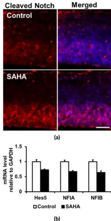
FIGURE 2An genically recognizable Notch ligand NICD was in-creased by SAHA treatment (red) (a) probably owing to the in-creased of IPC number in embryonic cortex. The expression of Notch signaling pathway downstream molecules includingHes5,
(a) (b) (c)
(d) (e) (f)
FIGURE 3SAHA increased cor cal interneuron in adult brains perhaps due to increased number of prolifera ng cells within the embryonic LGE and MGE. (a) Interneurons (green) originate from NSC located outside the embryonic cortex, in the LGE and MGE of ventral telen-cephalon. Interneurons migra ng into the cerebral wall from the MGE and LGE interact with NSC (red) and can exhibit changes in direc on of migra on a er contac ng NSCs. Interneurons can use glia fiber of NSCs as a scaffold upon which to migrate as they ascend to the cor cal plate (CP) or descend in the direc on of the ventricular zone (VZ). Once the interneurons invade the cortex from the MGE and LGE, differen-al interac ons between interneurons and NSC scaffold and locdifferen-alized mul direc ondifferen-al migra on of interneurons influenced by locdifferen-al guidance cues may facilitate interneuronal posi oning within dis nct domains of the developing cerebral cortex. Blue arrows indicate direc on of migra on. (b-c) Immunostaining (b) and the number of cells (c) that are prolifera ve (pH3+) during embryonic development in the LGE and MGE increased following SAHA applica on. (d-f) SAHA-treated adult cor ces showed increased Parvalbumin- and Somatosta n-expressing interneurons rela ve to Control ones. Ctx: Cortex.
3. Results and discussion
3.1. SAHA reduces cor cal astrocytogenesis
Considering the fact that NSCs lining the embryonic neu-ral tube give rise to the entire repertoire of neurons, astro-cytes, and oligodendrocytes of the adult CNS, we hypoth-esized that the decrease of NSCs in embryonic and adult SAHA-treated brains would influence the subsequent gen-eration of glial cells, astrocytes and oligodendrocytes that generally follows neurogenesis in the developing mam-malian brain. To determine the effect of in utero HDAC inhibition by SAHA on gliogenesis particularly astrocyto-genesis, we quantified astrocyte markers, gfap and S100β, expression of postnatal SAHA cortices using western blot and RT-PCR, respectively. Immunoblotting analysis and relative band density of gfap protein in SAHA-treated neo-cortex is reduced until 0.4 compared with the Control (Fig-ure 1a-b). mRNA level of S100β astrocytic gene con-firmed this phenotype (Figure 1c). Collectively, this re-sult indicates that SAHA repressed the production of cor-tical astrocyte. We assume that this phenotype is the con-sequence of the reduction of NSCs thus lead to the reduced number of astrocyte progenitors in SAHA-treated cortices.
Other possibility related to Notch signaling is the re-duction of NFIA expression by SAHA administration may cause decreased astrocytic differentiation potency of mid-gestational NSC (Figure2) since it has been suggested that NFIA, which bind to astrocytic gene promoters and act as downstream of the Notch signaling pathway to potentiate astrocytic differentiation of mid-gestational NSCs, accel-erated demethylation of astrocytic gene promoters by pre-venting DNMT1 from binding to them and thus allowed precocious astrocytic differentiation (Namihira et al. 2009; Juliandi et al. 2010b).
3.2. SAHA increased cor cal interneuron genera on
func-TABLE 1SAHA increased frequency of stretch a ends posture, grooming, freezing and decreased line crossing and center square dura on in open field test. Data represented in mean±SD (n=5).
Groups and Parameters Normal control Ethanol-induced brain disorder
control SAHA
Stretch a end posture 6.2±1.6 15.6±6.0 13.4±3.1∗
Line crossing 77.8±27.3 56.4±20.3 49.7±19.7∗
Grooming 30.6±12.2 76.2±26.0 65.6±17.4∗
Freezing 4.2±4.6 41.2±11.9 19.0±8.5∗
Center square dura on 13.4±10.5 3.0±3.3 2.4±3.1∗
Center square entries 3.0±2.5 0.8±0.8 2.0±2.6
Rearing 11.6±6.8 6.8±5. 16.8±13.9
∗p<0.05 significantly different rela ve to control.
TABLE 2SAHA changed social affilia on affect: reduced interac-on within the groups in social interac interac-on test. Data represented in mean±SD (n=5).
Frequency and
groups Frequency contactof mice with empty wire cup
Frequency contact of mice with intruder 1 Normal control 4.1±3.7 14.5±13.4 Ethanol-induced
brain disorder control 6.6±3.2 13.4±8.0 SAHA 6.9±2.5 11.3±4.9∗
∗p<0.05 significantly different rela ve to control.
TABLE 3SAHA changed social novelty: frequency contact of SAHA mice to intruder mice was less than normal control in social inter-ac on test. Data represented in mean±SD (n=5).
Frequency and
groups Frequency contactof mice with intruder 1
Frequency contact of mice with intruder 2 Normal control 13.3±5.7 11.2±4.4 Ethanol-induced
brain disorder control 12.8±4.1 7.5±1.6
SAHA 9.7±2.0 9.9±5.7
tion including cognition, sensory perception, and motor function. Thus, the constant fraction of cortical excita-tory:inhibitory (E:I) neurons circuit balance in 5:1 from near the start of cortical neurogenesis to adulthood ( Sa-hara et al. 2012) is necessary. The disruption of E:I circuit balance due to reduced expression of cortical GABAergic neurons such as GAD67, Reelin, Somatostatin, Parvalbu-min in the hippocampus, basal ganglia, or upper to layer 5 cortical layers of psychotic patients including schizophre-nia (SZ), autism spectrum disorder (ASD), and bipolar dis-order patients markedly reduces the effectiveness of the GABAergic inhibitory transmission that impinges on the dendrite and on the initial axon segments of excitatory neu-rons (Grayson et al. 2011). This deficit of inhibitory neu-rotransmitter disrupts the intermittent synchronization of excitatory neuron firing that is critical for normal neuronal function (Gonzalez-Burgos and Lewis 2008).
Our previous finding about the perturbation of postna-tal cortical lamination due to the increased and decreased production of excitatory neuron in UL and DL neocor-tex, respectively (Yuniarti et al. 2013), following midges-tational SAHA exposure might cause the inconstant E/I fraction thus lead to the alteration of excitatory neurons’ firing and induce the abnormality of neuronal function in the cortex.
During development, excitatory neurons and interneu-rons are born in separate locations. The excitatory neuinterneu-rons are generated from NSC in the ventricular zone (VZ) and subventricular zone (SVZ) of embryonic cortex and typi-cally migrate radially in an ‘inside-out’ sequence to form layers in the cortical plate (CP). Most, if not all, interneu-rons, however, are born from NSC located outside the cor-tex, in the lateral- and medial-ganglionic eminences (LGE and MGE) (Figure 3a) in ventral telencephalon, and mi-grate first tangentially from their birthplace to the proper cortical region, and then radially through the CP to reach their final laminar location. It seems that the LGE and MGE of the subcortical telencephalon also contribute cells to the neocortex and they are the primary sources of corti-cal interneurons in rodents (Anderson et al. 2002) includ-ing Parvalbumin- and Somatostatin-expressinclud-ing interneu-rons (Xu et al. 2004). MGE NSC give rise to early born and Parvalbumin- and Somatostatin-expressing interneu-rons, which distribute at higher densities in deep cortical layers (Butt et al. 2005;Fogarty et al. 2007).
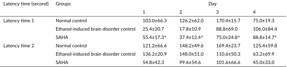
in-TABLE 4SAHA changed short- and long-term memory by reducing latency me 1 and 2 in passive avoidance test. Data represented in mean±SD (n=5).
Latency me (second) Groups Day
1 2 3 4
Latency me 1 Normal control 103.0±66.3 126.2±62.0 170.4±15.7 75.0±19.3 Ethanol-induced brain disorder control 25.4±20.7 17.8±10.9 88.8±69.0 106.0±84.4 SAHA 55.4±17.3∗ 37.4±12.4∗ 75.0±24.8∗ 88.8±14.7∗
Latency me 2 Normal control 121.2±66.6 148.2±49.6 169.4±23.7 125.4±59.8 Ethanol-induced brain disorder control 136.2±20.9 148.0±51.0 110.6±50.3 63.2±69.9 SAHA 54.8±42.3 99.6±54.6 101.6±66.6 45.0±33.0
∗p<0.05 significantly different rela ve to control.
creased percentage of cortical Parvalbumin+ cells (Fig-ure 3e–f). Immunoblotting supported the increased ex-pression of Parvalbum and Somatostatexpressing in-terneurons in SAHA-treated adult cortices (Figure3d).
Overall, the finding about the perturbation of postna-tal cortical lamination due to the increased and decreased production of excitatory neuron in UL and DL neocor-tex, respectively (Yuniarti et al. 2013), along with the in-creased number of cortical inhibitory interneurons follow-ing midgestational SAHA application might deregulate the normal ratio between excitatory and inhibitory neu-rons required for the precise regulation of physiological functions in the brain. To investigate the functional brain in SAHA mice after they reach adulthood, we performed behavior test that related to cortical functions in the reg-ulation of anxiety, learning and memory, and social in-teraction such as open field test, passive avoidance test, and social interaction test (Seo et al. 2013). SAHA mice at dose 50 mg/kg BW showed significantly increased de-pression, as shown in the increase in stretch attend pos-ture, grooming and freezing and decreased line crossing and center square duration in the open field test, decreased short term memory and changed long term memory in the passive avoidance test, and reduction in social interaction by reducing the capability of mice to interact within their groups (Table1–4).
The increased number of interneurons in the embryos exposed to HDACI SAHA in this study is in agreement withGrayson et al.(2011) by which treatment of neuronal progenitor cultured cells with various HDACIs (MS-275, VPA, TSA) led to a robust induction of interneuron mR-NAs such as reelin and GAD67 as well as DNMT in-hibitors. DNMT and HDAC inhibitors target DNMT1 and HDAC1 and facilitate the dissociation of DNMT-containing repressor complexes from interneuron (as pro-posed for the reelin and GAD67 genes) promoters led to DNA demethylation, histone acetylation, and a relaxation of chromatin allows the recruitment of specific transcrip-tion factors, and the general transcriptranscrip-tional machinery to the promoters.
Collectively, these results suggest that change in epi-genetic status that is histone acetylation of non-cancerous
cells by pharmacological inhibition of HDACs follow-ing anti-cancerous SAHA exposure durfollow-ing mid-pregnancy could alter fetal and adult brain structure. These support the precaution of SAHA as anticancer agent for pregnant patient by FDA, that is pregnancy category D meaning that SAHA has potential hazard to the fetus. All in all, these findings might contribute to the pharmaceutical therapy guideline not only for cancer treatment but also for treat-ment of other diseases employing HDACIs. Regarding that SAHA has a potential hazard to the fetus, it should not be prescribed to pregnant patient or to patient who planned to become pregnant while taking this drug.
4. Conclusions
The consequences of prenatal SAHA exposure in adult-hood are suppressed cortical astrocyte by downregulating
NFIAexpression thus decreased the potency of astrocyte differentiation, and increased cortical interneuron by in-creasing number of proliferating cells that might be NSC committed to PV and SST interneurons. This defect may alter the composition of cells within SAHA-treated cortex thus impact on brain function since SAHA mice at dose 50 mg/kg BW showed significantly increased anxiety, de-creased memory and long-term cognitive functions, and reduced social interaction.
Acknowledgments
This work was supported by Ministry of Education, Cul-ture, Sports, Science and Technology, Japan through NAIST Global COE Program (Frontier Biosciences: Strategies for survival and adaptation in a changing global environment) and Grant-in-Aid for Scientific Research on Innovative Area: Neural Diversity and Neocortical Orga-nization, and by the Government of the Republik Indone-sia through theHibah Kompetensi2016 Research Grant.
Authors’ contribu ons
con-tributed to sample analysis. BJ and KN supervised the work.
Compe ng interests
The authors declare that they have no significant compet-ing financial, professional or personal interests that might have influenced the performance or presentation of the work described in this manuscript.
References
Anderson SA, Kaznowski CE, Horn C, Rubenstein JLR, McConnell SK. 2002. Distinct origins of neocortical projection neurons and interneurons in vivo. Cereb Cortex 12(7):702–709. doi:10.1093/cercor/12.7.702. Butt SJB, Fuccillo M, Nery S, Noctor S, Kriegstein
A, Corbin JG, Fishell G. 2005. The temporal and spatial origins of cortical interneurons predict their physiological subtype. Neuron 48(4):591–604. doi:10.1016/j.neuron.2005.09.034.
Fogarty M, Grist M, Gelman D, Marín O, Pachnis V, Kessaris N. 2007. Spatial genetic pattern-ing of the embryonic neuroepithelium gener-ates GABAergic interneuron diversity in the adult cortex. J Neurosci. 27(41):10935–10946. doi:10.1523/JNEUROSCI.1629-07.2007.
Gonzalez-Burgos G, Lewis DA. 2008. GABA neu-rons and the mechanisms of network oscillations: implications for understanding cortical dysfunction in schizophrenia. Schizophr Bull. 34(5):944–961. doi:10.1093/schbul/sbn070.
Grayson D, Kundakovic M, Chen Y, Dong E, Guidotti A. 2011. Epigenetic regulation of GABAergic targets in psychiatry. In: A Petronis, J Mill, editors. Brain, be-havior and epigenetics. Berlin: Springer Science & Business Media. p. 23–40. doi: 10.1007/978-3-642-17426-1_2.
Hevner RF, Daza RAM, Rubenstein JLR, Stunnenberg H, Olavarria JF, Englund C. 2003. Beyond laminar fate: toward a molecular classification of cortical projec-tion/pyramidal neurons. Dev Neurosci. 25(2-4):139– 151. doi:10.1159/000072263.
Juliandi B, Abematsu M, Nakashima K. 2010a. Chro-matin remodeling in neural stem cell differen-tiation. Curr Opin Neurobiol. 20(4):408–415. doi:10.1016/j.conb.2010.04.001.
Juliandi B, Abematsu M, Nakashima K. 2010b. Epigenetic regulation in neural stem cell differentiation. Dev Growth Differ. 52(6):493–504. doi: 10.1111/j.1440-169X.2010.01175.x.
Kohyama J, Sanosaka T, Tokunaga A, Takatsuka E, Tsu-jimura K, Okano H, Nakashima K. 2010. BMP-induced REST regulates the establishment and main-tenance of astrocytic identity. J Cell Biol. 189(1):159– 170. doi:10.1083/jcb.200908048.
Marks PA. 2007. Discovery and development of SAHA as an anticancer agent. Oncogene 26(9):1351–1356. doi:10.1038/sj.onc.1210204.
Namihira M, Kohyama J, Abematsu M, Nakashima K. 2008. Epigenetic mechanisms regulating fate specification of neural stem cells. Philos Trans R Soc Lond B Biol Sci. 363(1500):2099–2109. doi:10.1098/rstb.2008.2262.
Namihira M, Kohyama J, Semi K, Sanosaka T, Deneen B, Taga T, Nakashima K. 2009. Committed neu-ronal precursors confer astrocytic potential on resid-ual neural precursor cells. Dev Cell 16(2):245–255. doi:10.1016/j.devcel.2008.12.014.
O’Connor OA, Heaney ML, Schwartz L, Richardson S, Willim R, MacGregor-Cortelli B, Curly T, Moskowitz C, Portlock C, Horwitz S, Zelenetz AD, Frankel S, Richon V, Marks P, Kelly WK. 2006. Clinical ex-perience with intravenous and oral formulations of the novel histone deacetylase inhibitor suberoylanilide hydroxamic acid in patients with advanced hemato-logic malignancies. J Clin Oncol. 24(1):166–173. doi:10.1200/JCO.2005.01.9679.
Okano H, Temple S. 2009. Cell types to order: temporal specification of CNS stem cells. Curr Opin Neurobiol. 19(2):112–119. doi:10.1016/j.conb.2009.04.003. Ramasamy S, Narayanan G, Sankaran S, Yu YH,
Ahmed S. 2013. Neural stem cell survival fac-tors. Arch Biochem Biophys. 534(1-2):71–87. doi:10.1016/j.abb.2013.02.004.
Sahara S, Yanagawa Y, O’Leary DDM, Stevens CF. 2012. The fraction of cortical GABAergic neurons is constant from near the start of cortical neuroge-nesis to adulthood. J Neurosci. 32(14):4755–4761. doi:10.1523/JNEUROSCI.6412-11.2012.
Seo TB, Cho HS, Shin MS, Kim CJ, Ji ES, Baek SS. 2013. Treadmill exercise improves behavioral outcomes and spatial learning memory through up-regulation of reelin signaling pathway in autistic rats. J Exerc Re-habil. 9(2):220–229. doi:10.12965/jer.130003. Vohora D, Pal SN, Pillai KK. 2000. Effect of locomotor
ac-tivity on the passive avoidance test for the evaluation of cognitive function. Indian J Pharmacol. 32(3):242– 245.
Xu Q, Cobos I, Cruz EDL, Rubenstein JL, Ander-son SA. 2004. Origins of cortical interneu-ron subtypes. J Neurosci. 24(11):2612–2622. doi:10.1523/JNEUROSCI.5667-03.2004.