Foot and mouth disease in and near the Maasai
Mara, Kenya
Amanda Karlsson
Uppsala 2017
Degree Project 30 credits within the Veterinary Medicine Programme ISSN 1652-8697
Examensarbete 2017:44
Faculty of Veterinary Medicine and Animal Science
Foot and mouth disease in and near the Maasai
Mara, Kenya
Mul- och klövsjuka i och runt Maasai Mara, Kenya
Amanda Karlsson
Supervisor: Johanna Lindahl, Department of Clinical Sciences
Assistant Supervisor (if any): Daniel Mutiso, International Livestock Research Institute
Examiner: Jonas Johansson Wensman, Department of Clinical Sciences
Degree Project in Veterinary Medicine
Credits: 30
Level: Second cycle, A2E Course code: EX0736 Place of publication: Uppsala Year of publication: 2017
Number of part of series: Examensarbete 2017:44 ISSN: 1652-8697
Online publication: http://stud.epsilon.slu.se
Key words: Foot and mouth disease, FMD, FMDV, Kenya, Livestock, Prevalence, Cattle, Maasai Mara Nyckelord: Mul- och klövsjuka, MK, Mul- och klövsjukevirus, Bokskap, Nötkratur, Prevalens, Kenya, Maasai
Mara
Sveriges lantbruksuniversitet
Swedish University of Agricultural Sciences Faculty of Veterinary Medicine and Animal Science Department of Clinical Sciences
SUMMARY
Livestock is very important to the sub-Saharan pastoralists, and the health and wellbeing of the cattle, which are a large portion of the livestock that the farmer keeps, can make or break a family’s livelihood. One important disease in cattle is foot and mouth disease (FMD), a highly contagious disease affecting many species of both cloven–hoofed wildlife and livestock such as cattle. The disease is caused by a virus of the genus Aphthovirus within the Picornaviridae
family and is characterized by the formation of vesicles and ulceration in the mouth, on the snout, interdigital space and on the teats. Classical clinical signs include anorexia, excessive salivation and lameness. It is one of the most financially important livestock diseases in the world. In the western world, it is one of the most feared diseases of livestock since it brings great financial losses and can limit the international trade of both livestock and their derived products.
This study investigated the prevalence of active foot and mouth disease in three villages (Lemek, Endoinyo Narasha and Mara Rianta) in and around the Maasai Mara national reserve in Kenya. Further, the possible risk factors for FMD transmission were assessed with the help of a questionnaire that was administered to farmers of each investigated herd. The selected villages were all at different distances from the national reserve and have adopted different animal husbandry practices. The proximity of Mara Rianta to the national reserve allow the pastoralists to graze in the parks while in Endonyio Narasha and Lemek, many farmers practice sedentary grazing in fenced lands. In order to find suspected cases of FMD a brief clinical examination of the animals was done. Three animals from each of the 75 farms (25 farms from each of the three different villages) was examined, a total of 225 animals. Animals with suspected active lesions were tested using a lateral flow device (Svanodip FMDV antigen test) to detect FMD viral antigen from vesicular fluid or epithelial cells. In total 27 animals (11.6%) with suspected active lesions were tested and out of these, one was found to be positive. There was no significant difference between the number of suspected active infections in the three different villages, therefore it was not possible to draw any conclusions regarding the potential risk factor of closeness to national park. More than 80% of the farmers were aware of FMD among neighbours. Analysis of the questionnaire yielded a statistically significant negative correlation between the likelihood of finding potential active infection and farmers to report having animals unwilling to walk or stand, which was reported in 19% of the farms. In contrary to that, it was more likely to find animals with suspected active FMD in farms where the farmer had reported seeing blisters in the mouth, snout or hooves, which was reported in 21% of the farms.
From this study one can conclude that a good question, in order to find animals with suspected FMD, is whether the farmer has any animals with blisters. This study confirms that FMD circulates in villages surrounding the national reserve Maasai Mara and concludes that more research is necessary which will help devise sustainable control strategies in the area.
SAMMANFATTNING
Boskap är väldigt viktigt för bönderna söder om Sahara. Djurens hälsa och välmående är av stor vikt för hela familjens försörjning. En av de viktiga sjukdomarna hos boskap är mul- och klövsjuka (FMD), som orsakas av ett virus tillhörande familjen Picornaviridae och karakteriseras av blåsor och erosioner i munnen, på mulen, i klövspalten och på juvret hos klövdjur. Symtom som associeras med sjukdomen är anorexi, ökad salivering och hälta. Mul- och klövsjuka är en viktig sjukdom hos tamboskap. I västvärlden är det en av de mest fruktade sjukdomarna som drabbar boskap då den har möjligheten att hindra ett land från handel med klövdjur.
Den här studien undersöker prevalensen av aktiv mul- och klövsjuka i tre byar i och kring nationalparken Maasai Mara i Kenya, samt möjliga riskfaktorer för sjukdomen med hjälp av ett frågeformulär. Byarna låg alla på olika avstånd från nationalparken och hade olika sätt att hålla djuren. För att finna misstänkta fall av FMD gjordes en översiktlig klinisk undersökning av djuren. Tre djur från vardera av de 75 gårdarna som ingick i studien undersöktes, totalt 225 djur. Då det återfanns lesioner som tydde på en infektion av mul- och klövsjukevirus testades djuren för sjukdomen med ett snabbtest (Svanodip FMDV antigen test). Det testet detekterar FMD antigen från blåsvätska eller epitelceller. Totalt misstänktes sjukdomen finnas hos 27 djur, endast ett av dessa testade positivt för mul- och klövsjuka.
Det var ingen signifikant skillnad mellan antalet misstänkta infektioner mellan de tre olika byarna, och därför var det inte möjligt att utvärdera riskfaktorn avstånd till nationalparken. Av alla gårdar rapporterade mer än 80% att de hade hört om FMD hos gårdar i närheten, 19% hade upplevt djur som hade svårt att gå, och 21% hade observerat blåsor. Analysen av frågeformuläret gav en negativ statistiskt signifikant relation mellan bönder som rapporterade att deras djur hade visat ovillighet att stå eller gå och närvaron av misstänkta FMD fall. I motsats till det återfanns en korrelation mellan bönder som rapporterade att de hade sett blåsor i munnen, på mulen eller i klövspalten på deras djur och närvaron av misstänkta fall.
Som ett resultat av den här studien kan man dra slutsatsen att den bästa frågan att ställa till bönder då man letar efter misstänkta fall av mul- och klövsjuka är om de har sett blåsor på sina djur. Studien bekräftar även att mul- och klövsjuka cirkulerar i området kring nationalparken och leder till slutsatsen att fler studier är nödvändiga för att hitta ett sätt att kontrollera sjukdomen i området.
CONTENT
Introduction ... 1
Literature review ... 2
Foot and mouth disease ____________________________________________ 2 Epidemiology ... 2
Clinical signs in cattle... 3
Pathogenesis of FMD in cattle ... 4
Vaccination ... 5
Consequences of foot and mouth disease ______________________________ 5 Diagnosis of FMD _________________________________________________ 6 Lateral flow device _________________________________________________ 6 Material and methods ... 7
Clinical examination and sampling for FMD _____________________________ 8 Pen side testing ... 8
Using the test ... 9
The questionnaire _________________________________________________ 9 Statistical analysis ________________________________________________ 10 Ethical considerations _____________________________________________ 10 Results... 10
Keeping of the cattle ______________________________________________ 10 FMDV testing ___________________________________________________ 10 Questionnaire ___________________________________________________ 12 Discussion ... 14
Conclusion ... 18
1 INTRODUCTION
For a pastoralist, livestock is of great importance, and in the sub-Saharan Africa there are over 166 million poor livestock keepers who depend upon livestock for their livelihood. Any disease that hurt the animals or lower their production is devastating for the people depending on them (Perry and Grace, 2009).
Foot and mouth disease (FMD) is a highly contagious viral disease of cloven-hooved animals, which, according to the World Organisation for Animal Health (Office Internacional de Epizootie, OIE), has the potential to cause severe economic loss (OIE, 2013). FMD is caused by one of the viruses of the Picornaviridae family, genus Aphtho-virus, and have seven different serotypes. It is the severity of the clinical signs generated by FMD, including loss in milk yield that lead to severe socio-economic consequences. This is one of the reasons that OIE has targeted FMD as one of three diseases to be eradicated (OIE, 2013). In many developed countries there are a big fear of an outbreak with FMD, since presence of the disease disables trading and affects the country’s or region’s possibility to move and sell livestock. In case of an outbreak in Europe, culling is used as a method to control and eradicate the disease. (OIE, 2013). Europe, North- and Central America, Asia, Australia, New Zeeland and the Pacific are currently FMD free regions (SVA, 2015).
In many regions of the world FMD is endemic, and the people and animals have to live with its effects. One of these regions is Africa, in particular the sub-Saharan Africa. In areas in Africa where farming and animal husbandry are the most important sources of income to families, an outbreak of this sort can lead to great consequences for an individual family or a community. The exact effect of FMD on farmers varies, but it is always a loss (Knight-Jones et al., 2016). The Food and Agricultural Organisation (FAO) claims in one of their information sheets that the burden of FMD in the sub-Saharan Africa will not ease as long as there are free movement of FMD infected African buffalo. This, in combination with the rise in international travel and trade, pose a threat to the FMD-free parts of the world (Food and Agricultural Organisation, 2004).
This study focuses on the prevalence of active FMD in cattle from three different areas, grazing in and near the Maasai Mara national reserve in Kenya, Africa, with the objective to investigate the prevalence of active FMD in three different areas in Narok County and analyse possible risk factors for infection.
2 LITERATURE REVIEW
Foot and mouth disease
Foot and mouth disease (FMD) is caused by a virus; foot and mouth disease virus (FMDV). It is one of two viruses of the Aphtho-virus genus of the Picornaviridae family. Foot and mouth disease virus is a positive-sense RNA virus with seven different serotypes, and within all serotypes there are additional variation. The serotypes are called A, O, C, Asia 1 and SAT (South African Territories) 1, 2, and 3 (Grubmanand Baxt, 2004). The seven serotypes together have more than 60 different strains, and infection with these different strains do not cause cross-immunity (Aftosa, 2014).
Foot and mouth disease virus primarily infect animals from the genus Artiodactyla, including different kinds of livestock, like cattle, water buffalo, pigs as well as sheep and goats. All cloven-hooved animals are susceptible and this means that even the wild cloven-hooved animals are at risk, such as wildebeest, African buffalo, giraffes and elephants as well as antelopes and gazelles (Aftosa, 2014). It has been suggested that FMD is a zoonosis (Prempeh et al., 2001). However, general consensus in veterinary medicine is that foot and mouth disease does not cause any disease in humans, but humans can transmit the virus on their clothes and with equipment (SVA, 2015).
Foot and mouth disease is endemic in most countries in the sub-Saharan Africa, excluding a few countries in the south (Gelaye et al., 2009). Vosloo et al. (2002) found a likely way of transmission of FMDV from African buffalo to livestock during two outbreaks in South Africa. Buffalo contact with previously not infected cattle was a likely cause of these outbreaks. By addressing this and fencing in wild buffalo in nature reserves, most southern countries in Africa have managed to control the disease and seldom have outbreaks (Gelaye et al., 2009).
Epidemiology
Foot and mouth disease is highly contagious, which combined with the virus’ ability to rapidly replicate and evolve, makes it a disease that has the potential for great economical loss (Thomson and Bastos, 2004). The high susceptibility of our modern high-producing cattle, combined with the risk of sub-clinical infections, makes it not only a financially damaging disease but also very hard to control in the case of an outbreak (Paton et al., 2009). The virus can travel several kilometers in the air inside droplets from infected animals which further increases its ability to spread (SVA, 2015).
The main route of infection with FMDV is through aerosols. Even direct contact with humans and/or objects that have been in contact with an infected animal is a possible way of infection (Alexandersen et al., 2003, OIE, 2015). If the skin barrier is damaged the virus can more easily infect the animal. In cattle this way of infection has been seen in animals feeding on thorny bushes as well as in lactating cows with damage to the teats (Alexandersen et al., 2003). In many parts of Africa, the production system of cattle contributes to the transmission of FMD. Animals are often kept together with several other species of livestock with lower grade of
3
clinically visible infections of FMD like goats and sheep, and share treks with livestock from other farms as well as wildlife. This is a possible way for FMDV to keep its endemic status in sub-Saharan Africa (Sahle et al., 2007).
In sub-Saharan Africa, FMDV can spread between wildlife and livestock. There is evidence that African buffalo can be sub-clinically infected, and when animals come into contact with each other, or when they share the same pathways, livestock can get infected. African buffalo is endemically infected with the SAT serotypes, at least in southern and eastern Africa (Sahle et al., 2007). This is believed to be an important route of infection in the east and south of Africa, as well as a contributing factor to FMD’s endemic status (Vooslo et al, 2002, Ayebazibwe et al., 2010). In a study in the national parks of Uganda over 80% of the buffalo tested were found to carry antibodies to one or more serotype of FMD, and were concluded to be an important part in the circulation of FMD in the country (Ayebazibwe et al., 2010). FMDV is resilient and can survive up to two weeks in dry fecal matter and 20 weeks in bedding (Merck veterinary manual, 2015).
Clinical signs in cattle
Clinical signs usually develop within 2-4 days after contact with the virus, the first signs are fever, anorexia and loss of milk production. Within a day of these first signs of illness the typical vesicles develop in the mouth and on the muzzle (SVA, 2015). In an experimental study of cattle infected with FMDV RNA, vesicles developed 48 h post infection in unvaccinated animals (Stenfeldt et al., 2015).
Alexandersen et al. (2003) propose that FMDV infection starts, like all infections, with an exposure to the virus, for example in the tonsils or soft palate. After that, a viremia with replication of virus in multiple organs follows and a gathering of virus in the cornified epithelial cells of for example the muzzle or coronary band. This is where the characteristic vesicle develops.
The signs caused by FMDV is due to tissue damage caused by the viruses’ ability to cause acute cytoplasmic infections (Paton et al., 2009). Infected animals usually develop fever and vesicles in the mouth, on the muzzle and around the hooves, and lactating animals can get vesicles on the teats (Grubman and Baxt, 2004). These vesicles usually rupture within a short period of time and cause ulcerations which due to their painfulness leads to the typical clinical signs of anorexia, unwillingness to move, and depression. Consequences of these clinical signs are loss in milk production, malformation of hooves and loss in condition of the affected animal (Grubman and Baxt, 2004).
For an animal keeper or veterinarian, the typical signs indicating foot and mouth disease include extensive drooling, smacking of the lips, nasal discharge, lameness and fever (OIE, 2015). The morbidity of FMD is high but the mortality in adult animals is low, approximately 1-5% (Afonsa, 2014). In youngsters the mortality is higher (20-50%), due to myocarditis; “tiger heart”. An infected animal usually recuperates quickly and is symptom-free within two weeks
4
if no secondary infections arise (OIE, 2015). In cattle, FMD is not clinically distinguishable from other vesicular diseases, for example vesicular stomatitis. Vesicular stomatitis is caused by another pathogen, a vesiculovirus, and infects apart from cattle, sheep, and pigs, also horses and some wildlife (SVA, 2016). Thus, to positively confirm diagnosis of FMD, laboratory testing is necessary (Alexandersen et al., 2003).
During acute FMD, virus is found in basically all organs of an infected animal. Thus all organs and secretions like milk, urine and blood should be considered contagious and disposed of properly to contain the virus (Alexandersen et al., 2003).
Pathogenesis of FMD in cattle
Foot and mouth disease virus mainly infect animals through air-borne droplets of secretions from infected animal. Virus is shed in high concentrations in saliva and nasal secretions as well as in milk, urine, semen, feces and blood. The main route of infection is through inhalation, although it is also possible, but not as likely, for animals to be infected from contact with the virus on broken skin and mucus membranes (Grubman and Brax, 2004).
The current general consensus is that the first phase of viral replication takes place in the lung and/or pharyngeal tissues, depending on the size of the aerosol infecting the animal and the viral strain. It is normally in this stage of the infection that pyrexia develops. Then the virus migrates to the oral and pedal epithelium causing vesicles that are filled with yellowish fluid. These vesicles ruptures, usually within 1-2 days, and cause erosions. The erosions start out as deep red and cover the entire surface of the former vesicles. During the healing process, that usually takes between 5-12 days, the erosion starts to get covered by epithelium (OIE, 2015, Alexandersen et al., 2003).
A guideline to dating lesions caused by FMDV was published by the Ministry of Agriculture, Fisheries and Food, United Kingdom, in 1986 and is cited by several sources including Alexandersen et al (2003). According to this guideline, with the start of clinical signs at day 0, vesicles develop from days 0-2. Vesicles rupture from day 1-3, and turn into sharply marginated erosions on days 2-3. The sharpness is lost from day 3. A serofibrinous exudate starts covering the erosion from days 4-6 and healing with fibrous tissue starts at day 7 (Alexandersen et al., 2003).
Around 3-4 days post infection the cattle have normally developed antibodies to FMDV which starts clearing the animal from the virus (Alexandersen et al., 2003). However, there are exceptions to this; some animals get persistently infected with FMDV (known as carrier-animals), and often the virus can be located in the pharynx of these animals. There is evidence that some African buffalo can stay persistently infected for up to 5 years, and that infection with a single strain of FMDV can circulate for up to 25 years in a heard of African buffalo. In cattle, persistent infection longer than 6 months is uncommon but some cases as long as 3 years have been seen (OIE, Terrestrial manual 2012, Alexandersen et al., 2003).
5
There is a difference in virus secretion depending upon the vaccination status of the infected animal. Normally the peak in virus secretion from an infected animal occurs 2 to 7 days post infection, both in vaccinated and non-vaccinated animals. The duration of virus excretion varies between 2.5 days for vaccinated animals and 10 days for non-vaccinated animals (Parthiban et al., 2015, Stenfeldt et al., 2015).
In a study by Parthiban et al. (2015) they found that after 21 days only 2.6% of the carrier-animals still excreted viral RNA in their nasal discharge. In the same study the authors also conducted an experiment with placing naïve sentinel animals with confirmed carriers of FMDV during 63 and 75 days respectively. This was done to test the potential infectiousness of the carrier animals to naïve cattle, and none of four sentinel animals contracted an FMDV infection during that time. Thus the authors concluded that there is a low risk of infection from carrier animals to non-vaccinated naïve cattle, at least in experimental settings (Parthiban et al., 2015). Vaccination
It is possible to vaccinate against FMD. The recommendation is to use a vaccine that is accepted by the OIE and made for the circulating serotype, and to follow the manufacturers’ vaccination program. A program of vaccination usually contains two starter vaccinations with 3-4 weeks in-between, followed by a booster vaccination every 6-12 months depending on the vaccine used and the burden of disease in the region (OIE, Terrestrial manual 2012).
Even though vaccination may not protect fully from infection, it may impact on the transmission. In an experimental study by Stenfeldt et al. (2015), all animals vaccinated against FMDV 14 days prior to infection with FMDV developed no clinical signs of FMD, nor any signs of viremia. In contrast all animals not vaccinated prior to exposure to FMDV developed clinical FMD and viremia (Stenfeldt et al., 2015).
Consequences of foot and mouth disease
During an acute infection with FMDV a lactating cow usually has a significant decrease in milk production. If in the early stages (day 0-50) of lactation the cow can have a drop of up to 95% in milk yield during a period of illness. A week prior to an outbreak investigated by Lyons et al. (2015) in Kenya a drop in milk yield was noticeable on a heard level. In general, the drop in milk production amongst cattle has a duration of two months (Lyons et al., 2015). High producing cattle breeds, like those found in developed countries, usually are more severally affected than traditional breeds (Afonsa, 2015).
Young cattle infected with FMDV can have a decreased weight gain during their growth period in early life. This can in some cases turn into a permanent state for the individual cattle and it can continue the trend of a low body weight the rest of its life (Jibat et al., 2013). Pregnant cows can miscarry their calf if infected during pregnancy. This leads to the farmer having to pay to keep an empty cow that will not produce milk until the next calf is born, with economic losses (Knight-Jones and Rushton, 2013). During an outbreak of FMD, milk-producing farmers generally experiences greatest loss of income due to the drastic drop in milk production in high-producing cows (Lyons et al., 2015).
6
Losses from FMD in endemic countries are ongoing and never ending. The disease keeps blossoming up in different herds and places and during those outbreaks the individual farmer as well as the entire community suffer big losses. This makes it even more difficult for the livestock sector to develop further (Knight-Jones and Rushton, 2013).
Diagnosis of FMD
An infection with FMDV is not clinically distinguishable from other vesicular diseases. Vesicular stomatitis, swine vesicular disease and vesicular erythema are the primary diseases that needs to be differentiated from FMD. There are some other diseases that have some signs that can be mistaken for FMD, such as rinderpest, mucosal disease, infectious bovine rhinotracheitis (IBR) and bluetongue (Epiwebb, 2016). This is why an early diagnosis is of great importance.
To test for FMD, and many other diseases, it is possible to use different test methods. The different tests can be roughly divided into two groups: antigen (ag) tests which detect a viral antigen, or antibody (ab) tests that detect antibodies to a pathogen (the body’s response to an infection that remains in the animal for several years to a lifetime after infection). When using an antibody test it is only possible to say that the animal has been exposed to the disease causing agent, it is not possible to, on basis of that test, say that the animal is currently infected. However, an antigen test confirms that the animal is currently carrying the disease-causing agent, i.e. is infected. This is the method chosen for this study.
The OIE has published guidelines regarding the diagnosis of FMDV infection in cloven-hooved animals in the Terrestrial Manual (2012). They say that testing should be conducted at a grade 4 laboratory to ensure that the virus is securely handled. Diagnosis is often made using an antigen Enzyme-Linked Immunosorbent Assay (antigen ELISA-test). This distinguishes all the seven serotypes of FMDV from each other and is today the preferred method for FMD diagnosis. Other tests that can be used is serological testing, that tests exposure to the virus not an actual infection, or Real-Time RT-PCR (rt-PCR) that, like an antigen ELISA-test, tests for the actual virus. These are the three tests that are validated and therefore recommended by the OIE (OIE, 2012).
The OIE also recognizes the lateral flow device as a method for diagnosis and detection of viral antigens, but this has not yet been included in the OIE test register (OIE, 2012).
Lateral flow device
A lateral flow device is a test used to detect antigen and it can be designed in a couple of different ways. The principle of the one used in this study is described below.
The device consists of four different areas. First is the sample application pad, where the sample is placed, and its main purpose is to smoothly and evenly transport the sample to the next stage of the test. It also helps pH regulation and separation of different components of the sample. This pad can be made of glass fibers or cellulose. The second area is the conjugate pad. It is here that the bio-markers are dispensed and bind to the antigen in a positive sample. The
7
conjugate can be of several different materials, one of the most common is gold attached to antibodies, due to gold’s good visibility and affinity to proteins. The third area of the lateral flow device is a membrane made by nitrocellulose. To this area the lines of disease specific antibodies are bound. It is to these antibodies that the antigen bound to the conjugate gold binds and form a visible line. In this area it is also a line of molecules that bind to the conjugate gold itself ad this works as a control line. If this line is not visible, the test has malfunctioned and a new test should be done (Sajid et al., 2015).
MATERIAL AND METHODS
The field study was conducted in the southwest of Kenya, in Narok County, during late September and early October in 2016. This region of the country was chosen because of its proximity to the Maasai Mara national reserve. The village selection was done by International Livestock Research Institute (ILRI) and was based on their proximity to the national parks, the land use type particularly the grazing patterns and their potential interaction with wildlife. Lemek village is approximately 55 km from the national reserve and farmers practice sedentary grazing graze within the village. Mara-Rianta village is approximately 1 km from the national reserve and farmers practice nomadic pastoralism by grazing their cattle in the parks and the surrounding Mara North conservancy where cattle freely intermingle and co-graze with wild cloven-hoofed animals like wildebeest, buffalos and gazelles. Such co-existence may expose livestock to pathogens of wildlife origin. The third village of Endoinyo Narasha is approximately 60 km from the national reserve and most of the farmers graze their cattle in fenced lands thus limiting contact with other herds. However, different cattle herds make contacts at shared watering points and also cross paths with wildlife on their way to watering points.
The project was conducted in collaboration with two other projects, also based at ILRI. From every village, 50 households were randomly selected from a list. In each household 3 heads of cattle were included. Cattle over one year of age were eligible for testing in this study but the big bulls and highly pregnant cows were excluded due to safety reasons. No specific individual was targeted, but rather the one that the hired men were able to get a noose around was the one cattle selected for testing. The age boundary was set so that no maternal antibodies would be present, since the other two projects focused on antibodies, and so that the animal would have had a risk of developing clinical FMD.
In the first village of Lemek we were able to visit 25 households, giving a total of 75 examined animals. After that, we limited ourselves to 25 households per village in Mara-Rianta and Endoinyo Narasha as well, this due to the time it took to examine every individual as well as the distance between farms. We were forced to tip the selected cattle and restrain them to be able to get blood for the other two projects, as well to make it possible for me to properly examine and sample suspected lesions. The same amount of farms and cattle was sampled in the remaining two villages as well.
8 Clinical examination and sampling for FMD
A clinical examination was made of sampled cattle to look for lesions consistent with an infection of FMDV, e.g. vesicles, ulcers or erosions in the mouth, on the muzzle, on the teats and hoof-margin as well as in the interdigital space. If any suspected lesions were found the individual animal had one lesion tested.
The vesical fluid was sampled by two local animal health workers, trained by the local veterinarian, this to adhere to the will of the local tribes. The extraction was made using an 18g needle and a 5-ml syringe with the aim of getting at least 0.1ml fluid to test. If there were no vesicles present in the animal suspected to be infected with FMDV, an ulceration or an erosion was tested using a cotton swab, this was done either by a local veterinarian or by myself. When testing with a cotton swab a lesion was chosen and carefully swiped, with some force, to make sure that enough material would be collected to do the test.
Pen side testing
Testing was done with the Svanodip FMD antigen test kit (SVANOVA, Uppsala, Sweden). The test was developed by Boehringer Ingelheim Svanova. The Svanodip FMDV antigen test is a lateral flow test where FMDV specific antibodies have been bound to colloid gold that binds the FMDV antigen in positive samples, and then it will travel through capillary force along the test. If antigen is present it will attach to the antibody line that is fixed in the test window. The colloid gold will form a visible red/purple line when it accumulates. If no antigen is present, the colloid gold will pass the antibody line and only attach to the control line (Boehringer Ingelheim Svanova, Manual number: 19-4100-13/06). This test is based on the 1F10 monoclonal antibody which function for detecting FMDV in epithelial cells has been validated and found to have a specificity of 99% and sensitivity of 83% for SAT1 and SAT3 serotypes. Its sensitivity to the SAT2 serotypes are lower and not fully validated (Ferris et al., 2009). According to the manufacturer, the Svanodip FMDV ag test can detect all seven serotypes of FMDV, but does not differentiate between them. If a vesicle, ulceration or lesion suspected to be caused by the FMDV is found on an animal this test can be used to give a direct answer if it is FMD or not. The test can only detect FMDV from lesions in acutely infected animals. Lesions older than approximately five days will in most cases show as negative due to the lack of antigen in the test site.
This method was chosen for this study in order to test for possibility to use the lateral flow device to detect ongoing outbreaks of the disease in the future. In complement to the lateral flow device used, antibody testing will be performed by other collaborators.
9 Using the test
Twelve droplets of the buffer solution were added to the test tube and mixed with either the vesicle fluid or the cotton swab. If a cotton swab was used it was pressed against the sides of the test tube in order to make sure that possible antigen is released into the buffer solution. After that the mixture was added to the sample well on the test kit, between 5-7 drops of the solution. After letting ten minutes pass the results were read. If nothing was visible the test was let to sit for another 20 minutes (a total of 30 minutes) and a second reading was performed. If an indication was present in the control area, but not in the test area, the test was considered negative. In the case of both a line in the control area as well as one in the test area were visible, the test was considered positive and the animal infected with FMDV.
The questionnaire
A questionnaire was administered to all the 75 households that participated in this study and data on possible FMD exposure factors was collected. The questionnaire was designed by myself, Sofie Enström (SLU) and Daniel Mutiso (PhD student, ILRI) to contain questions relevant to all our three projects. It was designed as a multiple-choice questionnaire, with some open questions The farmers were interviewed in both Swahili and the local Maasai language which was made possible by the help of a local guide/translator. The questions relevant to this study are presented below.
Do your cattle graze in the national park? a. Yes b. No
Do you sight wildlife near your livestock while grazing or on transhumance? a. Yes b. No
Which type of wildlife do you see near your cattle?
a. Ungulates b. Predators c. Monkeys d. Other How do you experience the contact with wildlife?
a. Positive b. Negative c. Indifferent
Did the neighboring farmers have FMD in the last one year? a. Yes b. No c. Unsure
Are there herds within the village that you know have FMD? a. Yes b. No
10
Have you noticed any of the following signs of illness in your cattle? (Choose as many as needed)
a. Fatigue b. Loss of pregnancy/abortion/stillbirth c. Decrease in milk production d. Mastitis/udder swelling and/or pain e. Unwillingness to walk/stand f. Fever g. Blisters in mouth, teats or hooves
The other questions posed in the questionnaire are not part of this study and is not analyzed here. The whole questionnaire is presented as an annex in the end of the report.
Statistical analysis
Due to the low number of positive samples, the data was analyzed to evaluate possible risk factors for FMD and indicators to suspect disease in the different farms. Odds ratio was calculated for the relevant variables that was suspected to be a risk factor. In complement to this Chi-square test was performed to determine the significance of the results.
Ethical considerations
The sampling of animals was approved by ILRI institutional animal care and use committee, approval number 2016.20. All participating farmers were informed about the study and asked for their informed consent before initiation.
RESULTS
Keeping of the cattle
The most common breed of cattle was Zebu (Bos indicus). In general, the cattle were kept in bomas, enclosures made of sticks or thorny bushes. The floor in the boma is covered in manure from the animals. Cows that are lactating are milked inside the boma or tied up outside of the boma. During the day cattle graze in the vicinity of the farmers’ home and when the sun is setting, the one herding the cattle take them back to the boma. All animals are kept together. Often no separate boma is available for sick or injured animals.
There is no separation between age-groups. The only separation witnessed is that of lactating cows being kept around the homestead if the other cattle are far away for grazing. This to be able to milk the cows once daily. The rest of the milk that the cow produces goes to her calf that suckles until the cow weans it herself.
FMDV testing
This study included a total of 75 farms and 225 heads of cattle. Out of all the 225 individuals examined approximately one fifth were found to have suspected old lesions from FMD, and in 27/225 (12%) animals the suspected lesions were sampled. Due to the limitation of the Svanodip FMD Ag-test, which can only detect lesions that are active and newer than approximately seven days, this study screened 27 cows that were found to have lesions consistent with an infection with FMDV no older than 7 days. Out of the 27 cows screened using the pen side kit, only one was found to be positive (Table 1). None of the lateral flow
11
devices malfunctioned during the study. The negatives were clearly negative and the one positive included in the study was a clear positive.
In the village of Lemek a total of 6 animals were tested, none of them were found to be positive. In Mara-Rianta 10 animals were tested and 1 was found to be positive. Finally, in Endoinyo narasha 11 animals were tested and none were found to be positive (Table 1).
Table 1: Number of cattle with suspected lesions and tested for foot and mouth disease (FMD) using a pen-side test (Svanodip FMD ag test) per village, out of three randomly selected animals in farms close to Maasai Mara, Kenya
Village No. of cattle examined per village
Cattle suspected with lesions and tested for FMD No. of cattle positive for FMD No. of cattle negative for FMD Lemek 75 6 (8%) 0 (0%) 6 (100%) Mara-Rianta 75 10 (13.3%) 1 (10%) 9 (90%) Endoinyo narasha 75 11 (14.7%) 0 (0%) 11 (100%)
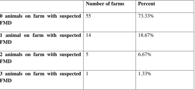
Most farms did not have any animals with suspected FMD, of the farms that had suspected cases one animal was the most common. Only one farm out of the 75 had all 3 animals selected tested for FMD (Table 2).
Table 2: Number of farms having cattle with suspected lesions and tested for foot and mouth disease (FMD) per farm, out of three randomly selected animals in farms close to Maasai Mara, Kenya
Number of farms Percent
0 animals on farm with suspected FMD
55 73.33%
1 animal on farm with suspected FMD
14 18.67%
2 animals on farm with suspected FMD
5 6.67%
3 animals on farm with suspected FMD
12 Questionnaire
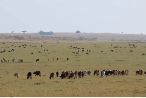
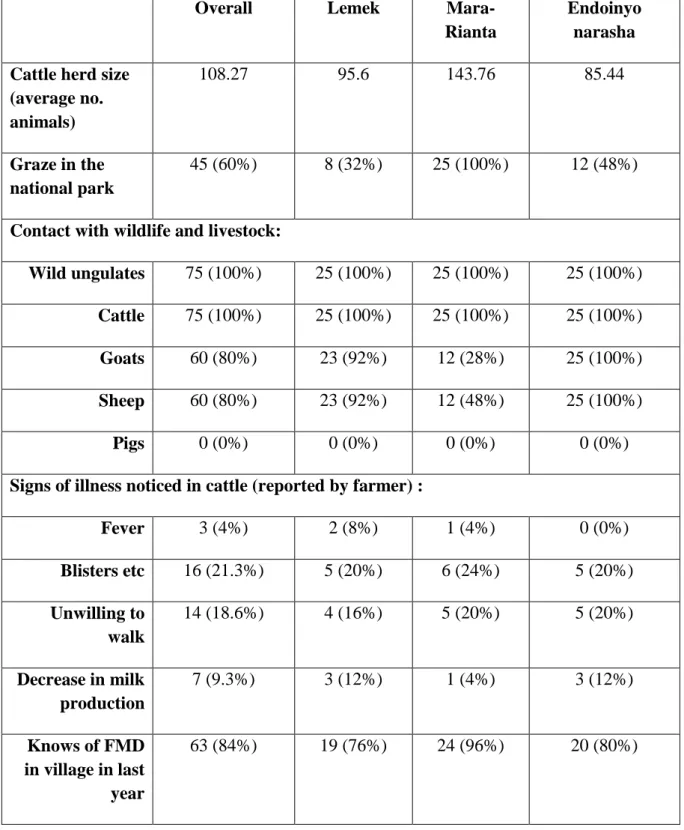
The herds varied in size between 5 and 400 heads of cattle, with an average of 108 animals and a median of 70 animals. The majority, 71 out of 75 herds, mixed with other herds during grazing, and 74 of the herds shared watering points with other herds. Out of the 75 farmers interviewed, 71 of them answered that their cattle grazed with at least one other herd. The number of herds that they grazed with varied between 1 and 200. All herds had direct or indirect contact with wild ungulates (Figure 1). All but one of the farmers saw the interaction with wildlife as something negative, the last one was indifferent. When asked, the majority of the farmers knew of farms in their villages that have had FMD in the last year (Table 3).
Figure 1: Cattle grazing in Mara-Rianta, Kenya, with wildebeest in the background.
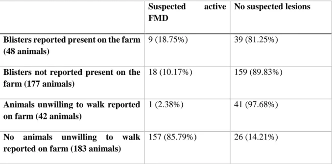
Comparing reported clinical signs and the odds of finding animals with suspected lesions was inconclusive for most clinical signs. The most interesting finding was farmers that had answered yes on having cattle that showed unwillingness to walk and stand was less likely to have cattle with suspected FMD lesions. A chi square test showed significant differences (p=0.03), and the odds ratio was 0.15 (95% confidence interval 0.02-1.12). The other analysis was whether the farmer had seen any blisters in the mouth, on the teats or on the hooves of his cattle. This question gave a p-value of 0.1 with a chi-square test, and the odds ratio was 2.04 (95% confidence interval 0.85-4.88) higher to find a suspected case in farms that reported this. (Table 4).
13
Table 3: Farms reporting possible risk factors for foot and mouth disease in three villages close to Maasai Mara, Kenya
Overall Lemek
Mara-Rianta
Endoinyo narasha Cattle herd size
(average no. animals) 108.27 95.6 143.76 85.44 Graze in the national park 45 (60%) 8 (32%) 25 (100%) 12 (48%)
Contact with wildlife and livestock:
Wild ungulates 75 (100%) 25 (100%) 25 (100%) 25 (100%)
Cattle 75 (100%) 25 (100%) 25 (100%) 25 (100%)
Goats 60 (80%) 23 (92%) 12 (28%) 25 (100%)
Sheep 60 (80%) 23 (92%) 12 (48%) 25 (100%)
Pigs 0 (0%) 0 (0%) 0 (0%) 0 (0%)
Signs of illness noticed in cattle (reported by farmer) :
Fever 3 (4%) 2 (8%) 1 (4%) 0 (0%) Blisters etc 16 (21.3%) 5 (20%) 6 (24%) 5 (20%) Unwilling to walk 14 (18.6%) 4 (16%) 5 (20%) 5 (20%) Decrease in milk production 7 (9.3%) 3 (12%) 1 (4%) 3 (12%) Knows of FMD in village in last year 63 (84%) 19 (76%) 24 (96%) 20 (80%)
14
Table 4: Number of animals per farms in two different groups with suspected FMD
Suspected active FMD
No suspected lesions
Blisters reported present on the farm (48 animals)
9 (18.75%) 39 (81.25%)
Blisters not reported present on the farm (177 animals)
18 (10.17%) 159 (89.83%)
Animals unwilling to walk reported on farm (42 animals)
1 (2.38%) 41 (97.68%)
No animals unwilling to walk reported on farm (183 animals)
157 (85.79%) 26 (14.21%)
DISCUSSION
A lower incidence of clinical disease than originally suspected was found. Since foot and mouth disease is considered a major problem in the area, we expected that we would find a large number of active infections. This study however, did not find more than one confirmed active case. Paton et al (2009) concludes that the modern breeds of cattle are more sensitive to infection with FMDV and therefor more likely than the native African species to get the disease. This is a possible explanation for the low occurrence rate of FMD in the area surrounding the Maasai Mara. The cattle that the farmers keep is mainly Zebu, a Bos indicus breed, which has been showed to be less sensitive than the modern high producing breeds of Europe.
Previous study in East Africa have studied occurrence of antibodies. One earlier study found that the seroprevalence of cattle in Narok county, where also this study took place, was 90.4%, and that the seroprevalence for FMD in the whole of Kenya was 52.5%, and the same study found that the likelihood of finding a sero-positive animal increased with age (Kibore et al., 2013). In the Somali Eco-system (south of the border to Somalia) another study found that the seroprevalence of FMD was 45.3% (Chepkwony et al., 2012). Unfortunately, no study using a lateral flow device in the same way this study did could be found for a comparison.
When taking the results of this study into consideration a trend was clearly visible. If a farmer has reported having cattle that have been unwilling to walk/stand it is less likely to find animals with suspected foot and mouth disease. However, if the farmer has reported seeing blisters in the mouth, on the teats or in the hooves it is more likely that we found animals with suspected FMD. It was not possible to investigate the potential risk factor; “contact with wild ungulates” and suspected infection with FMDV, this because every farmer reported contact with wild
15
ungulates. A study found that the density of cattle was positively correlated to the incidence of disease (Hegde et al., 2014), however in our study significant differences between the villages could not be found.
Several of the farms visited stated that they have had animals with blisters in their mouths and in the hooves. This was found to be correlated to the likelihood of finding an animal with lesions consistent with FMD. This is something that was expected. Generally, one would suspect that a farm that reports having seen blisters on their cattle would suffer from some sort of vesicular disease, and being in the area where FMD is common, FMD would be suspected. A retrospective study in India found evidence of cyclic behaviour of the disease, it seems to peak every 2-3 years. The authors propose two possible reasons for this, one being natural immunity that lasts about 2-3 years after infection. The other one being that a new generation susceptible animals being infected (Hegde et al., 2014). This is something that needs to be considered in the case of FMD in Kenya as well. Since the majority of the farmers have reported seeing clinical signs consisted with FMD in their cattle, as well as the fact that they claim that neighbouring farms have had FMD in the last year, this could indicate common occurrence of disease, which is consistent with the high seroprevalence observed by other authors (Kibore et al., 2013) and also consistent with our finding of low occurrence of clinical cases in animals above one year. To further investigate the possibility of FMD being cyclic, more research needs to be conducted.
On the other hand, the question if the farmer has had any animals showing unwillingness to walk or stand was shown to be a bad indicator for finding animals with suspected FMD lesions. A reason for this could be that since the cattle are generally not handled frequently, unless they are lactating, it would be hard for the farmer to find the signs of acute FMD infection that is only visible for a couple of days. Another possibility is that since many of the farmers have a large number of cattle it is easier to notice an animal that is lying down and not able to keep up with the herd. In addition, there can be many different reasons for lameness and unwillingness to walk, which may make it a poor indicator of FMD.
Since foot and mouth disease is a severe illness and can have major consequences for the village and farmer, proactive measures should be implied. To vaccinate all the animals in the area against all the serotypes in circulation in the Maasai Mara and surrounding villages is probably not a viable solution. The sheer amount of animals in the area and the way in which they are kept presents problems in any attempt to vaccinate the population. As long as the cattle walk long distances from their bomas to graze, and cross paths with other herds, infection cannot be stopped. This is due partly to the viruses’ ability to survive in nature for several weeks but also to the lack of barrier between different herds as well as the lack of isolation of diseased animals or infected farms. This leads to a never-ending cycle of infection, with animals that have just undergone infection being immune to that same serotype for a time, but still being able to transmit the virus through manure and mud in the hooves. This will keep the infection cycle going, infecting susceptible animals and the infected animals secreting high concentrations of virus in their body fluids. The study made in 2013 by Kibore et al. supports this theory. The
16
high number of sero-positive animals in Narok County is proof that FMD exists in the area, and that it is common, even though we did not find more than one active case in this study, possibly because of high levels of immunity among adult animals.
Vosloo et al (2002) found that two outbreaks in South Africa most likely originated from contact with infected buffalo. That is easier to suspect in South Africa due to the fact that FMD is not endemic in livestock. In Kenya, and especially in the area around the Maasai Mara, wildlife and livestock frequently mix, making it more suitable to consider it one epidemiological unit. In South Africa, the national parks are fenced, making contact between wildlife and livestock less common than it is in Kenya. This study could not evaluate any correlation between the contact with wild ungulates and the prevalence of foot and mouth disease, since all the farmers included in the study answered that the see wild ungulates around their cattle. Another possible risk factor, that did not yield any significant results, are whether the cattle graze in the national park or not. This is thought to be a possible way of keeping the infection cycle going between wildlife and livestock. The African buffalo is often given a lot of the blame for infecting cattle, however this study found no significant results to ether confirm or dismiss that theory.
The study had several limitations. The test kit used only detects active infections, and only three animals per farmer was tested, no matter how many animals the farmer owned. The planned random selection process had to be altered to the animals that could be caught safely, for both the animals and the handlers. This means that no big bulls were included in the sampling, nor was any highly pregnant cows or any animals showing signs of severe aggression. It would have been better to have a weighted random selection, sampling a certain percentage of the animals in all the farms, instead of a fixed number of 3 animals per farm. However, due to the time constraints and circumstances of this study this was not practically possible. It is likely that the low number of animals per farm caused us to miss some active infections. In addition, the restriction to animals above one year of age can also have made the sampling biased due to the risk of a clinical infection already may have passed the small window where this test can detect it, as well as the finding by Kibore et al. (2013) that sero-positivity increased with age. Another possible bias is that the questions in the questionnaire were translated from English into either Swahili or Maasai (Maa), and the interview performed by two different people. This makes it a possibility for either the questions to be posed differently, or the understanding of the farmer to differ depending on language and person during the interview. Also, there is the possibility of false statements in the interview.
The total number of animals with suspected FMD lesions that were sampled is approximately 12% out of the examined animals. Of these one was found to be positive. This low rate of positive animals is likely due to the limitations of the test with only being able to detect new infections. Another possible bias is that during the validation of the monoclonal antibody used in the LFD, the sensitivity and specificity of SAT serotypes was lower than the other serotypes. In East Africa, six out of the seven serotypes are endemic, only excluding the Asia 1 serotype (Food and Agricultural Organisation, 2004). It makes it a possibility that some of the negative
17
samples in fact could have been false negatives of the SAT2 serotype. It would have been possible to target animals with suspected FMD, but then this study would have been a disease investigation rather than a possibility to study the prevalence of active FMD in the region. Even though we during the sampling had several animals showing signs of painful lesions in their mouths and hooves, with clinical signs like drooling, lameness and unwillingness to eat, they were included in the study on the same selection criteria as all the other animals, and with the same chance of being selected. However, it is not possible to say, that it was in fact FMD these cattle suffered from, since this study did not target all animals, but only tested a random selection. Since FMD is not clinically distinguishable from other vesicular disease, like vesicular stomatitis, one would have to test for these diseases as well and rule them out before claiming that the cattle did have FMD. Taking all these other possibilities into consideration the data collected still leads to the belief that FMD is common in the investigated area and therefore due to its contagious status is likely to be circulating in the area.
Another likely possibility is that the high seroprevalence of FMD in Narok county is a sign that some of the animals could have a partial immunity towards FMD from a recent infection. This makes them less likely to develop clinical FMD from the same serotype in the near future. In order to find the clinical cases of FMD it is possible that young calves, under 1 year of age, are the best group to target. In one of the farms in Endoinyo Narasha we were in fact presented with an 8-month old male calf that the farmer suspected were infected with FMDV. After having done the sampling for the study this calf was examined. He had lesions that were consistent with FMD, and a test was done on swabs from lesions in the interdigital space. The test turned out positive. None of the 3 cattle that were a part of the study had any lesions that were consistent with FMD. Taking into consideration that this one calf had clinical FMD it is safe to assume that FMDV circulates in the herd. This was the scenario in several farms, the farmer said that they have had animals with FMD in the last year or so, but the random selection of three animals for us to test did not yield any clinically ill animals or the occasional animal with suspected old lesions that did not yield a positive result.
Though this study was not able to investigate the possible connection between wild ungulates and livestock infection with FMDV it cannot be ruled out. A connection between the finding of farmers reporting animals with blisters in their mouths, on snouts or in hooves and suspected FMD is clear. The reversed correlation between farmers reporting animals being unwilling to walk or stand and the presence of animals with suspected FMD is also clear, and quite surprising. This leads to the conclusion that to find farms with suspected FMD in their herds one should ask if the farmer have notices any animals with blisters, and not if the animals have been unwilling to follow along with the herd.
Controlling the infection is going to pose a great challenge to the animal health workers and government in Kenya. The high seroprevalence of the disease is a sign that the problem is extensive and that more research in the area is desperately needed to be able to find a way to combat this disease, if that is even possible.
18 CONCLUSION
As a result, this study can confirm that FMD is present in the livestock that graze inside the national park, but the prevalence of active infections seems low and we cannot draw any conclusion as to which serotype it is since that has not been tested. We cannot say for certain that the other suspected animals which were negative for FMD actually have had the disease and not another vesicular disease. From the questionnaires, we can see that the farmers are well aware of the presence of FMD and that a clear majority of them ether have had it in their herds or know of a neighbour that has had it in his herd in the last year. This leads to the conclusion that FMDV is a serious source of sickness in cattle in this area of Kenya, and that further investigation is needed to determine from where it originates and how to best control it.
19 REFERENSES
Alexandersen, S., Zhang, Z., Donaldson, A.., Garland, A.J.., 2003. The Pathogenesis and Diagnosis of Foot-and-Mouth Disease. J. Comp. Pathol. 129, 1–36.
Ayebazibwe, C., Mwiine, F.N., Tjørnehøj, K., Balinda, S.N., Muwanika, V.B., Okurut, A.R.A., Belsham, G.J., Normann, P., Siegismund, H.R., Alexandersen, S., 2010. The role of African buffalos (Syncerus caffer) in the maintenance of foot-and-mouth disease in Uganda. BMC Vet. Res. 6, 1.
Chepkwony, E.C., Giato, C.G., Muchemi, G.M., 2012. Seroprevalence of Foot and Mouth Disease in the Somali Eco-system in Kenya. International Journal of Animal and Veterinary Advances 4(3), 198-203, 2012.07.15
Ferris, N.P., Nordengrahn, A., Hutchings, G.H., Reid, S.M., King, D.P., Ebert, K., Paton, D.J., Kristersson, T., Brocchi, E., Grazioli, S., Merza, M., 2009. Development and laboratory validation of a lateral flow device for the detection of foot-and-mouth disease virus in clinical samples. J. Virol. Methods 155, 10–17.
Gelaye, E., Ayelet, G., Abera, T., Asmare, K., 2009. Seroprevalence of foot and mouth disease in Bench Maji zone, Southwestern Ethiopia. J. Vet. Med. Anim. Health 1, 5–10.
Grubman, M.J., Baxt, B., 2004. Foot-and-Mouth Disease. Clin. Microbiol. Rev. 17, 465–493. doi:10.1128/CMR.17.2.465-493.2004
Hegde, R., Gomes, A.R., Giridhar, P., Kowalli, S., Shivashankar, B.P., Sudharshana, K.J., Nagaraj, K., Sesharao, R., Mallinath, K.C., Shankar, B.P., Nagaraj, D., Seema, C.M., Khan, T.A., Nagaraj, G.V., Srikala, K., Dharanesh, N.K., Venkatesha, M.D., Renukaprasad, C., 2014. Epidemiology of foot and mouth disease in Karnataka state, India: a retrospective study. VirusDisease 25, 504– 509.
Jibat, T., Admassu, B., Rufael, T., Baumann, M.P., Pötzsch, C.J., 2013. Impacts of foot-and-mouth disease on livelihoods in the Borena Plateau of Ethiopia. Pastor. Res. Policy Pract. 3, 1. Kibore, B., Gitao, C.G., Sangula, A., Kitala, P., 2013. Foot and mouth disease sero-prevalence in cattle
in Kenya. J. Vet. Med. Anim. Health 5, 262–268.
Knight-Jones, T.J.D., McLaws, M., Rushton, J., 2016. Foot-and-Mouth Disease Impact on Smallholders - What Do We Know, What Don’t We Know and How Can We Find Out More? Transbound. Emerg. Dis.
Knight-Jones, T.J.D., Rushton, J., 2013. The economic impacts of foot and mouth disease – What are they, how big are they and where do they occur? Prev. Vet. Med. 112, 161–173. doi:10.1016/j.prevetmed.2013.07.013
Lyons, N.A., Alexander, N., Stärk, K.D., Dulu, T.D., Sumption, K.J., James, A.D., Rushton, J., Fine, P.E., 2015. Impact of foot-and-mouth disease on milk production on a large-scale dairy farm in Kenya. Prev. Vet. Med. 120, 177–186.
20
Parthiban, A.B.R., Mahapatra, M., Gubbins, S., Parida, S., 2015. Virus excretion from foot-and-mouth disease virus carrier cattle and their potential role in causing new outbreaks. PloS One 10, e0128815.
Paton, D.J., Sumption, K.J., Charleston, B., 2009. Options for control of foot-and-mouth disease: knowledge, capability and policy. Philos. Trans. R. Soc. B Biol. Sci. 364, 2657–2667.
Perry, B., Grace, D., 2009. The impacts of livestock diseases and their control on growth and development processes that are pro-poor. Philos. Trans. R. Soc. B Biol. Sci. 364, 2643–2655. Prempeh, H., Smith, R., Muller, B., 2001. Foot and mouth disease: the human consequences. Br. Med.
J. 322, 565–565.
Sahle, M., Dwarka, R.M., Venter, E.H., Vosloo, W., 2007. Study of the genetic heterogeneity of SAT-2 foot-and-mouth disease virus in sub-Saharan Africa with specific focus on East Africa. Onderstepoort J Vet Res 74, 289–299.
Sajid, M., Kawde, A.-N., Daud, M., 2015. Designs, formats and applications of lateral flow assay: A literature review. J. Saudi Chem. Soc. 19, 689–705.
Stenfeldt, C., Eschbaumer, M., Pacheco, J.M., Rekant, S.I., Rodriguez, L.L., Arzt, J., 2015. Pathogenesis of Primary Foot-and-Mouth Disease Virus Infection in the Nasopharynx of Vaccinated and Non-Vaccinated Cattle. PLOS ONE 10, e0143666.
SVA, Mul- och klövsjuka, http://www.sva.se/djurhalsa/epizootier/mul-och-klvsjuka, [19/09-2016]
SVA, Vesikulär stomatit, (2016-02-23) http://www.sva.se/djurhalsa/epizootier/vesikular-stomatit , [2016-11-08]
Epiwebb, Mul- och klövsjuka, differentialdiagnoser (2016-12-28) http://epiwebb.se/sjukdomar/mul-och-klovsjuka-mkfmd/differentialdiagnoser/ [2016-11-24]
Manual number: 19-4100-13/06, Boehringer Ingelheim Phone +46 18 65 49 00 Svanova, www.svanova.com
Food and Agricultural Organisation
http://www.fao.org/ag/againfo/commissions/docs/greece04/App22.pdf [08/10-2016]
Vonsloo et al. Annual new york acadamy of sience, 969, 187-190, (2002) Molecular epidemiological studies of Foot-and-Mouth disease virus in sub-Saharan Africa indicate the presence of large numbers of topotypes: implications for local and international control
Foot and mouth disase, Thomson and Bastos, 2004 pp 1325-1365 Infectious diseases of livestock with special reference to southern Africa, pages: 825-852
Oxford University Press Southern Africa Ministry of Agriculture, Fisheries and Food, Her Majesty’s Stationery Office, London, 1986
Terrestrial manual, Foot and mouth disease (infection with foot and mouth disease virus) Chapter 2.1.8, OIE, 2012