Inside the Lactating Breast: The Latest Anatomy Research
Donna T. Geddes, PhD
Although it is well recognized that a thorough understanding of the anatomy of an organ is essential to enable assessment of any abnormalities in that organ, there has been little investigation of the anatomy of the normal lactating breast since Sir Astley Cooper performed detailed dissections of the anatomy of the breast more than 160 years ago. Many mothers recognize that breast milk provides the ultimate nutrition and protection for the infant; however, a significant proportion of women experience difficulties breastfeeding, some of which lead to weaning the infant. Recently, a small number of studies have focused on the gross anatomy of the breast, and have found that the ductal system is comprised of fewer numbers of main ducts than previously thought. In addition, the ducts are compressible and do not contain large amounts of milk, the amount of fatty tissue in the breast is variable, and a proportion is situated within the glandular tissue. These findings add to our understanding of both the physiology and pathology of the lactating breast. J Midwifery Womens Health 2007;52:556 –563 © 2007 by the American College of Nurse-Midwives.
keywords:anatomy, breast, breastfeeding, lactation, mammary gland
INTRODUCTION
The human breast reaches its full functional capacity during lactation with the production of breast milk. In order to diagnose and treat breastfeeding problems and pathologies that arise during lactation, it is essential to have an extensive understanding of the normal anatomy and physiology of the breast. This review details the development of the breast and the most recent findings in breast anatomy. The effect breast anatomy has upon clinical practice as well as the importance of milk ejection is reviewed.
DEVELOPMENT OF THE BREAST
While it is undisputed that breast milk provides the optimal nutrition for the developing infant, breast milk also contains unique protective factors for the mother.1It
has been hypothesized that the mammary gland first evolved from the innate immune system as an inflamma-tory response to provide protection to the young, and that nutritional factors developed later.2,3 To date, nutrition
has assumed a position of dominance over the protective factors in considerations of the physiology of human lactation.
The human breast is a dynamic organ that does not go through all developmental stages unless a woman expe-riences pregnancy and childbirth. The course of breast development can be described in distinct phases begin-ning with the fetal phase and progressing through the neonatal/prepubertal and postpubertal phases. Develop-ment of the breast can then proceed through a number of lactation cycles (pregnancy, lactogenesis I, lactogenesis II, and involution; (see review by Kent on page 564).
BREAST DEVELOPMENT
Fetal Development
The human breast develops from a thickened ectodermal ridge (milk line) situated longitudinally along the ante-rior body wall from the groin to the axilla at about 6 weeks’ gestation. Regression of the ridge occurs except for the pectoral region (2nd– 6th rib), which forms the mammary gland. Supernumerary glands may develop anywhere along the ectodermal ridges, and in 2% to 6% of women, these glands either mature into mammary glands or remain as accessory nipples.4,5
During the 7th and 8th weeks of gestation, the mam-mary parenchyma invades the stroma, which appears as a raised portion called the mammary disc. Between the 10th and 12th weeks, epithelial buds form; parenchymal branching occurs during the 13th through 20th weeks. Between the 12th and 16th weeks of gestation, the smooth musculature of the areola and nipple are formed, and at approximately 20 weeks’ gestation, between 15 and 256 solid cords form in the subcutaneous tissue.
Branching continues, and canalization of the cords oc-curs, forming the primary milk ducts by 32 weeks’ gestation.6At 32 weeks’ gestation the ducts open onto the
area, which develops into the nipple.7The adipose tissue of
the mammary gland develops from connective tissue that has lost its capacity to form fibres, and it is considered necessary to further growth of the parenchyma.4
Shortly after birth, colostrum can be expressed from the infant’s mammary glands. This is attributed to the pro-lactation hormones present in the fetal circulation at birth. Regression of the mammary gland usually occurs by 4 weeks postpartum and coincides with a decrease in the secretion of prolactin from the anterior pituitary gland of the infant.4
Neonatal and Prepubertal Development
The ducts in the newborn breast are rudimentary and have small, club-like ends that regress soon after birth. Address correspondence to Donna T. Geddes, PhD, Department of
Bio-chemistry and Molecular Biology, School of Biomedical, Biomolecular and Chemical Sciences, Faculty of Life and Physical Sciences, M310 –The University of Western Australia, 35 Stirling Highway, Crawley WA 6009 Australia. E-mail: [email protected]
Before puberty, the growth of the breast is isometric. Allometric growth of both the stroma and epithelium begins with the onset of puberty (8 –12 years of age). Although the impact of obesity on breast development at this stage is unknown, it is of interest that ruminants fed a diet that is higher than their energy requirements have impaired mammary development and subsequent im-paired lactation performance.8,9
Puberty
At puberty, the increase in breast size is mainly caused by the increased deposition of adipose tissue within the gland. However, progressive elongation and branching of the ducts creates a more extensive ductal network.10The
major site of growth is the bud-like structures at the end of the ducts, and these form the terminal duct lobular units or acini.11 Although knowledge of the hormonal
regulation of mammary growth during puberty is not extensive, these maturational changes are associated with increased plasma concentrations of oestrogen, prolactin, luteinizing hormone, follicle stimulating hormone, and growth hormone.12,13
Menstrual Cycle Changes
During the follicular phase of the menstrual cycle, the lobules are small, with few alveoli, and there is low mitotic activity. During the luteal phase, the lobules and alveoli develop with open lumens and mitotic activity is at its greatest.14 From day 27 to menstruation, these
changes regress. However, the degeneration of the epi-thelial growth is not complete,15and some of the
follic-ular growth remains until the next cycle. With increasing years, there is a relative decrease in mitotic activity until about 35 years of age, when breast development pla-teaus.4
GROSS ANATOMY OF THE NON-LACTATING BREAST
For the past 160 years, the descriptions of the anatomy of the breast have changed little since Sir Astley Cooper’s16
meticulous dissections of breasts of women who were lactating when they died (Figure 1).
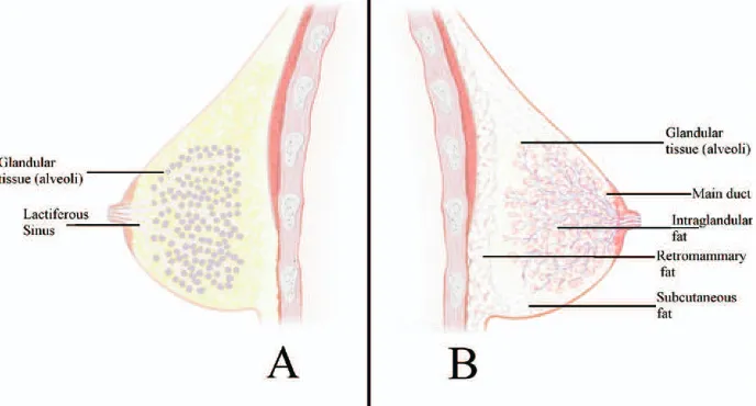
The breast is composed of glandular (secretory) and adipose (fatty) tissue, and is supported by a loose framework of fibrous connective tissue called Cooper’s ligaments. Traditional descriptions of breast anatomy describe the glandular tissue as consisting of 15 to 20 lobes that are comprised of lobules containing between 10 and 100 alveoli that are approximately 0.12 mm in diameter17(Figure 2). The size of each lobe is extremely
variable, and some lobes may differ by 20-to 30-fold.18
Although it is generally thought that each lobe is a single entity, a recent study that created three-dimensional reconstructions of the entire ductal system (16 lobes) of a mastectomized breast of a 69-year-old female was able to demonstrate two connections between lobes.19 It is
generally believed that 15 to 25 ducts drain the alveoli and merge into larger ducts that eventually converge into one main milk duct which dilates slightly to form the lactiferous sinus before narrowing as it passes through the nipple and opens onto the nipple surface (Figure 2). Recent histologic sections of mastectomy nipples have shown more than 17 ducts on average18,20; however, it is
not known whether these are all patent, and others suggest the average number of ducts is lower (5–9).21
The diameters of the main ducts in the non-lactating breast as measured by ultrasound are between 1.2 mm and 2.5 mm in diameter. Dilated ducts in the non-lactating breast may be caused by conditions such as polycystic ovarian disease22or ductal ectasia. The nipple
pores are 0.4 mm to 0.7 mm in diameter and are surrounded by circular muscle fibres.4,5
The heterogeneous distribution of glandular and adi-pose tissue in the breast has hindered measurement of these tissues. However, the ratio of glandular to adipose tissue estimated by mammography is 1:1 on average, and it is well documented that the proportion of glandular tissue declines with both advancing age23and increasing
breast size.24
Arterial Supply
The blood supply to the breast is provided mainly by the anterior and posterior medial branches of the internal Donna T. Geddes, PhD, is a research associate in the School of Biomedical,
Biomolecular and Chemical Sciences at The University of Western Australia.
Figure 1. Artist’s impression of the ductal system of the human lactating breast. The ductal system was injected with coloured wax and dissected. (Reproduced from Cooper.16
mammary artery (60%) and the lateral mammary branch of the lateral thoracic artery (30%).25Smaller sources of
arterial blood include the posterior intercostal arteries and the pectoral branch of the thoracoacromial artery.5
There is wide variation in the proportion of blood supplied by each artery between women,26 and little
evidence of symmetry between breasts. Moreover, the course of the arteries does not appear to be associated with the ductal system of the breast.4
Venous Drainage
The venous drainage of the breast is divided into the deep and superficial systems which are joined by short con-necting veins. Both systems drain into the internal thoracic, axillary, and cephalic veins. The deep veins are assumed to follow the corresponding mammary arteries, while the superficial plexus consists of subareolar veins that arise radially from the nipple and drain into the periareolar vein, which circles the nipple and connects the superficial and deep plexus. Symmetry of the super-ficial venous plexus is not apparent.25
Innervation
Cooper16 showed that the 2nd to 6th intercostal nerves
supply the breast. The distribution and course of these nerves are complex and variable. The anterior nerves take a superficial course in the subcutaneous tissues, while the lateral nerves travel a deep course through the breast. The nipple and areola are supplied by the anterior and lateral cutaneous branches of the 3rd to 5th intercos-tal nerves most commonly the 4th intercosintercos-tal nerve.27
Lymphatic Drainage
The drainage of lymph from the breast has been exten-sively investigated with particular reference to breast carcinoma, and there are two main pathways by which lymph is drained from the breast. The first is to the axillary nodes; the second is to the internal mammary nodes. The majority of the lymph from both the medial and lateral portions of the breast is drained to the axillary nodes (75%), whereas the internal mammary nodes receive lymph from the deep portion of the breast. However, as expected, there is a wide variation in the drainage of lymph from the breast, and less common pathways have been demonstrated.5
PREGNANCY
During the first half of pregnancy, extension and branch-ing of the ductal system occurs, along with intensified lobular–alveolar growth (mammogenesis). Growth of the mammary gland is influenced by a number of hormones, including oestrogen, progesterone, prolactin, growth hor-mone, epidermal growth factor, fibroblast growth factor, insulin-like growth factor,28,29 and parathyroid hormone–
related protein.30 Growth of the glandular tissue is
be-lieved to occur by invasion of the adipose tissue.4 By
mid-pregnancy, there is some secretory development, with colostrum present in the alveoli and milk ducts. In the last trimester, there is a further increase in lobular size.
While these changes typically lead to a marked in-crease in breast size during pregnancy, the proportion of growth varies greatly between women, ranging from little or no increase to a considerable increase in size. Figure 2. A, Traditional schematic diagram of the anatomy of the breast. The main milk ducts below the nipple are depicted as dilated portions or lactiferous sinuses, and the glandular tissue is deeper within the breast.B, Schematic diagram of the ductal anatomy of the breast based on the findings of Ramsay et al.38
While the major increase in breast size is usually com-pleted by week 22 of pregnancy, significant breast growth occurs during the last trimester of pregnancy in some women, and some women undergo significant breast growth postpartum. At the end of pregnancy, the volume of breast tissue had increased by 145 ⫾ 19 ml (mean ⫾ standard error of the mean; n ⫽ 13; range, 12–227 ml), with a further increase to 211⫾16 ml (n⫽
12; range, 129 –320 ml) by 1 month of lactation. The rate of growth of the mother’s breast during pregnancy is correlated with the increase in the concentration of human placental lactogen in the mother’s blood, which suggests that this hormone stimulates breast growth in women.31
During pregnancy, mammary blood flow approxi-mately doubles in volume. This increased blood flow is concomitant with both the increased metabolic activity and temperature of the breast. This elevation in blood flow persists during lactation and appears to decline to prepregnancy levels about 2 weeks after weaning.32
GROSS ANATOMY OF THE LACTATING BREAST
The breast reaches its full functional capacity at lactation, and as a result, several internal and external changes occur. During pregnancy, the areola darkens in colour, and the Montgomery glands, which are a combination of sebaceous glands and mammary milk glands, increase in size. The secretions of these glands, which number between 1 and 15,33 are thought to provide maternal
protection from both the mechanical stress of sucking and pathogenic invasion. In addition, it is also suspected the secretion may act as a means of communication with the infant via odor. In this connection, a recent study demonstrated that increased numbers of Montgomery glands is associated with increased infant weight gain in the first 3 days after birth, infant breastfeeding behaviour (increased latching speed and sucking activity), and decreased time to onset of lactation in primiparous mothers,33 suggesting that there is indeed a functional
role of the Montgomery glands during lactation. The standard descriptions of the human breast are based on Cooper’s16 magnificent cadaver dissections of
the breasts of women who were lactating at the time of death. Although imaging modalities have become more sophisticated, research has focused extensively on abnor-malities of the non-lactating breast. Mammography of the lactating breast is limited because of the increase in glandular tissue and the secretion of breast milk, which causes an increase in radiodensity, making images of the breast difficult to interpret.34
Galactography (the injection of radio-opaque contrast media into the duct orifice at the nipple and subsequent radiography) has illustrated only a portion of the ductal system, and few studies have examined lactating women. Of those that have looked at lactating breasts, some have
described the milk ducts as being significantly larger compared to those of the non-lactating breast. In contrast, Cardenosa and Eklund35have reported that the ducts do
not enlarge during lactation.
To date, both computed tomography and magnetic resonance imaging have had little to offer in elucidating mammary anatomy. However, in two recent studies that used magnetic resonance imaging to image the breast, one study was able to identify some central ducts in the breasts of lactating women,36 and another attempted to
quantify fatty and glandular tissue volumes in the breasts of non-lactating women.37 These findings suggest that
this modality may offer some new insights into the anatomy of the breast in future.
Our laboratory has recently reinvestigated the anatomy of the lactating breast using high-resolution ultrasound.38
Ultrasound is non-invasive and allows the structures of the breast to be examined without distortion. Compared with the quoted 15 to 25 ducts of conventional texts,4,5
fewer ducts were imaged with ultrasound (mean, 9; range, 4 – 8), which concurs with both Love and Bar-sky’s21observations of lactating women expressing milk
with a breast pump (mean, 5; range, 1–17) and Going and Moffatt’s39dissection of a nipple (4 patent ducts) from a
woman who was lactating. Interestingly, these are in agreement with Cooper,16who found 7 to 12 patent ducts in cadaver dissections of breast from a woman who was lactating before death, although he could cannulate up to 22 ducts.
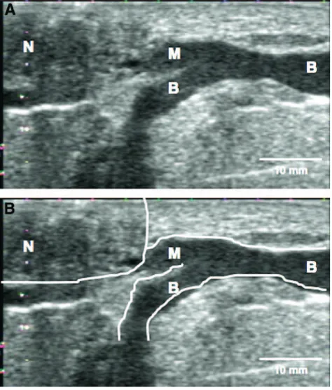
Ultrasound imaging has also elucidated other charac-teristics of the milk ducts, in that they are small (mean, 2 mm), superficial, and easily compressed. In addition, they do not display the typical sac like appearance of the “lactiferous sinus” originally thought to exist (Figure 3). Instead, branches drain glandular tissue located directly beneath the nipple and often merge into the main collecting duct very close to the nipple38 (Figure 3).
Furthermore, the milk ducts increase in diameter at milk ejection,40leading to the conclusion that it is likely that
the main function of the ducts is the transport rather than storage of milk. In addition, the actual course of the ducts from the nipple into the breast is erratic, and they are intertwined much like the roots of a tree38 (Figure 1),
making them difficult to separate surgically.5
either the storage capacity of the breast or milk produc-tion has been demonstrated.38
Nerve fibres have been identified in association with the major duct system in the lactating breast; however, they are sparse in the region of the smaller ducts, areola, and nipple.41 Although these nerves are sensory, apart
from a marked increase in areola and nipple sensitivity within the first 24 hours postpartum,42women tend not to
be particularly sensitive to breast changes associated with some abnormal conditions. For example, women with mastitis often experience influenza-like symptoms before becoming aware of breast tenderness or the presence of a blocked duct. In addition, the absence of motor innervation of the lactocyte and glandular tissue further supports that the production of milk is indepen-dent of neural stimulation.43It should be noted, however,
that as with the gross anatomy of the breast, the nerve supply of the breast has not been extensively investigated recently.
These new findings with regard to breast anatomy have several clinical implications (Table 1). For exam-ple, abnormalities of the lactating breast have not been extensively investigated in comparison to the pathologi-cal, non-lactating breast. Although ultrasound imaging is only semi-quantitative and can be subjective, it may provide information on the proportion of glandular tissue
in mothers with very low milk supply, breast hypoplasia (too little glandular tissue), or hyperplasia (overgrowth of glandular tissue). With rising rates of obesity, there is some concern about the effect of obesity on lactation, particularly with increasing reports that obese women are experiencing breastfeeding difficulties (see review by Jevitt et al. on page 606). Only a few studies have been carried out to investigate the effect of obesity on lacta-tion, and they have been difficult to perform because of known confounding factors such as the mode of delivery and parity. These studies show that pregnant women with a high body mass index are more likely to experience delayed lactogenesis II.44 While the cause of delayed
lactation is not clear, hormonal influences on milk production, increased difficulty attaining a successful infant latch to the breast, and socio-cultural factors have been suggested.45
Knowledge of the normal features of the ductal system is integral to diagnosing ductal abnormalities such as galactoceles and blocked ducts. A palpable lump and ultrasonic features of non-compressible ducts is indica-tive of a blocked duct and should not be considered “normal” for the lactating breast. Furthermore, the ultra-sound scan may identify the level of the blockage, providing useful information for treatment with thera-peutic ultrasound.
Mothers of premature and sick infants rely on breast pumps to initiate and maintain lactation. Clinically, it has been observed that larger shield sizes may optimize milk removal for some mothers. It is therefore feasible that compression of superficial ducts within the breast by the shield may indeed compromise milk flow. Further re-search is required to determine the effect of ductal anatomy on pumping performance in women.
Many women who have breast reduction surgery may be able to partially breastfeed their infant, but relatively few are able to exclusively breastfeed.52 This is likely
because of the codistribution of glandular and fatty tissue within both the lactating38 and non-lactating breast,53
making it difficult to preferentially remove fatty tissue. In addition, milk outflow is probably disrupted, because there are fewer numbers of ducts than previously thought.21,38 Furthermore, it is possible that the milk
ejection reflex may be inhibited if the nerve supply to the nipple is disturbed.
The absence of lactiferous sinuses or milk reservoirs leads one to reconsider the mechanism by which the infant removes milk from the breast. Generally, it is believed that the predominant action involved in remov-ing milk from the breast is peristalsis or a strippremov-ing action.54We have found that milk flows into the infant’s
mouth when its tongue is lowered and vacuum is applied to the breast. This finding suggests that the vacuum applied by the breastfeeding infant is a major component of milk removal.55 Indeed, it is evident that correct
positioning and attachment of the infant to the breast is Figure 3. AandB, Ultrasound image of a main milk duct (Toshiba, Aplio).
important for successful breastfeeding; however, the mechanism should be fully understood in order to diag-nose and manage infants with sucking abnormalities. Finally, the absence of the lactiferous sinuses further emphasises the critical nature of milk ejection for suc-cessful breastfeeding, because only small amounts of milk are available before the stimulation of milk ejec-tion.40,46
MILK EJECTION
Milk ejection is critical for successful lactation, because only small volumes of milk (1–10 mL) can be either expressed46 or removed by the breastfeeding infant40
before milk ejection. Failure to remove sufficient quan-tities of milk results in a decrease in milk production because of local control mechanisms.47 Stimulation of
the nipple initiates milk ejection via initiation of nervous impulses to the hypothalamus, which stimulates the posterior pituitary gland to release oxytocin into the bloodstream.48 Oxytocin causes the myoepithelial cells
surrounding the alveoli to contract, forcing milk into the ducts. This results in increased intraductal pressure,49
duct dilation40,50 (measured by ultrasound), and
conse-quently increased milk flow rate50(measured by
contin-uous weigh balance during breast expression). Multiple milk ejections almost always occur during breastfeed-ing40 (mean, 2.5; range, 0 –9) and breast expression50
(mean, 3– 6 for 15-minute expression period), and
al-though many women are able to sense the first milk ejection, few are able to sense subsequent ones.
While it is well known that stress can influence milk ejection—resulting in diminished amounts of milk re-moved by both the infant48 and breast pump51—it is
often the subtle stress which affects maternal confidence and subsequently milk ejection that is overlooked. There-fore, it is important to provide positive support to the mother during both breastfeeding and pumping. Another factor that may influence milk ejection and milk removal is the ductal anatomy of the breast. In a study of mothers expressing with an electric breast pump, ultrasound was used to image duct dilation in the breast that was not pumped. It was found that mothers with larger ducts expressed more milk during milk ejection and had longer milk ejections than mothers with smaller ducts.50
There-fore, the rate of milk removal for a mother may be influenced in part by her ductal anatomy.
CONCLUSION
New anatomy research has shown that the milk ducts of the breast are small, compressible, superficial, and closely intertwined. They do not display typical dilated “sinuses” and do not typically store large amounts of milk. In addition, the amount of adipose tissue in the breast is highly variable, particularly between the glan-dular tissue. This fundamental knowledge of the anatomy of the breast—particularly when it is fully functional— Table 1. Summary of Some of the Common Breastfeeding Conditions and Symptoms and the Connection With Breast Anatomy
Clinical Condition Symptoms Anatomical Relationship
Glandular anomaly
Hypoplasia Low milk production Possible deficiency of glandular tissue
Hyperplasia Excessive breast growth, lymphoderma, possible necrosis
Excess glandular tissue
Breast surgery Reduction
mammoplasty
Low milk production Large volume of glandular tissue removed, severing milk ducts (fewer in number than previously thought); possible nerve damage inhibiting milk ejection reflex
Breast augmentation Low milk production Possible compression of milk ducts by implant Possible deficiency in volume of glandular tissue Palpable mass
Blocked duct Mass (small or large)⫾pain Possible reduction in milk production
Compression of ducts: possible cause of blocked duct; if large lobe affected a significant reduction in milk production may occur; identification of the level of duct obstruction by ultrasound ensures treatment of entire affected area
Galactocele Mass (generally small) Possible ductal abnormality
Benign mass (cyst, fibroadenoma)
Mass Possible compression of ducts causing blocked duct; possible obstruction of milk flow in the area of attachment of the infant to the breast
Malignant mass Palpable non-resolving mass Irregular shaped mass that may be mistaken for a blocked duct or galactocele; ultrasound⫾mammography needed for diagnosis
Infant sucking mechanism
Ineffective suck Lack of milk sinuses and evidence that vacuum plays a major role in milk removal may alter intervention
Milk expression Differences in efficiency of pumping
Theorized that women with large milk ducts or duct dilations at milk ejection express milk quickly
Differences in effectiveness of pumping
Poor shield fit may result in compression of superficial ducts and inhibit milk flow
allows one to begin to understand and the myriad of problems that are experienced by women during lacta-tion. This knowledge will form a foundation for the development of appropriate treatments and interventions.
The author’s salary is funded by Medela AG.
REFERENCES
1. Lawrence RA, Lawrence RM. Breastfeeding: a guide for the medical profession. St Louis, MO: Mosby Inc, 2006.
2. Vorbach C, Capecchi MR, Penninger JM. Evolution of the mammary gland from the innate immune system? Bioessays 2006; 28:606 –16.
3. Offendal OT. The mammary gland and its origin during synapsid evolution. J Mammary Gland Biol Neoplasia 2002;7: 225–52.
4. Vorherr H. The breast: morphology, physiology and lacta-tion. London: Academic Press, 1974.
5. Bannister LH, Berry MM, Collins P, Dyson M, Dussek JE. Gray’s anatomy, 38th ed. New York: Churchill Livingstone, 1995: 417–24.
6. Hovey RC, Trott JF, Vonderhaar BK. Establishing a frame-work for the functional mammary gland: from endocrinology to morphology. J Mammary Gland Biol Neoplasia 2002;7:7–37.
7. Tobon H, Salazar H. Ultrastructure of the human mammary gland. II. Postpartum lactogenesis. J Clin Endocrinol Metab 1975; 40:834 – 44.
8. Sejrsen K, Purup S. Influence of prepubertal feeding level on milk yield potential of dairy heifers: a review. J Anim Sci 1997; 75:828 –35.
9. Shaw MA, Rasmussen KM, Meyers TR. Consumption of high-fat diet impairs reproductive performance in Sprague-Dawley rats. J Nutr 1997;127:64 –9.
10. Russo J, Russo IH. Development of the human mammary gland. In The mammary gland: development, regulation and func-tion. New York: Plenum Press, 1987.
11. Sternlicht MD, Kouros-Mer H, Lu P, Werb Z. Hormonal and local control of mammary branching morphogenesis. Differentia-tion 2006;74:365– 81.
12. Rose SR, Municchi K, Barnes KM, Kamp GA, Uriarte MM, Ross JL, et al. Spontaneous growth hormone secretion increases during puberty in normal girls and boys. J Clin Endocrinol Metab 1991;73:428 –35.
13. Ankarberg-Lindgren C, Elfving M, Wikland KA, Norjavaara E. Nocturnal application of transdermal estradiol patches produces levels of estradiol that mimic those seen at the onset of spontane-ous puberty in girls. J Clin Endocrinol Metab 2001;86:3039 – 44.
14. Potten CS, Watson RJ, Williams GT, Ticle S, Roberts SA, Harris M, et al. The effect of age and the menstrual cycle upon proliferative activity of the normal human breast. Br J Cancer 1988;58:163–70.
15. Longacre TA, Bartwo SA. A correlative morphologic study of human beast and endometrium in the menstrual cycle. Am J Surg Pathol 1986;10:382–93.
16. Cooper AP. Anatomy of the breast. London: Longman, Orme, Green, Browne and Longmans, 1840.
17. Hartmann PE. The breast and breast-feeding. In Scientific foundations of obstetrics and gynaecology, 4th ed. Oxford: But-terworth Heinemann, 1991.
18. Moffatt DF, Going JJ. Three dimensional anatomy of com-plete duct systems in the human breast: pathological and develop-mental implications. J Clin Pathol 1996;49:48 –52.
19. Ohtake T, Kimijima I, Fukushima T, Yasuda M, Sekikawa K, Takenoshita S, et al. Computer assisted complete three-dimen-sional reconstruction of the mammary ductal/lobular systems. Im-plications of ductal anastomoses for breast conserving surgery. Cancer 2001;91:2263–72.
20. Taneri F, Kurukahvecioglu O, Akyurek N, Tekin EH, Ilhan MN, Cifter C, et al. Microanatomy of milk ducts in the nipple. Eur Surg Res 2006;38:545–9.
21. Love SM, Barsky SH. Anatomy of the nipple and breast ducts revisited. Cancer 2004;101:1947–57.
22. Panaritis V, Despotidis P, Kyriadidis A. Diameter of mam-mary terminal ducts as an additional tool in evaluation of women with polycystic ovarium disease. Arch Gynecol Obstet 2004;270: 252– 4.
23. Jamal N, Ng KH, McLean D, Looi LM, Moosa F. Mammo-graphic breast glandularity in malaysian women: data derived from radiography. Am J Roentgenol 2004;182:713–7.
24. Cruz-Korchin N, Korchin L, Gonzalez-Keelan C, Climent C, Morales I. Macromastia. How much of it is fat? Plas Reconstr Surg 2002;109:64 – 8.
25. Cunningham L. The anatomy of the arteries and veins of the breast. J Surg Oncol 1977;9:71– 85.
26. Doughty JC, McCarter DHA, Kane E, Reid AW, Cooke TG, McArdle CS. Anatomical basis of intra-arterial chemotherapy for patients with locally advanced breast cancer. Br J Surg 1996;83: 1128 –30.
27. Schlenz I, Kuzbari R, Gruber H, Holle J. The sensitivity of the nipple-areola complex: an anatomic study. Plast Reconstr Surg 2000;105:905–9.
28. Oka T, Yoshimura M, Lavandero S, Wada K, Ohba Y. Control of growth and differentiation of the mammary gland by growth factors. J Dairy Sci 1991;71:2788 – 800.
29. Kelly PA, Bachelot A, Kedzia C, Hennighausen L, Ormandy CJ, Kopchick JJ, et al. The role of prolactin and growth hormone in mammary development. Mol Cell Endocrinol 2002;197:127–31.
30. Wysolmerski JJ, McCaughern-Carucc JF, Daifotis AJ, Broa-dus AE, Philbrick WM. Overexpression of parathyroid hormone related protein and parathyroid hormone in transgenic mice im-pairs branching morphogenesis during mammary gland develop-ment. Development 1995;121:3530 – 47.
32. Thoresen M, Wesche J. Doppler measurements of changes in human mammary and uterine blood flow during pregnancy and lactation. Obstet Gynecol Scand 1988;67:741–5.
33. Schaal B, Doucet S, Sagot P, Hertling E, Soussignan R. Human breast areolae as scent organs: morphological data and possible involvement in maternal-neonatal coadaptation. Dev Psy-chobiol 2005;48:100 –10.
34. Madjar H. The practice of breast ultrasound. Techniques, findings, differential diagnosis. New York: Thieme, 2000.
35. Cardeonosa G, Eklund GW. Ductography. In Interventional breast procedures. New York: Churchill Livingstone, 1996.
36. Espinosa LA, Dsaniel BL, Vidarsson L, Zakhour M, Ikeda DM, Herfkens RJ. The lactating breast: contrast-enhanced MR imaging of normal tissue and cancer. Radiology 2005;237:429 –36.
37. Lee NA, Rusinek H, Weinreb J, Chandra R, Toth H, Singer C, et al. Fatty and fibroglandular tissue volumes in the breasts of women 20 – 83 years old: comparison of x-ray mammography and computer-assisted MR imaging. Am J Roentgenol 1997;168:501– 6.
38. Ramsay DT, Kent JC, Hartmann RL, Hartmann PE. Anat-omy of the lactating human breast redefined with ultrasound im-aging. J Anat 2005;206:525–34.
39. Going JJ, Moffat D. Escaping from Flatland: clinical and biological aspects of human mammary duct anatomy in three dimensions. J Pathol 2004;203:538 – 44.
40. Ramsay DT, Kent JC, Owens RA, Hartmann PE. Ultrasound imaging of milk ejection in the breast of lactating women. Pedi-atrics 2004;113:361–7.
41. Montagna W, McPherson EE. Some neglected aspects of the anatomy of human breasts. J Invest Dermatol 1974;63:10.
42. Robinson JE, Sort RV. Changes in breast sensitivity at puberty, during the menstrual cycle, and at parturition. Br Med J 1977;1:1188 –91.
43. Hartmann PE, Ramsay DT. Mammary anatomy and physi-ology. In Feeding and nutrition in the preterm infant, London: Elsevier, Churchill Livingstone, 2005:53– 68.
44. Rasmussen KM, Hilson JA, Kjolhede CL. Obesity may impair lactogenesis II. J Nutr Sci 2001;131:3009S–11S.
45. Lovelady CA. Is maternal obesity a cause of poor lactation performance? Nutr Rev 2005;63:352–5.
46. Kent JC, Ramsay DT, Doherty D, Larsson M, Hartmann PE. Response of breasts to different stimulation patterns of an electric breast pump. J Hum Lact 2003;19:179 – 87.
47. Peaker M, Wilde CJ, Knight CH. Local control of the mammary gland. Biochem Soc Symp 1998;63:71–9.
48. Newton M, Newton NR. The let-down reflex in human lactation. Pediatrics 1948;33:698 –704.
49. Alekseev NP, Omel’yanyuk EV, Talalaeva NE. Dynamics of milk-ejection reflexes accompanying continuous rhythmic stimu-lation of the areola-nipple complex of the mammary gland. Russ J Physiol 2000;86:711–9.
50. Ramsay DT, Mitoulas LR, Kent JC, Cregan MD, Doherty DA, Larsson M, et al. Milk flow rates can be used to identify and investigate milk ejection in women expressing breast milk using an electric breast pump. Breastfeeding Med 2006;1:14 –23.
51. Yokoyama Y, Udea T, Irahara M, Aono T. Releases of oxytocin and prolactin during breast massage and suckling in puerperal women. Eur J Obstet Gynecol Reprod Biol 1994;53: 17–20.
52. Marshall DR, Callan PP, Nicholson W. Breastfeeding after reduction mammaplasty. Br J Plast Surg 1994;47:167–9.
53. Nickell WB, Skelton J. Breast fat and fallacies: more than 100 years of anatomical fantasy. J Hum Lact 2005;21:126 –30.
54. Woolridge MW. The anatomy of infant sucking. Midwifery 1986;4:164 –71.
55. Ramsay DT, Mitoulas LR, Kent JC, Hartmann PE. Ultra-sound imaging of the sucking mechanics of the breastfeeding infant. Abstract P53. Presented at the 12th International Confer-ence of the International Society for Research in Human Milk and Lactation (ISRHML). Queens’ College Cambridge, UK, Septem-ber 10 –14, 2004.